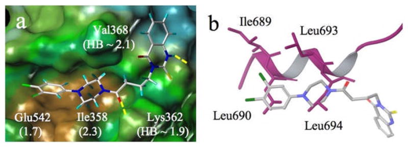
Figure 7.

a) Induced-fit docked pose for 1b at the coactivator binding site of the ERα receptor (PDB X-ray structure code 3ERD). Displacements of Glu542 and Ile358 side chains resulting from docking and H-bond distances are indicated in Å in parentheses; brown = hydrophobic; green = neutral; blue = polar; b) Alignment of 1b (stick) and the coactivator peptide (purple ribbon; 3ERD). Hydrophobic residues Leu690 and Leu694 are matched by ligand hydrophobes.
