Abstract
Purpose
A variety of templates are being used for mandible reconstruction with free flaps. We describe a simple and cost effective method using a ‘K’ wire template for the central segment reconstruction of the mandible with free fibula flap.
Materials and Methods
Over a period of 5 years from January 2005 to December 2009, there were a total of 386 mandible reconstructions with free fibula flaps of which 262 were central segment reconstructions. The number of osteotomies varied from 1 to 2 depending up on the length of the bone reconstructed. ‘V’ shaped closing wedge osteotomies were done and the segments were aligned with mini plates. In all the cases a ‘K’ wire was used in forming a template for the neo mandible from the resected specimen.
Results
The bone union was good in all the cases as determined by the orthopantomograms taken at the third month follow up. The most common complication was prognathism of the neo mandible.
Conclusion
‘K’ wire template is a simple and cost effective device to design the shape of the neo mandible with less osteotomies and align the osteotomised bone segments in an angle that gives a near normal shape.
Keywords: Mandible reconstruction, Central segment, Free fibula flap, ‘K’ wire template
Introduction
The complex oro mandibular defects or en bloc defects occur as a result of resection of T3 and T4 cancers and require reconstruction with composite vascularised free tissue transfer to provide the strength, stability, osseointegration of dental implants and to maintain the contour of the chin. Free fibula flap described by Hidalgo fulfils the above criteria and is the most common composite tissue used to reconstruct the middle third defects of the mandible. Templates are designs or patterns used in re-making of structures and a variety of templates are described in literature for mandible reconstruction with free fibula flaps. We present our experience with ‘K’ wire template which is simple, cost effective, easy to produce and enables to complete the osteotomies and alignment of the bone segments in relatively short time.
Materials and Methods
Three hundred and eighty six reconstruction were done over a period of 5 years from January 2005 to December 2009 of which 262 [164 male and 98 female] were central segment reconstructions. The age of the patients were ranging from 8 to 73 years. The most common tumour was squamous cell carcinoma at T3 or T4 stage. About 2 to 7 cm of bone was used for reconstructing the central segment and the numbers of osteotomies were ranging from 1 to 2 and majority [254] of them required two osteotomies. Miniplates and screws were used for fixing the segments. All the patients with malignant pathology underwent postoperative radiotherapy. Osseo integration of dental implants was done in 10 patients one year after completion of radiotherapy. The period of follow up was ranging from 3 months to 2 years. The post irradiation complications such as plate exposure, extrusion and oro cutaneous fistula were not included in the study. Mandible reconstruction plates were used only in cases where hemi mandibulectomy was done on one side with removal of additional bone on the other side. In these patients, cantilever reconstructions were done [5/386] and were not included in this study.
The Template
In this technique, a 15 cm long stainless steel ‘K’ wire was used which can be moulded to the shape of the central segment. The shape is determined from the resected specimen and a template is made by bending the ‘K’ wire to the desired shape (Fig. 1).
Fig. 1.
The ‘K’ wire template. A K-wire with a central segment of 3 cm and two lateral segments of 6 cm each. The central segment meets the lateral segments at an angle of 60*
Osteotomy and Bone Fixation
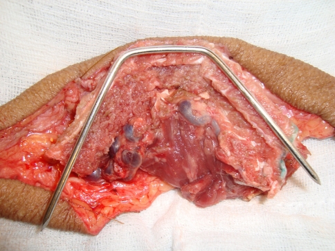
The osteotomies were planned from distal to proximal end of the fibula taking care of the septal perforators and excess bone is discarded from the proximal part of the bone so as to provide additional length of the peroneal vessels. To enable maximum contact between the segments and to maintain the contour, ‘V’ shaped osteotomy (Fig. 2a) were made with an oscillating saw after detaching the flap from the donor leg and marking the level of the septocutaneous perforators with methylene blue so as not to damage them during the osteotomy. The apex of the ‘V’ was placed over the peroneal surface of the fibula and the base was placed on the inner surface and thus the two limbs of the ‘V’ cutting across the anterior and posterior surfaces. The intact periosteum along with the blood vessels is separated from the segment of bone to be osteotomised and then the osteotomy is done (Fig. 2b). After the osteotomy was completed, the bone segments were fixed with mini plate and screws. Temporary inter maxillary fixation was done whenever possible and was removed after fixation of the neo mandible with the remnant mandible. This was done to avoid splaying of the ramus by adding extra length of bone. Following this, the reconstructed segments were placed in the mandibular defect and compared with the upper jaw so as to get the proper occlusion. Before incorporation into the remnant mandible, the anterior midline of the central segment and the upper incisors were made to fall in the same line so as to avoid chin deviation and prognathism. After this step, the excess bone was discarded from the proximal part of the fibula and this increases the pedicle length so as to facilitate tension free micro vascular anastomosis. Once this was done, the fibula resembles the central segment of the mandible (Fig. 3) and was then fixed into the native mandible (Fig. 4). If the central segment was found protruding beyond mid incisor line, then some more bone was removed from the lateral segments and the final fixation was done. If there was splaying of the ramus, malocclusion or prognathism, excess bone from the lateral segments was removed and occlusion was checked again.
Fig. 2.
a The marking for ‘V’ shaped osteotomy. b On the extreme left is the marking for osteotomy to discard the excess bone
Fig. 3.
After osteotomy and approximation of the closing angles the new central segment corresponds to the template
Fig. 4.
The new central segment is fixed into the remnant mandible
Results
All the osteotomised segments healed well as was seen in 3 month postoperative orthopantomograms (Fig. 5). There were no instances of plate fracture in the postoperative period. The common complications were prognathism (28 patients), deviation of chin (22 patients) and malocclusion (10 patients) (Fig. 6). Patients with malocclusion were managed with revision osteotomy after 1 year. Dental rehabilitation with osseous implants was done in 10 willing patients 1 year after irradiation. Mouth opening, jaw occlusion, oral competence, speech and swallowing were used to analyse the functional results. The results were good in 80% and acceptable in 20% of cases.
Fig. 5.
Three month postoperative X-rays showing good bone union
Fig. 6.
Prognathism of the reconstructed mandible seen in our earlier results
Discussion
Tumour ablative surgeries of the oral cavity results in severe defects of the facial skeleton. Depending up on the severity of the component loss Daniel [1] classified them accordingly and named those through and through defects of the oro mandibular region as en bloc defects or complex defects. Boyd [2] classified mandibular defects by dividing them into segments [HCL Classification]. Among the en bloc defects, the anterior defects or the central segment defects are difficult to reconstruct. This is because, they are associated with the loss of anterior skin and require a lip split incision [3] both of which interfere with the post operative appearance.
The free fibula flap described by Hidalgo [4] is the most common composite vascularised free tissue used for reconstruction of complex oro mandibular defects [1]. The techniques in flap design, harvest and osteotomy have undergone changes over the years [5]. The osteotomy of the fibula and the alignment of the osteotomised segments is a significant step in the challenging task of mandibular reconstruction. In order to simplify this step, a variety of mandibular templates are being used. The templates are important because they help in restoring the normal contour of the mandible especially the lower margin, help in planning the number of osteotomies thereby minimizing the periosteal stripping and reducing the vascular compromise. The advantages of using templates are well supported by Hidalgo [3] and Strackee [6]. These templates are made preoperatively from routine X-rays [7], orthopantomograms [8], three dimensional CT scan [9], acrylic jaw impressions [10] and lateral cephalogram [11]. Intra operatively, the templates can be made from the resected specimen [12], reconstruction plate [13], tongue blade template [14], aquaplast [15] template, by jigsaw [16] technique or by preplating systems [17]. Though making a template out of the radiological images are routine, converting the images into practical templates is very demanding and always an element of intuitive mind helps in making the correct osteotomy and there by the contour of the reconstructed mandible [3]. The intra operative methods of making the templates from the specimen or from the defect are more practical but have many disadvantages. Use of reconstruction plates gives good stability. The disadvantages are that there might be damage of the periosteum and thereby can compromise the vascularity of the bone, alter the shape of the neomandible and will require removal at a later date for dental rehabilitation [16]. Closing wedge osteotomies cannot be made in comparison with the tongue blade template [14] and using aquaplast [15] and jigsaw system [16] are not simple or cost effective and may require longer duration and thus prolong the duration of surgery.
The ‘K’ wire template was described by Wagner [17] and was his preferred method in mandibular reconstruction with fibula flaps. We modified this so as to apply for all the complex mandibular reconstructions. The complications in our study such as prognathism, malocclusion and deviation of chin were found in the earlier days. With increasing experience, our techniques refined and these were largely avoided in the recent years. Compared to the rest of the templates our technique is simple, cost effective and manage to provide and maintain the three dimensional contour of the mandible. Though this template is custom made, this can be used only for reconstructing the central segment defect of the mandible and not for the angle or ramus of the mandible.
Conclusion
The number of osteotomies and the alignment of the segments in proper angles are important in providing the shape and strength of the reconstructed mandible. 262 central segment defects out of a total of 386 mandible reconstructions over a 5 year period were done using this template with good results. The drawback seen during the initial stages were rectified as the technique was refined and the skills improved. Compared to all the other templates described so far, the ‘K’ wire template is simple, cost effective and enables the osteotomy and alignment of segments to be completed in quick time.
References
- 1.Daniel R. Mandibular reconstruction with vascularised iliac crest: a 10 years experience. Plast Reconstr Surg. 1988;82(5):802. doi: 10.1097/00006534-198811000-00012. [DOI] [PubMed] [Google Scholar]
- 2.Boyd JB, Gullane PJ, Rotstein LE, et al. Classification of mandibular defects. Plast Reconstr Surg. 1993;92(7):1266–1275. [PubMed] [Google Scholar]
- 3.Hidalgo DA. Aesthetic improvements in free flap mandible reconstruction. Plast Reconstr Surg. 1991;88(4):574. doi: 10.1097/00006534-199110000-00003. [DOI] [PubMed] [Google Scholar]
- 4.Hidalgo DA. Fibula free flap: a new method of mandible reconstruction. Plast Reconstr Surg. 1989;84:71. doi: 10.1097/00006534-198907000-00014. [DOI] [PubMed] [Google Scholar]
- 5.Cordeiro PG, Disa JJ, Hidalgo DA, Hu QY. Reconstruction of the mandible with osseous free flaps: a 10 year experience with 150 consecutive patients. Plast Reconcstr Surg. 1999;104:1314. doi: 10.1097/00006534-199910000-00011. [DOI] [PubMed] [Google Scholar]
- 6.Strackee SD, Kroon FHM, Spierings PTJ, Jaspers JEN. Development of modeling and osteotomy jig system for reconstruction of the mandible with a free vascularized fibula flap. Plast Reconcstr Surg. 2004;114(7):1851–1858. doi: 10.1097/01.PRS.0000142766.26117.5B. [DOI] [PubMed] [Google Scholar]
- 7.Giguere P, Demers G, Germain MA. Technique de transformation du perone en mandible. J Otolaryngol. 1995;24:9. [PubMed] [Google Scholar]
- 8.Flemming AFS, Brough MD, Evans ND, et al. Mandibular reconstruction using vascularised fibula. Br J Plast Surg. 1990;43:403. doi: 10.1016/0007-1226(90)90003-I. [DOI] [PubMed] [Google Scholar]
- 9.Serra JM, Paloma V, Mesa F, et al. The vasucalarised fibula graft in mandibular reconstruction. J Oral Maillofac Surg. 1991;49:244. doi: 10.1016/0278-2391(91)90213-6. [DOI] [PubMed] [Google Scholar]
- 10.Ueda K, Tajima S, Tanaka Y, et al. Mandibular reconstruction using computer generated three-dimensional solid models. J Oral Maxillofac Surg. 1994;10:291. doi: 10.1055/s-2007-1006597. [DOI] [PubMed] [Google Scholar]
- 11.Hidalgo DA. Fibula free flap mandibular reconstruction. Clin Plast Surg. 1994;21:25. [PubMed] [Google Scholar]
- 12.Soutar DA (1994) Mandibular reconstruction with vascularised bone. In: Souter DA, Rammohan MT (Eds) Excision and reconstruction in head and neck surgery, Churchill Livingstone, Newyork p 59
- 13.Futran ND, Urken MD, Buchbinder D. Rigid fixation of vascularised bone grafts in mandibular reconstruction. Arch Otolaryngol Head Neck Surg. 1995;121:70. doi: 10.1001/archotol.1995.01890010056010. [DOI] [PubMed] [Google Scholar]
- 14.Yap LH, Constantinides J, Butler CE. Tongue depressor template for free fibular flap osteotomies in mandibular reconstruction. Plast Reconstr Surg. 2008;122(6):209e–210e. doi: 10.1097/PRS.0b013e31818d2084. [DOI] [PubMed] [Google Scholar]
- 15.Kane WJ, Olsen KD. Enhanced bone graft contouring for mandibular reconstruction using intraoperatively fashioned templates. Ann Plast Surg. 1996;37:30. doi: 10.1097/00000637-199607000-00005. [DOI] [PubMed] [Google Scholar]
- 16.Marchetti C, Bianchi A, Mazzoni S, et al. Oromandibular reconstruction using fibula osteocutaneous free flap: four different ‘preplating’ techniques. Plast Reconstr Surg. 2006;118:643. doi: 10.1097/01.prs.0000233211.54505.9a. [DOI] [PubMed] [Google Scholar]
- 17.Wagner JD (1996) Pre operative assessment and preparation for mandible reconstruction. Oper Tech Plast Reconstr Surg 3(4):217–225


