Abstract
Objective
To reports ten Japanese surgical cases of verrucous carcinoma (VC) of the oral cavity.
Study design
The author reviewed histopathology of 10 cases of oral VC.
Results
Ten cases of oral VC were found in our pathology department in the last 10 years. During the 10 years, a total of 187 cases of oral malignancy were recognized. Therefore, the frequency of VC was 5.3% of all oral malignancies. The patients consisted of six women and four men. The age ranged from 52 to 84 years with a median of 68 years. The locations of VC were buccal mucosa in two cases, gingiva in three cases, hard palate in one case, tongue in three cases, and soft palate in one case. The presenting symptoms were oral discomfort in two cases and tumors in eight cases. All cases underwent surgical resection. Frozen sections were performed in three cases for margin check. Grossly, all cases showed verrucous lesions. The size ranged from 0.8 to 3.2 cm with a median of 1.3 cm. Histologically, tumor cells proliferated with verrucous or papillary features. The tumor cells had acidophilic, ample cytoplasm, and nuclear atypia was minimal. Individual keratinization, koilocytosis, basal cell mild atypia, and squamous pearl formation were recognized in all cases. Three cases showed microinvasion. One case had focal ordinary squamous cell carcinoma within the VC. Epithelial dysplasia in the mucosa was recognized in the vicinity of VC in two cases. One case showed multiple tumors of VC; the number was five. Lymphocytic infiltration in the dermis was recognized in seven cases. Immunohistochemically, p53 protein was positive in all the ten cases. Its location was accentuated near the basal cells and microinvasive parts. Ki-67 positive cells were also seen mainly in the basal cells and in the microinvasive areas, and the KI-67 labeling index ranged from 12 to 21%. Two patients recurred, and additional operations were performed. None show metastatic lesions. One patient died of other disease, and nine patients are now alive without tumors.
Conclusion
Clinicopathologic features of ten cases of oral VC were described.
Kewords: Verrucous carcinoma, Oral cavity, Histopathology
Introduction
Verrucous carcinoma (VC) is a rare type of low-grade, well differentiated squamous cell carcinoma, and develops mainly in the skin, genitalia, esophagus, and oral cavity. The pathogenesis of VC of the oral cavity is thought to be associated with human papilloma virus (HPV) [1–3], poor oral hygiene, chewing of tobacco, and use of snuff. Several studies of VC of the oral cavity have been reported [4–6], but they are rare in Japan. The author herein reports ten Japanese cases of VC of the oral cavity.
Materials and Methods
The author reviewed VC of the oral cavity in our pathology laboratory in the last 10 years. Ten cases of VC were found. During the periods 187 cases of malignancies of oral cavity were present. Thus, the frequency of VC was 5.3% of all oral malignancies. The author reviewed the HE slides of VC and collected clinical records. An immunohistochemical study was performed with the use of Dako Envision method (Dako Corp. Glostrup, Denmark), as previously reported [6, 7]. Microwave pretreatment for 5 min was employed. P53 protein (Dako, DO-7) and Ki-67 antigen (Dako, MIB-1) were examined to determine p53 mutation and cell proliferative activity.
Results
Clinically, the ten patients with VC of the oral cavity consisted of six women and four men. The age ranged from 52 to 84 years with a median of 68 years. The presenting symptoms were oral discomfort in two cases and tumors in eight cases. All patients were treated by surgical resection. Frozen sections were performed in three cases for margin check. Then, the seven patients received adjuvant chemoradiation. The length of follow-up ranged from 11 months to 9 years with a median of 4 years. The prognosis was good. Two patients recurred, and additional operations were performed. None show metastatic lesions. One patient died of other disease, and nine patients are now alive without tumors.
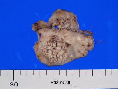
Grossly, the locations of VC were buccal mucosa in two cases, gingiva in three cases, hard palate in one case, tongue in three cases, and soft palate in one case. The size of the VC ranged from 0.8 to 3.2 cm with a median of 1.3 cm. The all cases showed verrucous lesions similar to cauliflower (Fig. 1).
Fig. 1.
Gross features of verrucous carcinoma. A polyp with cauliflower-like appearances is characteristic of verrucous carcinoma
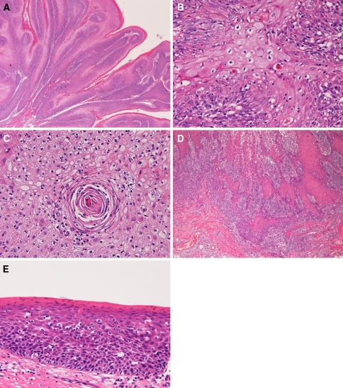
Histologically, VC was composed of squamous cell proliferations with verrucous or papillary features (Fig. 2a). The tumor cells had acidophilic, ample cytoplasm, and the nuclei were round and mildly hyperchromatic. The cellular atypia was mild or minimal (Fig. 2a, b). Individual keratinization (Fig. 2b), squamous pearl formation (Fig. 2c), koilocytosis (Fig. 2b, c) and basal cell mild atypia were recognized in all cases. Three cases showed microinvasion. One case had focal ordinary squamous cell carcinoma within the VC (Fig. 2d). Dysplasia of the surrounding squamous epithelium was found in two cases (Fig. 2e). One case showed multiple VCs; the number was five. Lymphocytic infiltration in the dermis was recognized in seven cases.
Fig. 2.
Histologic features of verrucous carcinoma. a Low power view of verrucous carcinoma. Verrucous proliferation of squamous epithelium is evident. The atypia is mild or minimal. HE, ×4. b Individual keratinization and koilocytosis are recognized in verrucous carcinoma. The atypia is mild. HE, ×200. c Squamous pearl is seen in the verrucous carcinoma. The cellular atypia is none. Koilocytosis is seen. HE, ×200. d Focal area of verrucous carcinoma shows apparent invasive squamous cell carcinoma. HE, ×100. e Epithelial dysplasia (moderate) in the vicinity of verrucous carcinoma. HE, ×200
Immunohistochemically, p53 protein was positive in all the ten cases. Its location was accentuated near the basal cells and microinvasive parts. Ki-67 positive cells were also seen mainly the basal cells in microinvasive parts, and the labeling index ranged from 12 to 21% with a median of 16%.
Discussion
Clinically, VC of the oral cavity is reported to be male-preponderance [4–6]. In the present series, male to female ratio was 6:4. The age with VC of oral cavity is reported to be high [4–9], as was the case of the present series. The presenting symptoms have not been reported in the literature. In the present study was oral discomfort in two cases and tumors in eight cases. The choice of treatment was surgical resection with or without adjuvant chemoradiation [4–6]. The prognosis of VC of the oral cavity was good, although recurrence is occasionally found [4–6], as in the present series.
The histology of the present series fulfills the criteria of VC. VCs of the present series are different from verrucous hyperplasia, proliferative verrucous leukoplakia, and well differentiated papillary squamous cell carcinoma from the standpoints of histology and immunohistochemistry [9]. The cases of the present series are not verrucous leukoplakia, a premalignant condition having a capacity to transform into squamous cell carcinoma. This entity had less atypia than VC. In addition, verrucous leukoplakia was not so verrucous, and was more superficial to the surrounding squamous epithelium [9].
The present cases showed koilocytosis of the VC, suggesting HPV infection. Of interest is that one case of the present series showed squamous cell carcinoma within VC. In this case, severe intraepithelial neoplasm were recognized next to the tumor. This phenomenon has not been reported in the literature, and suggests that ordinary squamous cell carcinoma can arise within VC. It is also suggested that VC may arise via dysplasia–carcinoma in situ sequence, and finally transforms into invasive squamous cell carcinoma. Also, of interest is that two cases of the present series showed dysplasia (squamous intraepithelial neoplasm) in the mucosa adjacent to the VC. This phenomenon has not been reported in the literature. This suggests that dysplasia may lead to VC, or that the mucosa of VC is prone to malignant transformation. One case of the present series showed multiple tumors of the VC. The number of the VC was five. This phenomenon is also not reported in the literature, and suggests that multiple VC of the oral cavity may develop in oral mucosa. Three cases of the present series showed microinvasion, suggesting that the atypical basal cells of VC underwent invasive characters. The expression of p53 protein and Ki-67 antigen in VC has been reported [10, 11]. The present cases showed p53 protein in all cases, and its expression was accentuated in the basal cells and invasive sites. Similar tendency was observed in Ki-67 antigen. The expression of p53 and relatively low Ki-67 labeling supports that VC is a low grade malignant tumor. The expression of both protein in the basal part may indicate basal part of VC have relatively higher p53 mutation and cell proliferative capacity than non-basal part of VC.
Conflict of interest
The author has no conflict of interest.
References
- 1.Fijita S, Senba M, Kumatori A, Hayashi T, Ikeda T, Toriyama K. Human papillomavirus infection in oral verrucous carcinoma: genotyping analysis and inverse correlation with p53 expression. Pathobiology. 2008;75:257–264. doi: 10.1159/000132387. [DOI] [PubMed] [Google Scholar]
- 2.Stankiewicz E, Kudahetti SC, Prowse DM, Ktori E, Cuzick J, Ambroisine L, Zhang X, Watkin N, Corbishley C, Berney DM. HPV infection and immunohistochemical detection of cell-cycle markers in verrucous carcinoma of the penis. Mod Pathol. 2009;22(9):1160–1168. doi: 10.1038/modpathol.2009.77. [DOI] [PubMed] [Google Scholar]
- 3.Shroyer KR, Greer RO, Fankhouser CA, McGuirt WF, Marshal R. Detection of human papilloma DNA in oral verrucous carcinoma by polymerase chain reaction. Mod Pathol. 1993;6:669–672. [PubMed] [Google Scholar]
- 4.McCoy JM, Waldron CA. Verrucous carcinoma of the oral cavity: a review of forty nine cases. Oral Surg Oral Med Oral Pathol. 1981;52:623–629. doi: 10.1016/0030-4220(81)90081-5. [DOI] [PubMed] [Google Scholar]
- 5.Medina JE, Dichtel W, Luna MA. Verrucous-squamous cell carcinoma of the oral cavity: a clinicopathologic study of 104 cases. Arch Otolaryngol. 1984;110:437–440. doi: 10.1001/archotol.1984.00800330019003. [DOI] [PubMed] [Google Scholar]
- 6.Kamath VV, Varma RR, Gadewar DR, Muralidhar M. Oral verrucous carcinoma: a analysis of 37 cases. J Craniomaxillofac Surg. 1989;17:309–314. doi: 10.1016/s1010-5182(89)80059-9. [DOI] [PubMed] [Google Scholar]
- 7.Terada T, Kawaguchi M. Primary clear cell adenocarcinoma of the peritoneum. Tohoku J Exp Med. 2005;206:271–275. doi: 10.1620/tjem.206.271. [DOI] [PubMed] [Google Scholar]
- 8.Terada T, Kawaguchi M, Furukawa K, Sekido Y, Osamura Y. Minute mixed ductal-endocrine carcinoma of the pancreas with predominant intraductal growth. Pathol Int. 2002;52:740–746. doi: 10.1046/j.1440-1827.2002.01416.x. [DOI] [PubMed] [Google Scholar]
- 9.Murrah VA, Batsakis JG. Proliferative verrucous leukoplakia and verrucous hyperplasia. Ann Otol Rhinol Laryngol. 1994;103:660–663. doi: 10.1177/000348949410300816. [DOI] [PubMed] [Google Scholar]
- 10.Saito T, Nakajima T, Mogi K. Immunohistochemical analysis of cell-cycle-associated proteins p16, pRb, p53 and Ki-67 in oral cancer and precancer with special reference to verrucous carcinoma. J Oral Pathol Med. 1999;28:226–232. doi: 10.1111/j.1600-0714.1999.tb02029.x. [DOI] [PubMed] [Google Scholar]
- 11.Drachenberg CB, Blanchaert R, Ioffe OB, Ord RA, Padadimitriou JC. Comparative study of invasive squamous cell carcinoma and verrucous carcinoma of the oral cavity; expression of bcl-2, p53, and HER2/neu, and indexes of cell turnover. Cancer Detect Prev. 1997;21:483–489. [PubMed] [Google Scholar]


