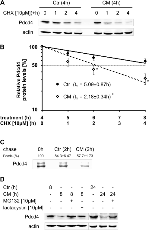
Fig. 3.
CM attenuates Pdcd4 protein expression by increasing proteasomal degradation. (A) MCF7 cells were incubated with Ctr or CM for 4 h. Then, CHX [10 μM] was added to block translation and incubations continued for 1, 2 or 4 h. (B) Protein levels determined in (A) were quantified densitometrically and expression was normalized to actin. Data are presented relative to 4 h pretreated samples (n > 3, *P < 0.05). (C) MCF7 cells were subjected to a pulse-chase experiment, in the presence of Ctr or CM during the 2 h chase period. Pdcd4 was immunoprecipitated and detected by autoradiography. The autoradiographic picture is representative for at least three independent experiments and the remaining labeled Pdcd4 levels are presented relative to 0 h (n = 3). (D) MCF7 cells were incubated with Ctr or CM for 8 or 24 h. MG132 [10 μM] or lactacystin [10 μM] were added for the last 8 h of the incubations to block proteasomal degradation. Whole-cell extracts were subjected to western blot analysis and probed with the indicated antibodies. Blots are representative of at least three independent experiments.

