Abstract
The prenatal and perinatal periods of brain development are especially vulnerable to insults by environmental agents. Early life exposure to cigarette smoke (CS), which contains both genotoxicants and oxidants, is considered an important risk factor for both neurodevelopmental and neurodegenerative disorders. Yet, little is known regarding the underlying pathogenetic mechanisms. In the present study, neonatal Swiss ICR (CD-1) albino mice were exposed to various concentrations of CS for 4 weeks and the brain examined for lipid peroxides, DNA damage, base-excision repair (BER) enzymes, apoptosis, and levels of the microtubule protein tau. CS induced a dose-dependent increase in both malondialdehyde and various types of DNA damage, including single-strand breaks, double-strand breaks, and DNA-protein cross-links. However, the CS-induced DNA damage in the brain returned to basal levels 1 week after smoking cessation. CS also modulated the activity and distribution of the BER enzymes 8-oxoguanine-DNA-glycosylase (OGG1) and apyrimidinic/apurinic endonuclease (APE1) in several brain regions. Normal tau (i.e., three-repeat tau, 3R tau) and various pathological forms of tau were also measured in the brain of CS-exposed neonatal mice, but only 3R tau and tau phosphorylated at serine 199 were significantly elevated. The oxidative stress, genomic dysregulation, and alterations in tau metabolism caused by CS during a critical period of brain development could explain why CS is an important risk factor for both neurodevelopmental and neurodegenerative disorders appearing in later life.
Keywords: cigarette smoke, brain, neonatal mice, DNA damage, base-excision repair, tau, neurodegenerative disorders
Estimates indicate that 1 in 10 pregnant mothers in the United States smoke cigarettes and approximately 16% of them continue to smoke after delivery (Baler et al., 2008). Maternal smoking has been linked to many deleterious effects that put the fetus and newborn at increased risk for adverse health outcomes including impaired growth and neurobehavioral development. Exposure to environmental tobacco smoke during pregnancy is dose dependently associated with low neonatal birth weight and length and reduced head circumference (Perera et al., 2005). Since reduced growth and DNA damage are significantly correlated in neonates of mothers who were exposed to environmental tobacco smoke during pregnancy (Tsui et al., 2008), DNA damage may also be responsible for the brain injury and behavioral problems induced by the genotoxicants in cigarette smoke (CS).
The developing brain is especially vulnerable to environmental genotoxic agents and the ensuing brain injury is dependent upon two critical factors: the type of DNA damage and the efficiency of DNA repair (Kisby et al., 2009b). According to the “Barker hypothesis,” early life exposure to toxic agents is an important risk factor for the development of late-life diseases, such as respiratory, neurodevelopmental disorders, and neurodegenerative disease (Landrigan et al., 2005). In support, we previously demonstrated that the sudden transition from the maternal-mediated respiration of the fetus to the autonomous pulmonary respiration of newborn mice results in a significant increase, in the lung, of bulky DNA adducts and oxidative DNA lesions (e.g., 8-hydroxy-2′-deoxyguanosine, 8-oxo-dGuo). This DNA damage was attenuated by the upregulation of a number of genes having adaptive functions (e.g., antioxidant enzymes, oxidative DNA repair). Interestingly, prenatal administration of the antioxidant N-acetylcysteine (NAC) reduced the DNA damage and prevented all of the transcriptional changes in the neonatal lung (Izzotti et al., 2003) suggesting important links between oxidative stress, DNA damage, and tissue function.
Based on these findings, we demonstrated that mice exposed early in life to CS are particularly susceptible to the induction of lung tumors (Balansky et al., 2007) and alterations of intermediate biomarkers, notably DNA damage and oxidative stress (De Flora et al., 2008). The ability of antioxidants (e.g., NAC) to prevent CS-induced lung tumors in mice (Balansky et al., 2009) and to protect against oxidative stress–induced neuronal death (Arakawa and Ito, 2007) suggests that oxidative stress–mediated mechanisms contribute to CS-induced brain injury.
Although epidemiological studies indicate that the association between CS and tumors of the central nervous system is controversial (International Agency for Research on Cancer, 2004), there is substantial evidence linking perinatal and postnatal exposure to CS with neurodevelopmental and behavioral disorders (Herrmann et al., 2008) and chronic neurodegenerative diseases, including amyotrophic lateral sclerosis (Gallo et al., 2009), multiple sclerosis (Healy et al., 2009), and Alzheimer disease (Cataldo et al., 2010). Mainstream CS and sidestream CS are complex mixtures of over 4000 compounds including genotoxicants (e.g., tobacco-specific nitrosamines, polycyclic aromatic hydrocarbons, etc.) as well as high levels of oxidants and reactive chemical species, also resulting from endogenous processes, such as inflammatory processes. The developing brain is particularly vulnerable to oxidative stress because of its high metabolic activity, concentration of unsaturated fatty acids, high rate of oxygen consumption, and low concentration of antioxidants (Chen et al., 2007). Oxidative insult of the brain also leads to the accumulation of DNA damage, an effect that could have long-term consequences on neuronal function in later life (Rao, 2009).
The goal of the present study was to determine whether CS induces oxidative stress and DNA damage and perturbs DNA repair and tau proteins in the brain of neonatal mice. Oxidative stress was assessed by examining the brain for the levels of malondialdehyde (MDA). Because oxidative stress generates DNA lesions such as apurinic (AP) sites and 8-oxoguanine, the regional distribution and activities of the base-excision repair (BER) enzymes 8-oxoguanine-DNA-glycosylase (OGG1) and apyrimidinic/apurinic endonuclease (APE1) were also evaluated. The BER pathway handles a wide spectrum of base modifications including alkylation and oxidative DNA damage. The comet assay was used to examine the brain for the induction and persistence of multiple types of DNA lesions including single-strand breaks (SSBs), double-strand breaks (DSBs), and DNA-protein cross-links (DPCs). The brain was also examined for changes in the levels of normal tau (i.e., tau isoforms) and pathological forms of tau (tau oligomers, truncated tau, phosphotau). Tau is a microtubule-associated protein that plays a major role in the development and morphology of neurons by promoting the assembly and stability of microtubules, whereas the abnormal posttranslational modification of tau (e.g., phosphorylation) is a pathological hallmark of neurodegenerative disorders such as tauopathies (Wang and Liu, 2008). The results show that CS has a significant influence on the developing brain by causing oxidative stress induced injury (lipid peroxidation, DNA damage, BER activity) and associated changes in tau, a cytoskeletal protein that plays an important role in maintaining neuronal function.
MATERIALS AND METHODS
Mice.
Healthy 8-week-old Swiss ICR (CD-1) albino mice, on the 15th day of pregnancy, were supplied by Harlan Italy (San Pietro al Natisone, Udine, Italy) were maintained on standard rodent chow (Teklad 2018; Harlan Italy) and tap water ad libitum, at a temperature of 23 ± 2°C, relative humidity of 55%, 15 air renewal cycles per h, and a 12-h day/night cycle. Housing and treatments of mice were in accordance with our national and institutional guidelines and were approved by the Italian Ministry of Health.
Treatments.
Mainstream CS was generated by burning Kentucky 2R4F reference cigarettes (University of Kentucky, Lexington, KY), having a declared content of 9.4 mg tar, 0.73 mg nicotine, and 12 mg CO each. Exposure of mice to CS was achieved by using a whole-body system (model TE-10a; Teague Enterprises, Davis, CA). This system was equipped with a peristaltic pump that delivered CS continuously into the exposure chambers, made of teflon-painted aluminum with plexiglass front, connected to an air exhaust system. Each exposure chamber has a 70 l capacity, with a flow rate of 17.5 l/min that results in 15 air renewal changes per hour. Each cigarette burns completely in 4 min and generates a volume of smoke of 1020 ml. Therefore, the CS produced by 15 consecutive cigarettes was delivered in 1 h. The exposure started within 12 h after birth and continued daily, 1 h/day, for 4 weeks.
Six dams generated 65 newborn mice, which were divided into six experimental groups, each group corresponding to a litter. The groups included (A) sham-exposed mice, kept in filtered air; (B) mice exposed to the CS generated from a single filter cigarette; (C) mice exposed to the CS generated from a single cigarette, defiltered before use; (D) and (E) mice exposed to the CS generated from two defiltered cigarettes burning at one time; and (F) mice exposed to the CS generated from four defiltered cigarettes burning at one time. The average total suspended matter (TPM) was 119, 292, 438, and 631 mg/m3 in the exposure chambers accommodating the mice belonging to Groups B, C, D/E, and F, respectively.
Collection of brains.
After 4 weeks, the mice belonging to Groups A–D were fasted overnight, deeply anesthetized, and euthanized by cervical dislocation. The mice belonging to Group E were similarly terminated 1 week later. The whole heads from five mice in Groups A and C were each immersed in ice-cold Hibernate medium supplemented with B27 (Invitrogen, Carlsbad, CA). The right hemisphere from all other mice was immersion fixed in formalin, embedded in paraffin, and used for measuring apoptotic neurons. The left hemispheres from two mice in Groups A and C were fixed in formalin and shipped, together with the heads immersed in Hibernate medium, from Genoa to Portland, OR. The remaining left hemispheres were used for single cell gel electrophoresis (SCGE) analyses.
SCGE assay.
The brains were analyzed by SCGE (comet) assay as described by Fairbairn et al. (1995). SCGE was performed in either a neutral or an alkaline environment in order to detect DSBs and SSBs, respectively. In order to discriminate between DPCs and DNA-DNA cross-links under alkaline conditions, we performed a proteinase K (PK) treatment by preincubating the samples for 1 h at 37°C with either PK or its buffer. Images of at least 100 randomly selected cells from each mouse, derived from two slides, were acquired and analyzed on an automated imaging system (CASP or Comet Assay Software Project, http://www.casp.sourceforge.net). SSBs and DSBs were quantified in terms of tail moment, which is the product of the tail length and the fraction of total DNA in the tail, by analyzing ∼100 comets/sample.
Determination of lipid peroxidation products.
Brain tissue levels of MDA and other thiobarbituric acid reactive substances were measured as markers of lipid peroxidation as described by Ohkawa et al. (1979), with slight modifications. Briefly, brain tissue homogenates were mixed with a solution containing 8.1% SDS, 20% acetic acid (pH 3.5), 0.8% thiobarbituric acid, and the samples heated at 95°C for 1 h. After cooling in ice-cold water, the samples were extracted with 4 ml n-butanol and pyridine (15:1, vol/vol), centrifuged at 950 × g for 10 min, and the absorbance of the organic phase measured at a wavelength of 532 nm. Sample concentrations were derived from a standard curve using 25, 50, 100 and 150mM MDA and expressed as nanomole MDA equivalent per milligram protein.
Evaluation of apoptotic neurons.
Apoptotic neurons in the cerebral cortex were detected by the terminal deoxynucleotidyl transferase–mediated nick end labeling (TUNEL) method using the NeuroTACS In Situ Apoptosis detection staining kit (Trevigen, Gaithersburg, MD). Briefly, sagittal brain tissue sections (5 μm) were placed on glass slides, the sections probed with the NeuroTACS kit as described by the manufacturer and then each slide scored at a 400× magnification by counting 500 cells from three serial sections per mouse. The results were expressed as percentage of apoptotic neurons.
Detection of BER proteins.
The brains from sham- and CS-exposed mice were cryoprotected in sucrose (10–30%), rapidly frozen in Tissue-Tek, and sagittal brain tissue sections (10 μm) placed on slides as previously described (Kisby et al., 2009b). The slides were incubated overnight at 4°C with blocking solution (0.2% TX-100 in PBS) or blocking solution containing rabbit polyclonal antibodies to either OGG1 (NB100-106), APE1 (NB100-101; Novus Biologicals, Littleton, CO) as previously described (Kisby et al., 1997). After removal of the primary antibody, the sections were washed with PBS and the proteins detected using a Vectastain Elite peroxidase kit and NovaRed (Vector Labs, Burlingame, CA). Adjacent brain sections were stained with cresyl violet to label Nissl substance. The sections were examined by light microscopy using a Zeiss Axioskop 2 microscope.
Determination of oxidative DNA repair activities.
APE1 and OGG1 activities were determined by measuring the amount of cleavage product from the 32P-labeled 5′–5′ oligonucleotide substrate containing a tetrahydrofuran or 8-oxoguanine and a complementary strand, as previously described (Kisby et al., 2009a). The lesion containing strand was 5′-end labeled with [γ-32P]ATP (PerkinElmer Life Sciences, Boston, MA) using T4 polynucleotide kinase (New England Biolabs, Ipswich, MA).
APE1 reaction was performed in 20 μl reaction buffer (10mM NaCl, 10mM Tris, 2mM MgCl2, pH 8.0) containing 100 fmol of labeled substrate and 50 ng of brain tissue homogenate and was incubated for 15 min at 37°C. OGG1 reaction was performed in 20 μl of reaction buffer (75mM NaCl and 10mM Tris, pH 8.0) containing 100 fmol of labeled substrate and 5 μg of brain tissue homogenate and was incubated for 1 h at 37°C. The product and substrate were separated on a 12.5% denaturing polyacrylamide gel and analyzed for band intensity using a Typhoon phosphoImager (Amersham Biosciences, Piscataway, NJ).
Analysis of tau proteins.
Tissue extracts were prepared from the cortex, hippocampus, midbrain, and brainstem of sham- and CS-exposed mice and examined for the levels of three-repeat tau (3R tau), tau oligomers, truncated tau, and various phosphorylated forms of tau by ELISA assay (Woltjer et al., 2009). Briefly, protein extracts from the soluble fractions of each mouse brain (n = 4–5) were diluted in 100mM Tris, pH 7.4, with 0.05% azide and 0.002% bromopheonl blue, 100 μl of the sample, containing 200 ng protein, spotted onto 96-well plates, and then the plates incubated overnight at RT in a humidified chamber. Plates were subsequently washed twice with PBS, blocked with 1% bovine serum albumin in PBS with 0.05% sodium azide, and washed again with PBS. ELISAs were performed as described by Harlow and Lane (1988) and developed using tetramethylbenzidine with absorbances determined at 405 nm in a UV-VIS microplate reader. Primary antibodies were monoclonal antibodies to 3R tau (RD3; Upstate/Millipore), tau oligomers (MC-1, ALZ-50; gift from P. Davies, Albert Einstein College of Medicine, Bronx, NY), truncated tau (cleaved tau, Asp421, clone C3; Upstate/Millipore), and various epitopes of phosphorylated tau (phosphoserine 202, CP13; gift from P. Davies and phosphothreonine 231 and phosphoserine 199; Upstate/Millipore). Secondary antibody/alkaline phosphatase conjugates were from Amersham. All antibodies were diluted 2000 times from the initial stock concentration before use in the detection assay. The values for 3R tau and other modified forms of tau were corrected for the background absorbance of wells without protein (10 per plate).
Statistical analysis.
The data for each evaluated endpoint are expressed as means ± SE within each experimental group. The overall statistical significance was evaluated by ANOVA, followed by nonparametric Mann-Whitney test for comparisons between groups.
RESULTS
Survival and Body Weights of Mice
The dam exposed to the highest CS dose, which was initially kept together with its litter, died after 3 days of treatment. Accordingly, neonatal mice belonging to this group were discarded. Thereafter, the dams were removed during exposure periods in order to exclude any influence on lactation. All other mice survived until the end of the experiment. The overall body weight of mixed gender neonatal mice was 1.2 ± 0.03 g (means ± SE). Table 1 reports the body weights of the mice belonging to Groups A–E at 2, 3, and 4 weeks after birth. The loss of body weights recorded in Groups C–E was statistically significant at all measurement times.
TABLE 1.
Body Weights, SCGE Data, Generated Either in Neutral or Alkaline Environment and Either in the Presence (PK+) or in the Absence (PK–) of Proteinase K, and TBARS in the Mixed Cell Brain Population, and Apoptotic Neurons (TUNEL+) in the Brain Cortex of Mice Exposed to Varying Doses of MCS
| Group | TPM | No. of | Body weight (g ) |
SCGE (TM) |
SCGE (TM) |
TBARS | TUNEL+ | ||||
| (mg/m3) | mice | 14 days | 21 days | 28 days | Neutral | Alkaline | PK– | PK+ | (nmol/mgprotein) | neurons(%) | |
| A | 0 | 12 | 10.0 ± 0.26 | 13.1 ± 0.30 | 23.1 ± 0.63 | 5.4 ± 0.29 | 48.6 ± 2.02 | 31.9 ± 1.23 | 33.9 ± 1.63 | 0.5 ± 0.12 | 1.4 ± 0.51 |
| B | 119 | 10 | 12.3 ± 0.37 | 14.6 ± 0.38 | 25.4 ± 1.22 | 6.3 ± 0.40 | 67.8 ± 2.41** | 27.1 ± 1.62 | 41.6 ± 2.20*** | 0.7 ± 0.11 | 1.6 ± 1.52 |
| C | 292 | 13 | 6.3 ± 0.35** | 9.3 ± 0.39** | 18.8 ± 0.42** | 12.1 ± 0.50** | 68.6 ± 2.48** | 38.3 ± 1.46 | 49.0 ± 1.86*, *** | 0.7 ± 0.15 | 2.0 ± 1.22 |
| D | 438 | 9 | 6.9 ± 0.31** | 10.2 ± 0.29** | 17.4 ± 0.50** | 15.6 ± 0.92** | 79.2 ± 2.84** | 59.7 ± 2.71* | 75.8 ± 2.61**, *** | 1.2 ± 0.16* | 3.2 ± 1.68 |
| Ea | 438 | 11 | 6.4 ± 0.28** | 9.7 ± 0.53** | 25.5 ± 1.06 | NT | NT | 28.5 ± 1.51 | 29.8 ± 2.27 | NT | NT |
Note. All data are means ± SE within each experimental group. NT, not tested. *p < 0.05 and **p < 0.001, as compared with the corresponding controls (dose 0 of TPM); ***p < 0.001, as compared with the corresponding PK–.
a The mice in this group were sacrificed one week after discontinuation of exposure to MCS rather than the day after.
Smoke-Induced Double-Strand and Single-Strand DNA Breaks
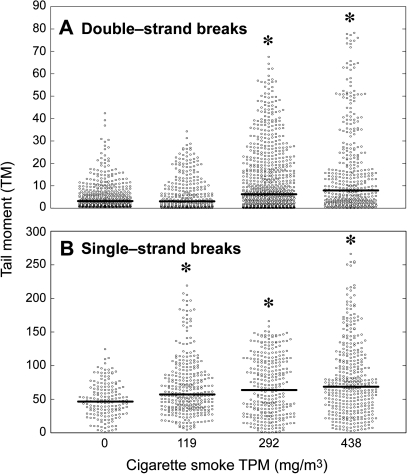
Initial studies examined cells from the brain of CS-exposed mice for DNA damage using the SCGE assay. Figure 1 shows the distribution of SCGE data, generated in either a neutral environment (DSBs, Fig. 1A) or alkaline environment (SSBs, Fig. 1B), as related to CS dose. The corresponding levels of DSBs and SSBs are reported in Table 1. Due to the small size of subgroups and to interindividual variability, there were no significant gender-related differences in the SCGE data. The basal levels of SSBs (48.6 ± 2.02) were ninefold higher than those of DSBs (5.4 ± 0.29). Under both experimental conditions, there was a dose-dependent increase in DNA strand breaks, which was statistically significant for DSBs at 292 and 438 mg/m3 TPM and for SSBs at all TPM doses tested. The maximum increases in DSBs and SSBs in CS-exposed mice were 2.9- and 1.6-fold, respectively.
FIG. 1.
Distribution of SCGE data generated either in neutral (A) or in alkaline (B) environment, as related to the dose of whole-body CS exposure of mice. The data were generated by testing 100 nuclei per brain sample obtained from five mice in Group A, eight mice in Group B, eight mice in Group C, and five mice in Group D. The horizontal bold lines indicate the medians within each experimental group. *p < 0.01, as compared with the corresponding sham.
Smoke-Induced DNA-Protein Cross-links
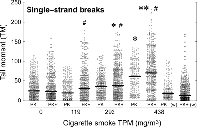
Figure 2 shows the levels of DPCs in the brain of CS-exposed mice, generated by evaluating alkaline SCGE data either in the absence (PK−) or in the presence (PK+) of proteinase K. The corresponding DPCs levels are reported in Table 1. In the absence of PK, the observed increase in DPCs over the corresponding sham was statistically significant (p < 0.05) only at the highest CS dose. In the presence of PK, the observed increases over the corresponding sham were significant at 292 and 438 mg/m3 TPM (p < 0.05 and p < 0.01, respectively). At all CS doses, the values recorded in the presence of PK were significantly higher (p < 0.001) than those recorded in the absence of PK. However, at the highest dose tested, DPCs had returned to basal levels 1 week after discontinuation of CS exposure (Table 1, last line).
FIG. 2.
Distribution of SCGE data generated in alkaline environment, either in the absence (PK−) or presence (PK+) of proteinase K, as related to the dose of whole-body CS exposure of mice. The horizontal bold lines indicate the medians within each experimental group. Columns identified with (w) refer to mice that were sacrificed 1 week after discontinuation of CS exposure rather than the day after. *p < 0.05 and **p < 0.001, as compared with the corresponding sham. #p < 0.001, as compared with PK−.
Smoke-Induced Lipid Peroxides and Apoptosis
As shown in Table 1, brain tissue levels of lipid peroxides were significantly increased only at the highest CS dose. The occurrence of TUNEL-positive cells, identified by a brown precipitate over the nucleus, was rare in the cerebral cortex of CS-exposed mice. At the highest dose tested, the increase of TUNEL-positive neurons (Table 1) approached statistical significance (p = 0.06) in mixed gender mice and was statistically significant (p < 0.05) in female mice (data not shown).
Distribution and Activity of BER Proteins
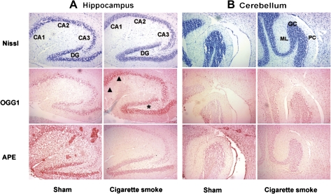
The influence of CS on oxidative DNA repair in the brain was assessed by examining brain tissue sections and extracts for the BER proteins OGG1 and APE1. Figure 3 shows representative photomicrographs of the hippocampus (A) and cerebellum (B) from sham-exposed mice and mice exposed to CS, at a TPM dose of 438 mg/m3. Nissl staining showed that CS did not induce remarkable loss of pyramidal or granule neurons in either the hippocampus (Fig. 3A) or the cerebellum (Fig. 3B). A similar staining pattern was observed in other brain regions (data not shown). The regional distribution of OGG1 and APE1 proteins was strongly affected by CS treatment but with opposite effects. In the hippocampus, CS induced OGG1 in all three pyramidal cell layers (CA1–CA3) and the granule cell layer in the dentate gyrus. CS induced a similar pattern of staining in the granule cell and Purkinje cell layers of the cerebellum. In contrast, APE1 staining was reduced in the same regions of the hippocampus and cerebellum of CS-exposed mice.
FIG. 3.
Nissl staining and immunohistochemical detection of OGG1 and APE1 in the hippocampus (A) and cerebellum (B) of a sham-exposed mouse and a CS-exposed mouse. CA1, CA2 and CA3 indicate the three hippocampal pyramidal cell layers and DG indicates the dentate gyrus. Note the heavy staining of OGG1 in the CA1 (arrowheads) and DG (star) regions of the hippocampus. GC, granule cell; ML, molecular layer; PC, Purkinje cell.
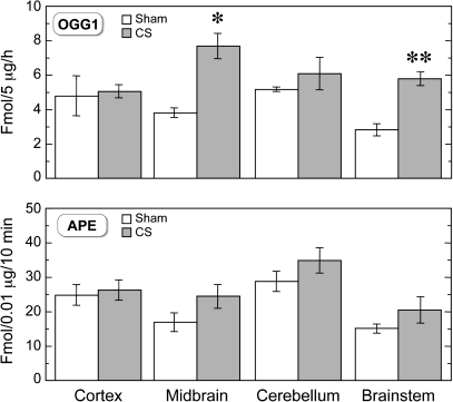
OGG1 and APE1 activities were evaluated in the brainstem, cerebellum, cerebral cortex, and midbrain of sham- and CS-exposed mice. The hippocampus was not examined because of an insufficient amount of tissue. As shown in Figure 4, OGG1 activity was significantly increased in the brainstem and midbrain of CS-exposed mice, whereas no changes were observed in APE1 activity.
FIG. 4.
OGG1 and APE1 activities in various brain regions of sham exposed (empty columns, n = 5) and mice exposed to CS, at a TPM of 438 mg/m3 (full columns, n = 3–5). *p < 0.01 and **p < 0.001, as compared with sham.
Tau Proteins
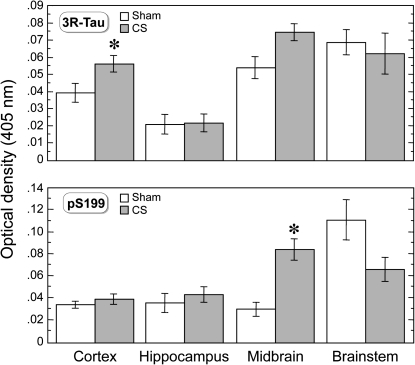
The goal of the next experiments was to determine if CS-induced oxidative stress is associated with changes in tau, a structural protein that plays an important role in maintaining neuronal function during development and is perturbed in neurodegenerative disorders. As shown in Figure 5, 3R tau levels were significantly increased in the cortex, whereas the levels of phosphoserine 199 (pS199) were significantly increased in the midbrain of CS-exposed mice as compared with sham. All other tau proteins, including tau oligomers, truncated tau (Tau-421), and the phosphorylated tau species phosphothreonine 231, were not significantly affected by CS in any brain region examined (data not shown).
FIG. 5.
3R tau and pS199 levels in various brain regions of sham exposed (empty columns, n = 5) and mice exposed to CS, at a TPM of 438 mg/m3 (full columns, n = 3–5). *p < 0.05, as compared with sham.
DISCUSSION
Exposure of mice to CS during the first 4 weeks of life, a time of extensive synaptogenesis, myelinogenesis, gliogenesis, and neurogenesis in the developing brain (Watson et al., 2006), induced oxidative stress, DNA damage, oxidative DNA repair, and significant tau changes in the brain. These findings are consistent with previous work demonstrating that CS induces lipid peroxidation, DNA damage, and oxidative DNA repair in nonneural tissues of CS-exposed mice (De Flora et al., 2008). Such changes might explain how prenatal exposure to CS leads to neurobehavioral changes in children (Herrmann et al., 2008) and increases the risk of developing late-life neurodegenerative disorders (Cataldo et al., 2010; Gallo et al., 2009; Healy et al., 2009).
CS induces lipid peroxidation in a variety of rodent organs (De Flora et al., 2008). MDA, reactive oxygen species, and oxidized proteins were significantly elevated in the brain of CS-exposed adult mice (Tuon et al., 2010) and the levels were significantly higher than in any other organ (Rueff-Barroso et al., 2010). The present study is the first to demonstrate that CS, at high doses, also increases MDA levels in the immature mouse brain. Increased levels of lipid peroxides are particularly relevant due to the abundance of fatty acids in the immature brain, which are critical for both infant and childhood brain development (Ryan et al., 2010).
CS induces multiple types of DNA lesions and alters OGG1 expression in several nonneural tissues of neonatal mice (De Flora et al., 2008). The present study demonstrates that CS also induces multiple types of DNA lesions (i.e., SSBs, DSBs, DPCs) and alters the level and activity of OGG1 in the brain of neonatal mice. The higher level of SSBs may be due to the formation of alkali labile DNA lesions (i.e., apurinic sites) following the repair of CS-induced oxidative DNA lesions by OGG1 (Fortini et al., 2003). In the alkaline comet assay, the repair of oxidative DNA damage or potentially other forms of DNA damage may be a major contributor to SSBs. Interestingly, DNA damage returned to basal levels 1 week after CS exposure was discontinued. This suggests that the immature brain efficiently repairs these types of DNA lesions and that smoking cessation has beneficial effects toward these endpoints. However, a significant amount of oxidative DNA lesions (e.g., 8-oxo-dGuo) would have escaped repair due to the low level of OGG1 in the neonatal brain. Therefore, it is likely that 8-oxo-dGuo might persist in the brain of CS-exposed mice because these DNA lesions accumulate in the brain with age (Larsen et al., 2006).
Environmental tobacco smoke has been shown to stimulate apoptotic pathways in a variety of cells, tissues, and organs, including the brain (Fuller et al., 2010). The ability of a high CS dose to induce apoptosis in mouse cortical neurons, as detectable by TUNEL, is certainly consistent with the results of the neutral comet assay. Apoptosis leads to the death of damaged cells, including postmitotic neurons in the developing brain (D'Agostini et al., 2005). Consistent with this conclusion, smoking was associated with reduced cortical gray matter in the Alzheimer-diseased brain (Almeida et al., 2008).
The CNS is equipped with the machinery to repair DNA damage. In mammals, BER is the primary pathway for repairing oxidative DNA damage (Fortini et al., 2003). The BER enzyme OGG1 plays a pivotal role in protecting the brain from oxidative DNA damage (Larsen et al., 2006). APE1, an enzyme that repairs the DNA lesions produced by OGG1, also plays an important role in protecting neurons from oxidative damage (Meira et al., 2001; Vasko et al., 2005). The present results show that CS alters both BER enzymes in a region- and cell-specific manner in the immature brain. The levels of APE and OGG1 vary considerably in the human, primate, and rodent brain. In the mouse brain, APE1 is very abundant, whereas OGG1 is substantially lower. This is reflected in the amount of protein we used to assay APE1 (50 ng) versus OGG1 (5.0 mg) activity in the neonatal mouse brain. Consequently, changes to OGG1 are more easily observed by immunohistochemical methods than APE1. The increased expression of OGG1 in murine brain tissue sections was also consistent with an increase in the regional activity of OGG1. This might explain why we primarily observed significant changes in OGG1 and not in APE1.
In the murine brain, nuclear and mitochondrial OGG1 activities are heterogeneous (Imam et al., 2006). Because there is a good correlation between regional OGG1 activity and the repair of oxidative DNA damage (Sava et al., 2006), this could explain why CS induced OGG1 activity only in those brain regions with low activity (i.e., brainstem, midbrain). In contrast, APE activity is fairly uniform in the murine brain (Imam et al., 2006), which might explain why APE1 activity did not change in the neonatal brain after CS exposure. However, CS did induce changes in both OGG1 and APE1 in the hippocampus, a brain region that is especially vulnerable to oxidative stress (Martone et al., 2000).
Tau isoforms and phosphorylated tau play an important role in regulating the dynamic microtubule network during periods of high plasticity in the developing brain. A shift from the fetal three-repeat (3R) tau isoform to the adult four-repeat (4R) tau isoform occurs during brain development and this coincides with the formation of synapses and the appearance of stable microtubules (Bullmann et al., 2009). However, 3R tau and 4R tau are approximately equally expressed in the mature mammalian brain. Tau phosphorylation is also required during periods of intense axonal outgrowth and synaptogenesis in the developing brain (Brion et al., 1994). 3R tau levels were significantly elevated in the cerebral cortex of CS-exposed mice and a similar trend was observed in the midbrain. The phosphorylation of tau was also increased by CS but only in the midbrain and at certain sites (i.e., Ser199). These studies demonstrate that CS perturbs the tau isoform ratio and alters the phosphorylation of tau during a critical period of brain development. Because the tau isoform ratio is essential for maintaining the proper dynamics of neuronal microtubules (e.g., axonal transport) and preventing abnormal tau filament assembly (Adams et al., 2010), an imbalance in this ratio could lead to neuronal dysfunction and possibly behavioral changes like those reported in the offspring of mothers who smoked during pregnancy (Herrmann et al., 2008). If the effect of CS on 3R tau is persistent, such changes could lead to increased tau phosphorylation, tau aggregation, and possibly neurodegeneration like that observed in mice that overexpress the fetal tau isoform (Ishihara et al., 1999) or various tauopathies (Espinoza et al., 2008). The ability of elevated brain levels of 3R tau to induce an age-dependent increase in tau-immunoreactive inclusions that contain tau phosphorylated at Ser199/202 as well as other sites is certainly consistent with such a mechanism.
In conclusion, early life exposure to CS induces genomic dysregulation in the immature mouse brain through an oxidative stress–mediated mechanism and possibly other mechanisms triggered by CS components. The identification of several types of DNA lesions and increased levels of cytoskeletal proteins (i.e., tau) in the immature brain suggests that these early events may be responsible for the long-term effects of smoking on human brain function. Because DNA repair was also altered in specific brain regions, the CS-induced DNA damage in the immature brain might also be selective. We have recently shown that pulmonary alveolar macrophages and lung cells from the same CS-exposed mice used in the present study exhibit dose-dependent cytogenetic alterations (Balansky, D’Agostini, Micale, La Maestra, Steele, and De Flora, in preparation) as well as increased bulky DNA adducts, 8-oxo-dGuo lesions, and dysregulated microRNA expression (Izzotti et al., 2010).
While DNA damage in mitotic tissues activates cellular processes that may lead to tumorigenesis, DNA damage in postmitotic tissues might activate similar cellular processes that disrupt normal development or lead to degenerative diseases (Behrens et al., 2009; De Flora et al., 1996; Staropoli, 2008). This hypothesis is consistent with the observed CS-induced alterations of tau, a cytoskeletal protein with an important role in both brain development and function. Hence, we suggest that early life exposure to CS-induced oxidative stress and DNA damage in the brain during early life may be a risk factor for neurodevelopmental disorders and neurodegenerative disease. This hypothesis is in agreement with the conclusions of a recent study in which prenatal exposure of mice to environmental tobacco smoke modulated, in the hippocampus, the expression of genes with important roles in axon guidance, cell cycle, DNA damage, DNA repair, and oxidative stress (Mukhopadhyay et al., 2010). Moreover, a recent study by Kisby et al. (2011) provides evidence that the DNA damage produced by the environmental genotoxin methylazoxymethanol in the underdeveloped mouse brain is linked to changes in the expression of genes in cell signaling pathways associated with cancer, human neurodegenerative disease and neurodevelopmental disorders. These findings suggest that environmental genotoxins target common pathways involved in neurodegeneration, neurodevelopment, and cancer, the outcome depending on whether the cell can divide (cancer) or not (neurodegeneration and neurodevelopmental delay). If confirmed in longer term studies, this would have important health implications for the long-term effects of maternal smoking on the neurological development of the fetus or neonate.
FUNDING
U.S. National Cancer Institute (NO1-CN53301, CA90860); Department of Defense (DAMD17-98-1-8625).
Acknowledgments
We thank Dr Ilaria Righi for skillful assistance in preparation of the manuscript.
References
- Adams SJ, DeTure MA, McBride M, Dickson DW, Petrucelli L. Three repeat isoforms of tau inhibit assembly of four repeat tau filaments. PLoS One. 2010;5:e10810. doi: 10.1371/journal.pone.0010810. [DOI] [PMC free article] [PubMed] [Google Scholar]
- Almeida OP, Garrido GJ, Lautenschlager NT, Hulse GK, Jamrozik K, Flicker L. Smoking is associated with reduced cortical regional gray matter density in brain regions associated with incipient Alzheimer disease. Am. J. Geriatr. Psychiatry. 2008;16:92–98. doi: 10.1097/JGP.0b013e318157cad2. [DOI] [PubMed] [Google Scholar]
- Arakawa M, Ito Y. N-acetylcysteine and neurodegenerative diseases: basic and clinical pharmacology. Cerebellum. 2007;6:308–314. doi: 10.1080/14734220601142878. [DOI] [PMC free article] [PubMed] [Google Scholar]
- Balansky R, Ganchev G, Iltcheva M, Steele VE, D'Agostini F, De Flora S. Potent carcinogenicity of cigarette smoke in mice exposed early in life. Carcinogenesis. 2007;28:2236–2243. doi: 10.1093/carcin/bgm122. [DOI] [PubMed] [Google Scholar]
- Balansky R, Ganchev G, Iltcheva M, Steele VE, De Flora S. Prenatal N-acetylcysteine prevents cigarette smoke-induced lung cancer in neonatal mice. Carcinogenesis. 2009;30:1398–1401. doi: 10.1093/carcin/bgp128. [DOI] [PMC free article] [PubMed] [Google Scholar]
- Baler RD, Volkow ND, Fowler JS, Benveniste H. Is fetal brain monoamine oxidase inhibition the missing link between maternal smoking and conduct disorders? J. Psychiatry Neurosci. 2008;33:187–195. [PMC free article] [PubMed] [Google Scholar]
- Behrens MI, Lendon C, Roe CM. A common biological mechanism in cancer and Alzheimer’s disease. Curr. Alzheimer Res. 2009;6:196–204. doi: 10.2174/156720509788486608. [DOI] [PMC free article] [PubMed] [Google Scholar]
- Brion JP, Octave JN, Couck AM. Distribution of the phosphorylated microtubule-associated protein tau in developing cortical neurons. Neuroscience. 1994;63:895–909. doi: 10.1016/0306-4522(94)90533-9. [DOI] [PubMed] [Google Scholar]
- Bullmann T, Holzer M, Mori H, Arendt T. Pattern of tau isoforms expression during development in vivo. Int. J. Dev. Neurosci. 2009;27:591–597. doi: 10.1016/j.ijdevneu.2009.06.001. [DOI] [PubMed] [Google Scholar]
- Cataldo JK, Prochaska JJ, Glantz SA. Cigarette smoking is a risk factor for Alzheimer's Disease: an analysis controlling for tobacco industry affiliation. J. Alzheimers Dis. 2010;19:465–480. doi: 10.3233/JAD-2010-1240. [DOI] [PMC free article] [PubMed] [Google Scholar]
- Chen L, Lee HM, Greeley GH, Jr, Englander EW. Accumulation of oxidatively generated DNA damage in the brain: a mechanism of neurotoxicity. Free Radic. Biol. Med. 2007;42:385–393. doi: 10.1016/j.freeradbiomed.2006.11.009. [DOI] [PMC free article] [PubMed] [Google Scholar]
- D'Agostini F, Izzotti A, Balansky RM, Bennicelli C, De Flora S. Modulation of apoptosis by cancer chemopreventive agents. Mutat. Res. 2005;591:173–186. doi: 10.1016/j.mrfmmm.2005.03.034. [DOI] [PubMed] [Google Scholar]
- De Flora S, D'Agostini F, Balansky R, Camoirano A, Cartiglia C, Longobardi M, Travaini G, Steele VE, Pesce C, Izzotti A. High susceptibility of neonatal mice to molecular, biochemical and cytogenetic alterations induced by environmental cigarette smoke and light. Mutat. Res. 2008;659:137–146. doi: 10.1016/j.mrrev.2007.11.004. [DOI] [PubMed] [Google Scholar]
- De Flora S, Izzotti A, Randerath K, Randerath E, Bartsch H, Nair J, Balansky R, van Schooten F-J, Degan P, Fronza G, et al. DNA adducts and chronic degenerative diseases. Pathogenetic relevance and implications in preventive medicine. Mutat. Res. 1996;366:197–238. [PubMed] [Google Scholar]
- Espinoza M, de Silva R, Dickson DW, Davies P. Differential incorporation of tau isoforms in Alzheimer's disease. J. Alzheimers Dis. 2008;14:1–16. doi: 10.3233/jad-2008-14101. [DOI] [PMC free article] [PubMed] [Google Scholar]
- Fairbairn DW, Olive PL, O'Neill KL. The comet assay: a comprehensive review. Mutat. Res. 1995;339:37–59. doi: 10.1016/0165-1110(94)00013-3. [DOI] [PubMed] [Google Scholar]
- Fortini P, Pascucci B, Parlanti E, D’Errico M, Simonelli V, Dogliotti E. The base excision repair: mechanisms and its relevance for cancer susceptibility. Biochimie. 2003;85:1053–1071. doi: 10.1016/j.biochi.2003.11.003. [DOI] [PubMed] [Google Scholar]
- Fuller BF, Gold MS, Wang KK, Ottens AK. Effects of environmental tobacco smoke on adult rat brain biochemistry. J. Mol. Neurosci. 2010;41:165–171. doi: 10.1007/s12031-009-9316-2. [DOI] [PMC free article] [PubMed] [Google Scholar]
- Gallo V, Bueno-De-Mesquita HB, Vermeulen R, Andersen PM, Kyrozis A, Linseisen J, Kaaks R, Allen NE, Roddam AW, Boshuizen HC, et al. Smoking and risk for amyotrophic lateral sclerosis: analysis of the EPIC cohort. Ann. Neurol. 2009;65:378–385. doi: 10.1002/ana.21653. [DOI] [PubMed] [Google Scholar]
- Harlow E, Lane D. Antibodies: A Laboratory Manual. Cold Spring Harbor, NY: Cold Spring Harbor Laboratory Press; 1988. [Google Scholar]
- Healy BC, Ali EN, Guttmann CR, Chitnis T, Glanz BI, Buckle G, Houtchens M, Stazzone L, Moodie J, Berger AM, et al. Smoking and disease progression in multiple sclerosis. Arch. Neurol. 2009;66:858–864. doi: 10.1001/archneurol.2009.122. [DOI] [PMC free article] [PubMed] [Google Scholar]
- Herrmann M, King K, Weitzman M. Prenatal tobacco smoke and postnatal secondhand smoke exposure and child neurodevelopment. Curr. Opin. Pediatr. 2008;20:184–190. doi: 10.1097/MOP.0b013e3282f56165. [DOI] [PubMed] [Google Scholar]
- Imam SZ, Karahalil B, Hogue BA, Souza-Pinto NC, Bohr VA. Mitochondrial and nuclear DNA-repair capacity of various brain regions in mouse is altered in an age-dependent manner. Neurobiol. Aging. 2006;27:1129–1136. doi: 10.1016/j.neurobiolaging.2005.06.002. [DOI] [PubMed] [Google Scholar]
- International Agency for Research on Cancer. Tobacco Smoke and Involuntary Smoking. IARC Monographs on the Evaluation of Carcinogenic Risks to Humans. Vol. 83. IARC, Lyon, France: 2004. [PMC free article] [PubMed] [Google Scholar]
- Ishihara T, Hong M, Zhang B, Nakagawa Y, Lee MK, Tojanowski JQ, Lee VM. Age-dependent emergence and progression of a tauopathy in transgenic mice overexpressing the shortest human tau isoform. Neuron. 1999;24:751–762. doi: 10.1016/s0896-6273(00)81127-7. [DOI] [PubMed] [Google Scholar]
- Izzotti A, Balansky RM, Camoirano A, Cartiglia C, Longobardi M, Tampa E, De Flora S. Birth related genomic and transcriptional changes in mouse lung. Modulation by transplacental N-acetylcysteine. Mutat. Res. 2003;544:441–449. doi: 10.1016/j.mrrev.2003.05.004. [DOI] [PubMed] [Google Scholar]
- Izzotti A, Larghero P, Longobardi M, Cartiglia C, Camoirano A, Steele VE, De Flora S. Dose-responsiveness and persistence of microRNA expression alterations induced by cigarette smoke in mouse lung. Mutat. Res. 2010 doi: 10.1016/j.mrfmmm.2010.12.008. Advance Access published on December 24, 2010; doi: 10.1016/j.mrfmmm.2010.12.008. [DOI] [PubMed] [Google Scholar]
- Kisby GE, Fry RC, Lasarev MR, Bammler TK, Beyer RP, Churchwell M, Doerge DR, Meira LB, Palmer VS, Ramos-Crawford A-L, et al. The cycad genotoxin MAM modulates brain cellular pathways involved in neurodegenerative disease and cancer in a DNA damage-linked manner. PLoS One. 2011 doi: 10.1371/journal.pone.0020911. 6, e20911. [DOI] [PMC free article] [PubMed] [Google Scholar]
- Kisby GE, Milne J, Sweatt C. Evidence of reduced DNA repair in amyotrophic lateral sclerosis brain tissue. Neuroreport. 1997;8:1337–1340. doi: 10.1097/00001756-199704140-00004. [DOI] [PubMed] [Google Scholar]
- Kisby GE, Muniz JF, Scherer J, Lasarev M, Koshy M, Kow YW, McCauley L. Oxidative stress and DNA damage in agricultural workers. J. Agromedicine. 2009a;14:206–214. doi: 10.1080/10599240902824042. [DOI] [PubMed] [Google Scholar]
- Kisby GE, Olivas A, Park T, Churchwell M, Doerge D, Samson LD, Gerson SL, Turker MS. DNA repair modulates the vulnerability of the developing brain to alkylating agents. DNA Repair (Amst.) 2009b;8:400–412. doi: 10.1016/j.dnarep.2008.12.002. [DOI] [PMC free article] [PubMed] [Google Scholar]
- Landrigan PJ, Sonawane B, Butler RN, Trasande L, Callan R, Droller D. Early environmental origins of neurodegenerative disease in later life. Environ. Health Perspect. 2005;113:1230–1233. doi: 10.1289/ehp.7571. [DOI] [PMC free article] [PubMed] [Google Scholar]
- Larsen E, Reite K, Nesse G, Gran C, Seeberg E, Klungland A. Repair and mutagenesis at oxidized DNA lesions in the developing brain of wild-type and Ogg1−/− mice. Oncogene. 2006;25:2425–2432. doi: 10.1038/sj.onc.1209284. [DOI] [PubMed] [Google Scholar]
- Martone ME, Hu BR, Ellisman MH. Alterations of hippocampal postsynaptic densities following transient ischemia. Hippocampus. 2000;10:610–616. doi: 10.1002/1098-1063(2000)10:5<610::AID-HIPO12>3.0.CO;2-E. [DOI] [PubMed] [Google Scholar]
- Meira LB, Devaraj S, Kisby GE, Burns DK, Daniel RL, Hammer RE, Grundy S, Jialal I, Friedberg EC. Heterozygosity for the mouse Apex gene results in phenotypes associated with oxidative stress. Cancer Res. 2001;61:5552–5557. [PubMed] [Google Scholar]
- Mukhopadhyay P, Horn KH, Greene RM, Pisano MM. Prenatal exposure to environmental tobacco smoke alters gene expression in the developing murine hippocampus. Reprod. Toxicol. 2010;29:164–175. doi: 10.1016/j.reprotox.2009.12.001. [DOI] [PMC free article] [PubMed] [Google Scholar]
- Ohkawa H, Ohishi N, Yagi K. Assay for lipid peroxides in animal tissues by thiobarbituric acid reaction. Anal. Biochem. 1979;95:351–358. doi: 10.1016/0003-2697(79)90738-3. [DOI] [PubMed] [Google Scholar]
- Perera FP, Rauh V, Whyatt RM, Tang D, Tsai WY, Bernert JT, Tu YH, Andrews H, Barr DB, Camann DE, et al. A summary of recent findings on birth outcomes and developmental effects of prenatal ETS, PAH, and pesticide exposures. Neurotoxicology. 2005;26:573–587. doi: 10.1016/j.neuro.2004.07.007. [DOI] [PubMed] [Google Scholar]
- Rao KS. Free radical induced oxidative damage to DNA: relation to brain aging and neurological disorders. Indian J. Biochem. Biophys. 2009;46:9–15. [PubMed] [Google Scholar]
- Rueff-Barroso CR, Trajano ET, Alves JN, Paiva RO, Lanzetti M, Pires K, Bezerra FS, Pinho RA, Valenca SS, Porto LC. Organ-related cigarette smoke-induced oxidative stress is strain-dependent. Med. Sci. Monit. 2010;16:BR218–B226. [PubMed] [Google Scholar]
- Ryan AS, Astwood JD, Gautier S, Kuratku CN, Nelson EB, Salem N., Jr. Effects of long-chain polyunsaturated fatty acid supplementation on neurodevelopment in childhood: a review of human studies. Prostaglandins Leukot. Essent. Fatty Acids. 2010;82:305–314. doi: 10.1016/j.plefa.2010.02.007. [DOI] [PubMed] [Google Scholar]
- Sava V, Reunova O, Velasquez A, Song S, Sanchez-Ramos J. Neuroanatomical mapping of DNA repair and antioxidative responses in mouse brain: effects of a single dose of MPTP. Neurotoxicology. 2006;27:1080–1093. doi: 10.1016/j.neuro.2006.05.023. [DOI] [PubMed] [Google Scholar]
- Staropoli JF. Tumorigenesis and neurodegeneration: two sides of the same coin? Bioessays. 2008;30:719–727. doi: 10.1002/bies.20784. [DOI] [PubMed] [Google Scholar]
- Tsui HC, Wu HD, Lin CJ, Wang RY, Chiu HT, Cheng YC, Chiu TH, Wu FY. Prenatal smoking exposure and neonatal DNA damage in relation to birth outcomes. Pediatr. Res. 2008;64:131–134. doi: 10.1203/PDR.0b013e3181799535. [DOI] [PubMed] [Google Scholar]
- Tuon T, Valvassori SS, Lopes-Borges J, Fries GR, Silva LA, Kapczinski F, Quevedo J, Pinho RA. Effects of moderate exercise on cigarette smoke exposure-induced hippocampal oxidative stress values and neurological behaviors in mice. Neurosci. Lett. 2010;475:16–19. doi: 10.1016/j.neulet.2010.03.030. [DOI] [PubMed] [Google Scholar]
- Vasko MR, Guo C, Kelley MR. The multifunctional DNA repair/redox enzyme Ape1/Ref-1 promotes survival of neurons after oxidative stress. DNA Repair (Amst.) 2005;4:367–379. doi: 10.1016/j.dnarep.2004.11.006. [DOI] [PubMed] [Google Scholar]
- Wang JZ, Liu F. Microtubule-associated protein tau in development, degeneration and protection of neurons. Prog. Neurobiol. 2008;85:148–175. doi: 10.1016/j.pneurobio.2008.03.002. [DOI] [PubMed] [Google Scholar]
- Watson RE, Desesso JM, Hurtt ME, Cappon GD. Postnatal growth and morphological development of the brain: a species comparison. Birth Defects Res. B Dev. Reprod. Toxicol. 2006;77:471–484. doi: 10.1002/bdrb.20090. [DOI] [PubMed] [Google Scholar]
- Woltjer RL, Sonnen JA, Sokal I, Rung LG, Yang W, Kjerulf JD, Klingert D, Johnson C, Rhew I, Tsuang D, et al. Quantitation and mapping of cerebral detergent-insoluble proteins in the elderly. Brain Pathol. 2009;19:365–374. doi: 10.1111/j.1750-3639.2008.00190.x. [DOI] [PMC free article] [PubMed] [Google Scholar]


