Abstract
The study aimed to assess the accuracy and reproducibility of occlusal caries depth measurements obtained from different imaging modalities. The study comprised 21 human mandibular molar teeth with occlusal caries. Teeth were imaged using film, CCD, two different cone-beam computerized tomography (CBCT) units and a microcomputer tomography (micro-CT). Thereafter, each tooth was serially sectioned, and the section with the deepest carious lesion was scanned using a high-resolution scanner. Each image set was separately viewed by three oral radiologists. Images were viewed randomly, and each set was viewed twice. Lesion depth was measured on film images using a digital caliper, on CCD and CBCT images using built-in measurement tools, on micro-CT images using the Mimics software program, and on histological images using AxioVision Rel. 4.7. Intra- and inter-rater reliabilities were assessed according to the Bland/Altman method by calculating Intraclass Correlation Coefficients (ICCs). Mean/median values obtained with intraoral systems were lower than those obtained with 3-D and histological images for all observers and both readings. Intra-observer ICC values for all observers were highest for histology and micro-CT. In addition, intra-observer ICC values were higher for histology and CBCT than for histology and intra-oral methods. Inter-observer ICC values for first and second readings were high for all observers. No differences in repeatability were found between Accuitomo and Iluma CBCT images or between intra-oral film and CCD images. Micro-CT was found to be the best imaging method for the ex vivo measurement of occlusal caries depth. In addition, both CBCT units performed similarly and better than intra-oral modalities.
Keywords: Occlusal caries, Depth measurement, CBCT, Micro-CT, Radiography
Introduction
Diagnosis of occlusal caries is an important issue for the clinician since occlusal fissures are the most susceptible sites for primary carious lesions requiring restorative treatment [1, 2]. In order to overcome difficulties during diagnosis and enable better detection of occlusal caries, authors have recommended that traditional methods like visual examination and probing [3] that may cause enamel defects [4] be combined with other diagnostic aids such as radiography, laser or light fluorescence-based methods, or electrical impedance measurements [5–9]. Conventional intraoral film, solid-state detectors and photostimulable phosphor plates are the most commonly preferred and available modalities for diagnosing occlusal caries in conjunction with visual and clinical examination in routine dental practice. These modalities also make it possible to measure caries depth, which can be useful in the diagnosis, treatment planning, management, and follow-up of dental caries. However, because intra-oral systems can only provide two-dimensional information about dental tissue and disease, their ability to measure caries depth is affected by beam angulation, imaging settings, and patient-related factors [10–15]. Cone-beam computerized tomography (CBCT) was introduced in response to the high demand for a technique that could provide three-dimensional data at the tooth level. CBCT offers a number of potential advantages over medical tomography, including easier image acquisition, higher image accuracy, reduced artifacts, lower effective radiation doses, faster scan times, and greater cost-effectiveness [16, 17]. Previous research has examined the effectiveness of CBCT in measuring proximal caries depth using dedicated measurement tools [18] as well as the effectiveness of CBCT in detecting occlusal caries [19] and occlusal caries mixed with proximal caries without the use of measurement tools [20, 21].
While histological evaluation of hard tissue sections is commonly accepted as the gold standard for the detection and measurement of caries lesions [22], microcomputer tomography (micro-CT) represents an innovative and non-invasive technique that offers the possibility of studying human dental tissue without the need to destroy the tissue specimen [23]. High-resolution micro-CT produces three-dimensional reconstructions using cubic voxels and isotropic resolution. While it is technically possible to produce ultra-high resolutions of 1 μm ex vivo, the high radiation dose is incompatible with living organisms [24, 25].
In view of the importance of depth in the diagnosis and treatment of occlusal caries, the present study aimed to assess the accuracy and reproducibility of occlusal caries depth measurements obtained from intraoral film and digital images, CBCT images, and micro-CT images by comparing them to histological measurements.
Materials and Method
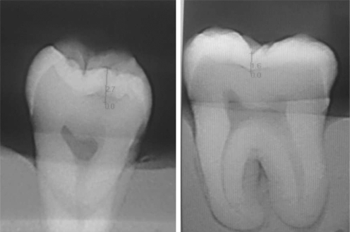
The present study sample was comprised of 21 human mandibular molar teeth with no restorations and with pit-fissure occlusal caries that were extracted due to periodontal loss or pericoronitis. Informed consent was obtained for the use of all extracted teeth. Teeth were cleaned of calculus and debris, disinfected in 2% sodium hypochlorite solution for 20 min, and stored in distilled water. Each tooth was embedded in a block of silicone impression material. All teeth were imaged using analog X-ray film, CCD digital X-ray, two different CBCT units and a micro-CT unit. Pilot studies were conducted to determine image acquisition exposure parameters, with visibility of the pulpal root canal, dentine, and enamel used as an indication of optimal image quality for all images. Intra-oral radiographs were taken using Size 2 E/F Kodak Insight Film (Eastman Kodak Co, Rochester, NY) and an RVG 5.0 CCD (Trophy, Marne la Valle, France) with a size 1 sensor and 10–15 lp mm spatial resolution. All intra-oral images were exposed under reproducible conditions using an Evaluation X 3000-2C X-Ray unit (New Life Radiology Srl, Grugliasco, Torino, Italy) at 70 kVp, 8 mA, and a focus-receptor distance of 30 cm behind a plexiglass soft-tissue equivalent. Exposure times were 0.4 s for film and 0.1 s for CCD. Films were automatically processed on the same day as exposure using an Extra-x Velopex (Medivance Instruments Ltd., London, England) and fresh chemicals (Hacettepe, Ankara, Turkey) in accordance with the manufacturer's instructions. Intraoral images obtained with RVG Trophy CCD sensor are shown in Fig. 1.
Fig. 1.
Intraoral images obtained with RVG Trophy CCD sensor
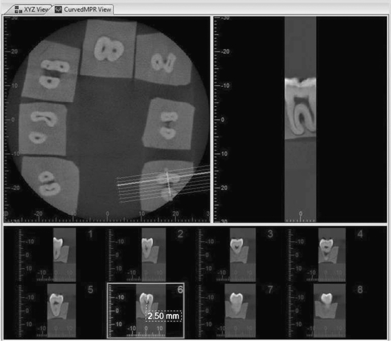
CBCT images were taken using Iluma (3M Imtec, Oklahoma, USA) and 3D Accuitomo 170 (3D Accuitomo; J Morita MFG. Corp, Kyoto, Japan) systems. Iluma images were obtained at 120 kV, 3.8 mA, and an exposure time of 40 s with a 24.4 × 19.5 cm amorphous silicon flat-panel image detector and reconstructed using a voxel size of 0.2 mm3. Accuitomo images were obtained at 63 kV, 2 mA, an exposure time of 30.8 s, and a voxel size of 0.125 mm using 6 cm × 6 cm field of view (FOV). Axial scans and multiplanar reconstructions were obtained for both CBCT systems, and volumetric data was reconstructed using the systems' own software programs to provide serial coronal and sagittal sections along each tooth plane. CBCT images obtained with Accuitomo 170 unit are shown in Fig. 2.
Fig. 2.
CBCT images obtained with Accuitomo 170 unit
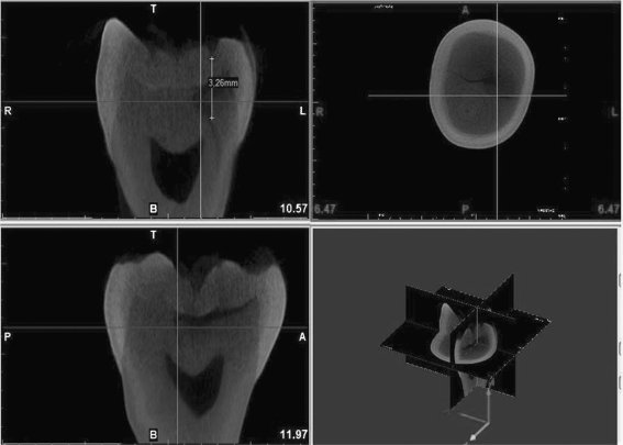
Micro-CT images of tooth crowns were obtained using a SkyScan 1174 (SkyScan, Kontich, Belgium) at 50 kV, 800 μA and a pixel size of 18-μm. Axial images were exported as bitmap (bmp) files to the Mimics software program (Materialise, Leuven, Belgium) in order to obtain serial coronal and sagittal images. While every scanning procedure, micro-CT system created a .log file which has whole scanning parameters in internationally calibrated metric system. Additionally, when uploading files to Mimics software, we also aligned and adjusted the sections according to parameters displayed in software interface. Micro-CT images obtained with SkyScan 1174 unit are shown in Fig. 3.
Fig. 3.
Micro-CT images obtained with SkyScan 1174 unit
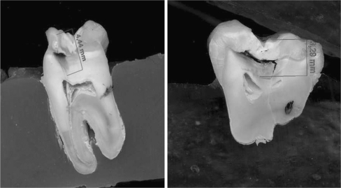
Following imaging, teeth were embedded in acrylic and serially sectioned mesio-distally in parallel to the long axis using a water-cooled diamond saw at low speed. For each tooth, the section with the deepest carious lesion was scanned using a high-resolution scanner (Epson Perfection V750-M Pro Scanner) at 600 dpi resolution and ×15 magnification and viewed using the AxioVision Rel. 4.7 image software (Carl Zeiss Imaging Solutions, Göttingen, Germany). Histological sections obtained from the teeth are shown in Fig. 4.
Fig. 4.
Histological sections obtained from the teeth
Images were evaluated separately by three oral radiologists experienced in image interpretation. Each image set (film, CCD, Iluma CBCT, Accuitomo CBCT, micro-CT, and histology) was viewed in a random order in a different session at 1-week intervals. Intra-observer agreement was assessed by having each observer view all image sets twice, with a 7-week interval between viewing to eliminate memory bias. Conventional film radiographs were evaluated using a light box and magnifier (×2), and a digital caliper (Absolute Digimatic, Mitutoyo Corp., Kawasaki, Japan) was used to measure lesion depth, defined as the distance from the enamel surface to the innermost part of the lesion. Digital 2-D and 3-D images were viewed on a 22" LG Flatron monitor (LG, Seoul, South Korea) at a 1,440 × 900 pixel screen resolution and 32-bit color depth. CCD and CBCT measurements of the largest dimensions of occlusal caries lesions were performed using the built-in measurement tools provided by the imaging software. Micro-CT measurements were obtained from the available sections using the Mimics software measurement tools, and histological measurements were performed using the AxioVision Rel. 4.7 imaging software.
Intra- and inter-rater reliability was assessed with the Bland/Altman method by calculating Intraclass Correlation Coefficients from residual variances of one-way ANOVA (with reading as the main variable) and two-way ANOVA (with the variables reading, observer and the interaction between the two). One-way ANOVA for each observer was also used to calculate a within-variability for each of them. Statistical analysis was performed using SAS 9.0 (PROC GLM), and Bland/Altman plots were drawn with the limits of agreement using MedCalc.
Results
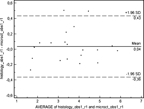
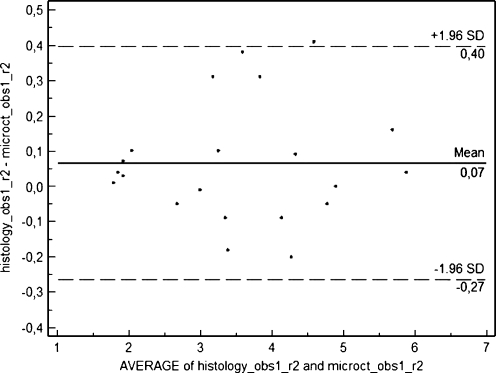
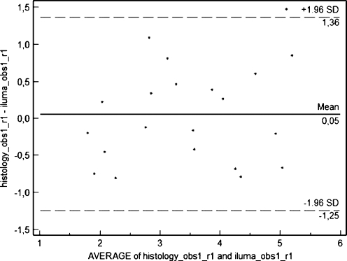
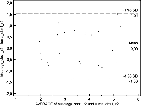
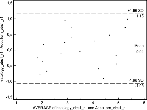
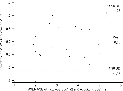
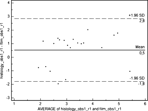
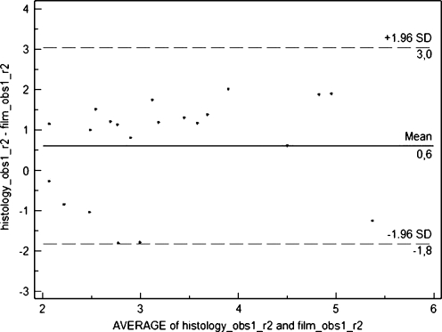
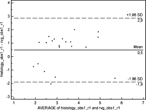
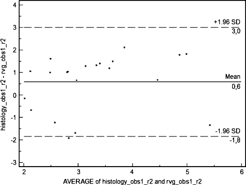
Occlusal caries depth measurements by modality, observer and reading are given in Table 1. In general, mean/median values were lower for intra-oral systems than for 3-D modalities and histology for all observers and both readings. ICC values for each observer are given in Table 2. Intra-observer ICC values for all observers were highest for histology and micro-CT. In addition, intra-observer ICC values were higher for histology and CBCT than for histology and intra-oral methods. Inter-observer ICC values for first and second readings were high for all observers (Tables 3 and 4). Bland-Altman plots (Figs. 5, 6, 7, 8, 9, 10, 11, 12, 13, and 14) showed very close agreement between micro-CT and histological measurements. (Given the high inter-observer ICC values for both readings, Bland/Altman plots were drawn for observer 1 only.) Repeatability of measurements for each modality is given in Table 5. No differences in repeatability were found between Accuitomo CBCT and Iluma CBCT images or between intra-oral film and CCD images.
Table 1.
Descriptive statistics for caries depth measurements obtained using different methods are given in millimeters
| Observer | Reading | Histology | Micro CT | CBCT (Iluma) | CBCT (Accuitomo) | Film | CCD (Rvg) | |
|---|---|---|---|---|---|---|---|---|
| 1 | 1 | |||||||
| Mean | 3.5257 | 3.4905 | 3.4714 | 3.4875 | 3.0014 | 3.0448 | ||
| Median | 3.5000 | 3.4100 | 3.6400 | 3.2550 | 2.8000 | 2.8100 | ||
| SD | 1.25313 | 1.23278 | 1.09188 | 1.12962 | .98717 | 1.07908 | ||
| Minimum | 1.54 | 1.81 | 1.90 | 1.95 | 1.51 | 1.66 | ||
| Maximum | 5.84 | 5.86 | 5.37 | 5.15 | 5.94 | 6.50 | ||
| 1 | 2 | |||||||
| Mean | 3.5748 | 3.5090 | 3.4862 | 3.5065 | 2.9629 | 2.9900 | ||
| Median | 3.3300 | 3.4000 | 3.5600 | 3.6000 | 2.8000 | 2.7000 | ||
| SD | 1.25116 | 1.23745 | 1.11685 | 1.16207 | 1.03450 | 1.04113 | ||
| Minimum | 1.80 | 1.79 | 1.80 | 1.90 | 1.50 | 1.60 | ||
| Maximum | 5.90 | 5.86 | 5.45 | 5.25 | 6.00 | 6.10 | ||
| 2 | 1 | |||||||
| Mean | 3.6095 | 3.5176 | 3.4929 | 3.4329 | 3.0376 | 3.0510 | ||
| Median | 3.7800 | 3.3400 | 3.4000 | 3.2000 | 2.9000 | 2.8700 | ||
| SD | 1.15680 | 1.12881 | 1.06354 | 1.07624 | 1.01788 | 1.02167 | ||
| Minimum | 1.90 | 1.84 | 1.90 | 1.90 | 1.60 | 1.70 | ||
| Maximum | 5.95 | 5.90 | 5.39 | 5.19 | 5.99 | 6.00 | ||
| 2 | 2 | |||||||
| Mean | 3.6333 | 3.6048 | 3.4429 | 3.4700 | 2.9195 | 2.9476 | ||
| Median | 3.5000 | 3.4800 | 3.5500 | 3.7000 | 2.8900 | 2.8700 | ||
| SD | 1.19814 | 1.16154 | 1.05910 | 1.05714 | .93118 | 1.03974 | ||
| Minimum | 1.76 | 1.80 | 1.70 | 1.80 | 1.50 | 1.40 | ||
| Maximum | 5.89 | 5.90 | 5.01 | 5.08 | 5.00 | 5.80 | ||
| 3 | 1 | |||||||
| Mean | 3.5581 | 3.5705 | 3.4900 | 3.5029 | 2.8129 | 2.9062 | ||
| Median | 3.8000 | 3.8000 | 3.2000 | 3.3000 | 2.8000 | 2.8000 | ||
| SD | 1.23774 | 1.23973 | 1.08835 | 1.07052 | .95900 | 1.08435 | ||
| Minimum | 1.50 | 1.40 | 2.00 | 2.20 | 1.40 | 1.35 | ||
| Maximum | 5.90 | 5.99 | 5.80 | 5.78 | 5.00 | 6.00 | ||
| 3 | 2 | |||||||
| Mean | 3.5667 | 3.5443 | 3.3748 | 3.3995 | 2.9910 | 2.9524 | ||
| Median | 3.5000 | 3.5000 | 3.0000 | 3.2500 | 2.8000 | 2.8000 | ||
| SD | 1.23077 | 1.23520 | 1.02609 | 1.01945 | 1.19872 | 1.16860 | ||
| Minimum | 1.80 | 1.80 | 1.90 | 2.00 | 1.60 | 1.60 | ||
| Maximum | 5.90 | 5.90 | 5.24 | 5.25 | 6.20 | 6.00 | ||
Table 2.
ICC values for intra-observer variability
| Method | Observer 1 | Observer 2 | Observer 3 |
|---|---|---|---|
| Histology and micro-CT | 0.989 | 0.982 | 0.997 |
| Histology and CBCT (Iluma) | 0.823 | 0.734 | 0.639 |
| Histology and Accuitomo | 0.882 | 0.794 | 0.669 |
| Histology and film | 0.431 | 0.317 | 0.254 |
| Histology and CCD (Rvg) | 0.440 | 0.296 | 0.294 |
Table 3.
ICC values for inter-observer variability for first readings
| Method | Obs1–obs2 | Obs1–obs3 | Obs2–obs3 |
|---|---|---|---|
| Histology | 0.942 | 0.958 | 0.920 |
| Micro-CT | 0.952 | 0.937 | 0.914 |
| CBCT (Iluma) | 0.963 | 0.910 | 0.916 |
| CBCT (Accuitomo) | 0.965 | 0.912 | 0.892 |
| Film | 0.959 | 0.895 | 0.932 |
| CCD (Rvg) | 0.950 | 0.918 | 0.956 |
Table 4.
ICC values for inter-observer variability for second readings
| Method | Obs1–obs2 | Obs1–obs3 | Obs2–obs3 |
|---|---|---|---|
| Histology | 0.964 | 0.962 | 0.920 |
| Micro-CT | 0.969 | 0.971 | 0.935 |
| CBCT (Iluma) | 0.975 | 0.956 | 0.948 |
| CBCT (Accuitomo) | 0.970 | 0.947 | 0.944 |
| Film | 0.968 | 0.932 | 0.896 |
| CCD (Rvg) | 0.979 | 0.912 | 0.926 |
Fig. 5.
Difference versus average of values measured by micro-CT and histology with 95% limits of agreement for the first reading of observer 1
Fig. 6.
Difference versus average of values measured by micro-CT and histology with 95% limits of agreement for the second reading of observer 1
Fig. 7.
Difference versus average of values measured by Iluma and histology with 95% limits of agreement for the first reading of observer 1
Fig. 8.
Difference versus average of values measured by Iluma and histology with 95% limits of agreement for the second reading of observer 1
Fig. 9.
Difference versus average of values measured by Accuitomo and histology with 95% limits of agreement for the first reading of observer 1
Fig. 10.
Difference versus average of values measured by Accuitomo and histology with 95% limits of agreement for the second reading of observer 1
Fig. 11.
Difference versus average of values measured by film and histology with 95% limits of agreement for the first reading of observer 1
Fig. 12.
Difference versus average of values measured by film and histology with 95% limits of agreement for the second reading of observer 1
Fig. 13.
Difference versus average of values measured by RVG and histology with 95% limits of agreement for the first reading of observer 1
Fig. 14.
Difference versus average of values measured by RVG and histology with 95% limits of agreement for the second reading of observer 1
Table 5.
Repeatability for each method
| Method | Obs1 | Obs2 | Obs3 |
|---|---|---|---|
| Histology | 0.30 | 1.05 | 1.00 |
| Micro-CT | 0.20 | 0.81 | 0.90 |
| CBCT (Iluma) | 0.36 | 0.73 | 1.28 |
| CBCT (Accuitomo) | 0.43 | 0.79 | 1.24 |
| Film | 0.25 | 0.71 | 1.25 |
| CCD (Rvg) | 0.33 | 0.61 | 1.25 |
Discussion
The present research mainly focused on observers' measurement reliability conducted on images obtained by using different X-ray systems. Observers performed occlusal caries measurements which were defined in millimeters. There are also various diagnostic tests for occlusal caries detection which are of clinical interest. DIAGNOdent (KaVo, Biberach, Germany), a quantitative laser fluorescence (LF) unit, is currently used as an aid in the diagnosis of occlusal carious lesions. LF was found to be capable of monitoring de-/re-mineralization process in early caries lesions in situ [26]. Electrical resistance measurement (ERM) which is proposed for occlusal caries detection uses a probe with a coaxial airflow and depends on the permeability changes due to caries demineralization of the tissues [27]. A study evaluated the performance of four observers by using ERM adjunct to visual inspection in diagnosing small dentinal lesions at one site per occlusal surface and the performance of ERM was also compared with that of radiographic examination. The ERM had the highest sensitivity, and visual inspection had the highest specificity. ROC analysis showed no statistically significant differences between the performance of the observers when using visual inspection and ERM (P > 0.05). One examiner performed statistically significantly better by measuring the electrical resistance of enamel than by radiographic examination (P < 0.05) [28]. Another in vitro study [27] compared two methods of ERM and found CRM (cariometer) prototype to perform similarly compared with ECM (electronic caries monitor). CRM (cariometer) prototype was proposed as a candidate for further development, owing to its simplicity and ease of usage [27]. In addition, thermal imaging was shown to be a promising technology for occlusal caries detection [29].
According to the findings of the present study, micro-CT and histological measurements of occlusal caries depth are highly comparable, suggesting that micro-CT may be used as a non-destructive ex vivo imaging modality. Previous studies by the authors found similarly favorable results in the ex-vivo imaging and assessment of teeth and teeth pathology using micro-CT images [24, 25]. However, while micro-CT has been established as an accurate gold standard for determining the presence or absence of caries [21], its effectiveness in caries depth measurement had not been previously assessed.
This study found intra-oral analog X-ray film and digital X-ray modalities performed similarly in the detection of occlusal caries depth. Accuracy of both intra-oral analog X-ray film and digital X-ray sensor measurements is limited by the two-dimensional nature of the technology, the superimposition of anatomical structures and patient-related factors; however, because of the lower radiation doses associated with intra-oral sensors and the built-in measurement tools of digital systems, digital is preferable to film technology. In the present study, calibration of intra-oral images through inclusion of an object with a known diameter within the image was not performed. Occlusal caries depth measurements from intra-oral images were lower when compared with histological assessment, suggesting that clinicians must be cautious when assessing caries depth using intra-oral radiography in routine clinical practice. However, micro-CT images were calibrated during importing process to Mimics software.
Our study found both CBCT units performed similarly and better than both intra-oral modalities in measuring occlusal caries depth. These results are analogous to those of a previous study that found CBCT performed better than intra-oral modalities in millimetric proximal caries depth measurement [18]. Another recent study found CBCT to be superior to intra-oral imaging in the detection of deep enamel occlusal caries, superficial dentine caries and deep dentine caries, but found no differences between modalities in the detection of superficial enamel occlusal caries or healthy subjects [19]. The present study included dentine caries lesions only.
CBCT systems offer a variety of different image acquisition settings. In this study, the Accuitomo 170 CBCT images were acquired at 6 × 6 cm (0.125 mm3 voxel resolution), and the Iluma CBCT images were reconstructed using a voxel size of 0.2 mm3, since they are more applicable in routine applications in terms of reconstruction times. It is possible that measurement accuracy would have increased by using the smallest voxel sizes available (4 × 4 FOV/0.08 mm3 voxel resolution with the Accuitomo CBCT system and 0.1 mm3 voxel size with the Iluma CBCT system).
In clinical situations, the quality of CBCT images can be seriously affected by patient motion and by beam hardening and scatter caused by metallic restorations; however, neither of these issues were factors in the present study, which was conducted ex vivo and did not include teeth with restorations.
CBCT is not considered suitable for use in routine clinical dental practice because of the high effective radiation doses. A recent study reported effective doses for the Accuitomo CBCT to vary between 11 and 77 μSv, depending on FOV, exposure parameters, and region examined [30]. According to ICRP 2007 tissue weights, Iluma CBCT images acquired at 120 kVp and 3.8 mA (as in the present study) result in an effective dose of 498 μSv [31]. Current research is being directed towards offering higher-definition 3-D images with similar or lower effective doses than film. Although micro-CT is an innovative device offering high-definition dental images, the ultra-high radiation dose, high cost, and long scanning and image reconstruction times associated with micro-CT make it unsuitable for clinical use. However, in view of the results of the present study, it can be recommended as a non-destructive ex vivo method for dental caries depth measurement.
Conclusion
This study found micro-CT to be the best imaging method for use in the ex vivo measurement of occlusal caries depth. In addition, CBCT units were found to perform similarly and better than intra-oral modalities.
References
- 1.Poorterman JHG, Weerheijm KL, Groen HJ, Kalsbeek H. Clinical and radiographic judgment of occlusal caries in adolescents. Eur J Oral Sci. 2000;108:93–98. doi: 10.1034/j.1600-0722.2000.00791.x. [DOI] [PubMed] [Google Scholar]
- 2.Dowker SEP, Elliott JC, Davis GR, Wilson RM, Cloetens P. Three-dimensional study of human dental fissure enamel by synchrotron X-ray microtomography. Eur J Oral Sci. 2006;114(Suppl. 1):353–359. doi: 10.1111/j.1600-0722.2006.00315.x. [DOI] [PubMed] [Google Scholar]
- 3.Shi XQ, Welander U, Angmar-Mansson B. Occlusal caries detection with KaVo DIAGNOdent and radiography: an in vitro comparison. Caries Res. 2000;34:151–158. doi: 10.1159/000016583. [DOI] [PubMed] [Google Scholar]
- 4.Kühnisch J, Dietz W, Stösser L, Hickel R, Heinrich-Weltzien R. Effects of dental probing on occlusal surfaces—a scanning electron microscopy evaluation. Caries Res. 2007;41:43–48. doi: 10.1159/000096104. [DOI] [PubMed] [Google Scholar]
- 5.Souza-Zaroni WC, Ciccone JC, Souza-Gabriel AE, Ramos RP, Corona SAM, Palma-Dibb RG. Validity and reproducibility of different combinations of methods for occlusal caries detection: an in vitro comparison. Caries Res. 2006;40:194–201. doi: 10.1159/000092225. [DOI] [PubMed] [Google Scholar]
- 6.Hamilton JC, Gregory WA, Valentine JB. DIAGNOdent measurements and correlation with the depth and volume of minimally invasive cavity preparations. Oper Dent. 2006;31:291–296. doi: 10.2341/05-47. [DOI] [PubMed] [Google Scholar]
- 7.Jablonski-Momeni A, Stachniss V, Ricketts DN, Heinzel-Gutenbrunner M, Pieper K. Reproducibility and accuracy of the ICDAS-II for detection of occlusal caries in vitro. Caries Res. 2008;42:79–87. doi: 10.1159/000113160. [DOI] [PubMed] [Google Scholar]
- 8.Rodrigues JA, Hug I, Diniz MB, Lussi A. Performance of fluorescence methods, radiographic examination and ICDAS II on occlusal surfaces in vitro. Caries Res. 2008;42:297–304. doi: 10.1159/000148162. [DOI] [PubMed] [Google Scholar]
- 9.Abalos C, Herrera M, Jimenez-Planas A, Llamas R. Performance of laser fluorescence for detection of occlusal dentinal caries lesions in permanent molars: an in vivo study with total validation of the sample. Caries Res. 2009;43:137–141. doi: 10.1159/000209347. [DOI] [PubMed] [Google Scholar]
- 10.Hintze H, Wenzel A. Clinical and laboratory radiographic caries diagnosis. A study same teeth. Dentomaxillofac Radiol. 1996;25:115–118. doi: 10.1259/dmfr.25.3.9084258. [DOI] [PubMed] [Google Scholar]
- 11.Kositbowornchai S, Basiw M, Promwang Y, Moragorn H, Sooksuntisakoonchai N. Accuracy of diagnosing occlusal caries using enhanced digital images. Dentomaxillofac Radiol. 2004;33:236–240. doi: 10.1259/dmfr/94305126. [DOI] [PubMed] [Google Scholar]
- 12.Ricketts DNJ, Ekstrand KR, Martignon S, Ellwood R, Alatsaris M, Nugent Z. Accuracy and reproducibility of conventional radiographic assessment and subtraction radiography in detecting demineralization in occlusal surfaces. Caries Res. 2007;41:121–128. doi: 10.1159/000098045. [DOI] [PubMed] [Google Scholar]
- 13.Pereira AC, Eggertsson H, Martinez-Mier EA, Mialhe FL, Eckert GJ, Zero DT. Validity of caries detection on occlusal surfaces and treatment decisions based on results from multiple caries-detection methods. Eur J Oral Sci. 2009;117:51–57. doi: 10.1111/j.1600-0722.2008.00586.x. [DOI] [PubMed] [Google Scholar]
- 14.Kühnisch J, Ifland S, Tranaeus S, Heinrich-Weltzien R. Comparison of visual inspection and different radiographic methods for dentin caries detection on occlusal surfaces. Dentomaxillofac Radiol. 2009;38:452–457. doi: 10.1259/dmfr/34393803. [DOI] [PubMed] [Google Scholar]
- 15.Kamburoğlu K, Şenel B, Yüksel SP, Özen T. A comparison of the diagnostic accuracy of in vivo and in vitro photostimulable phosphor digital images in the detection of occlusal caries lesions. Dentomaxillofac Radiol. 2010;39:17–22. doi: 10.1259/dmfr/91657756. [DOI] [PMC free article] [PubMed] [Google Scholar]
- 16.Scarfe WC, Farman AG. What is cone-beam CT and how does it work? Dent Clin North Am. 2008;52:707–730. doi: 10.1016/j.cden.2008.05.005. [DOI] [PubMed] [Google Scholar]
- 17.Tyndall DA, Rathore S. Cone-beam CT diagnostic applications: caries, periodontal bone assessment, and endodontic applications. Dent Clin North Am. 2008;52:825–841. doi: 10.1016/j.cden.2008.05.002. [DOI] [PubMed] [Google Scholar]
- 18.Akdeniz BG, Gröndahl HG, Magnusson B. Accuracy of proximal caries depth measurements: comparison between limited cone beam computed tomography, storage phosphor and film radiography. Caries Res. 2006;40:202–207. doi: 10.1159/000092226. [DOI] [PubMed] [Google Scholar]
- 19.Kamburoğlu K, Murat S, Yüksel SP, Cebeci ARİ, Paksoy CS. Occlusal caries detection by using a cone-beam CT with different voxel resolutions and a digital intraoral sensor. Oral Surg Oral Med Oral Pathol Oral Radiol Endod. 2010;109:e63–e69. doi: 10.1016/j.tripleo.2009.12.048. [DOI] [PubMed] [Google Scholar]
- 20.Haiter-Neto F, Wenzel A, Gotfredsen E. Diagnostic accuracy of cone beam computed tomography scans compared with intraoral image modalities for detection of caries lesions. Dentomaxillofac Radiol. 2008;37:18–22. doi: 10.1259/dmfr/87103878. [DOI] [PubMed] [Google Scholar]
- 21.Young SM, Lee JT, Hodges RJ, Chang TL, Elashoff DA, White SC. A comparative study of high-resolution cone beam computed tomography and charge-coupled device sensors for detecting caries. Dentomaxillofac Radiol. 2009;38:445–451. doi: 10.1259/dmfr/88765582. [DOI] [PubMed] [Google Scholar]
- 22.Jablonski-Momeni A, Ricketts DNJ, Stachniss V, Maschka R, Heinzel-Gutenbrunner M, Pieper K. Occlusal caries: evaluation of direct microscopy versus digital imaging used for two histological classification systems. J Dent. 2009;37:204–211. doi: 10.1016/j.jdent.2008.11.014. [DOI] [PubMed] [Google Scholar]
- 23.Schwass DR, Swain MV, Purton DG, Leichter JW. A system of calibrating microtomography for use in caries research. Caries Res. 2009;43:314–321. doi: 10.1159/000226230. [DOI] [PubMed] [Google Scholar]
- 24.Kamburoğlu K, Barenboim SF, Arıtürk T, Kaffe I. Quantitative measurements obtained by micro-computed tomography and confocal laser scanning microscopy. Dentomaxillofac Radiol. 2008;37:385–391. doi: 10.1259/dmfr/57348961. [DOI] [PubMed] [Google Scholar]
- 25.Neves Ade A, Coutinho E, Cardoso MV, Jaecques SV, Meerbeek B. Micro- CT based quantitative evaluation of caries excavation. Dent Mater. 2010;26:579–588. doi: 10.1016/j.dental.2010.01.012. [DOI] [PubMed] [Google Scholar]
- 26.Spiguel MH, Tovo MF, Kramer PF, Franco KS, Alves KMPR, Delbem ACB. Evaluation of laser fluorescence in the monitoring of the initial stage of the de-/remineralization process: an in vitro and in situ study. Caries Res. 2009;43:302–307. doi: 10.1159/000218094. [DOI] [PubMed] [Google Scholar]
- 27.Kühnisch J, Heinrich-Weltzien R, Tabatabaie M, Stösser L, Huysmans MCDNJM. An in vitro comparison between two methods of electrical resistance measurement for occlusal caries detection. Caries Res. 2006;40:104–111. doi: 10.1159/000091055. [DOI] [PubMed] [Google Scholar]
- 28.Verdonschot EH, Wenzel A, Truin GJ, König KG. Performance of electrical resistance measurements adjunct visual inspection in the early diagnosis of occlusal caries. J Dent. 1993;21:332–337. doi: 10.1016/0300-5712(93)90003-9. [DOI] [PubMed] [Google Scholar]
- 29.Zakian CM, Taylor AM, Ellwood RP, Pretty IA. Occlusal caries detection by using thermal imaging. J Dent. 2010;38:788–795. doi: 10.1016/j.jdent.2010.06.010. [DOI] [PubMed] [Google Scholar]
- 30.Lofthag-Hansen S, Thilander-Klang A, Ekestubbe A, Helmrot E, Gröndahl K. Calculating effective dose on a cone beam computed tomography device: 3D Accuitomo and 3D Accuitomo FPD. Dentomaxillofac Radiol. 2008;37:72–79. doi: 10.1259/dmfr/60375385. [DOI] [PubMed] [Google Scholar]
- 31.Ludlow JB, Ivanovic M. Comparative dosimetry of dental CBCT devices and 64-slice CT for oral and maxillofacial radiology. Oral Surg Oral Med Oral Pathol Oral Radiol Endod. 2008;106:106–114. doi: 10.1016/j.tripleo.2008.03.018. [DOI] [PubMed] [Google Scholar]


