Abstract
Bone hemangiomas are benign and infrequent lesions. At Tc-99m bone scintigraphy they show variable degrees of radiotracer uptake and even absence of it. At fluordeoxy-glucose (FDG) Positron Emission Tomography-Computed Tomography (PET/CT), hemangioma is one of the causes of “cold” vertebrae, apart from postexternal radiotherapy. We present a woman diagnosed of breast carcinoma, with a photopenic defect at a thoracic vertebrae at Tc-99m bone scan. In order to rule out bone lytic metastasis, a FDG PET/CT was performed showing a “cold” vertebrae too. Findings were highly suggestive of vertebral hemangioma, that was confirmed by magnetic resonance imaging.
Keywords: Bone hemangioma, cold vertebrae, fluordeoxy-glucose, positron emission tomography/computed tomography, photopenic
INTRODUCTION
Bone haemangiomas are benign and infrequent lesions. They may be erroneously labelled as metastases on bone scan in a known case of primary malignancy with predilection for skeletal (lytic) metastases. Authors describe a case of breast carcinoma with vertebral haemangioma posing diagnostic difficulty. Scintigraphic findings on bone scan and FDG PET- CT scans were confirmed with magnetic resonance imaging.
CASE REPORT
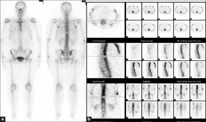
We present a 47-year-old woman who had been diagnosed of infiltrating lobular breast carcinoma and had undergone right mastectomy and lymphadenectomy. Before starting chemotherapy treatment, a bone scan with Tc-99m methyl diphosphonate (MDP) was performed. A “cold” defect was detected at the ninth thoracic (T9) vertebra at both anterior and posterior projections [Figure 1a], which was confirmed by tomographic images. Transversal, sagittal and coronal slices are shown in Figure 1b.
Figure 1.

A “cold” defect was detected at the ninth thoracic (T9) vertebra at both anterior and posterior projections (a), which was confirmed by tomographic images. Transversal, sagittal and coronal slices are shown in (b)
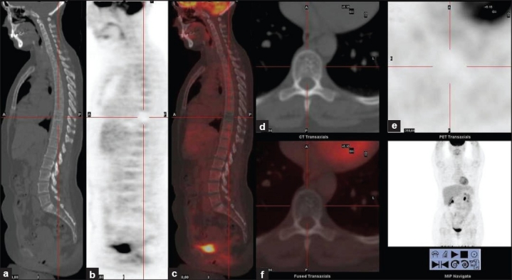
Because of her personal history of cancer, the vertebral finding was highly suggestive of lytic bone metastases. So, an F-18 fluordeoxy-glucose (FDG) positron emission tomography/computed tomography (PET/CT) study was performed. In Figure 2 are shown CT (a), PET (b) and PET/CT (c) sagittal slices, and CT (d), PET (e) and PET/CT (f) transversal slices. The only pathological finding at the F-18 FDG PET/CT study was an ametabolic area at the T9 vertebra on PET images. CT showed thickened vertical trabeculae on sagittal images and punctate sclerotic foci on transversal images at the body of T9. Findings were highly suggestive of vertebral hemangioma [Figure 2].
Figure 2.

Computed tomography (CT) (a), positron emission tomography (PET) (b) and PET/CT (c) sagittal slices, and CT (d) PET (e) and PET/CT (f) transversal slices. The only pathological finding at the F-18 FDG PET/CT study was an ametabolic area at the T9 vertebra on PET images. CT shows thickened vertical trabeculae on sagittal images and punctate sclerotic foci on transversal images at the body of T9. Findings are highly suggestive of vertebral hemangioma
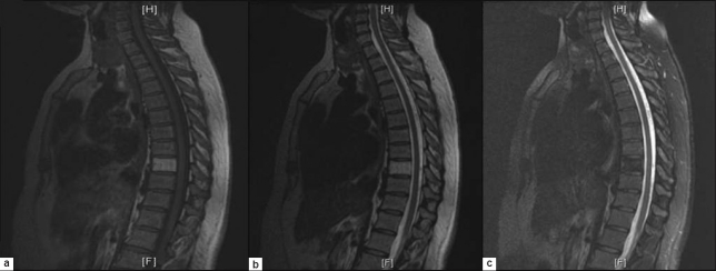
A magnetic resonance was performed in order to support the diagnosis. The body of T9 appeared as an area of high signal intensity on T1-T2-weighted images [Figure 3a and b] and fat suppression at T2-weighted FatSat images [Figure 3c]. Magnetic resonance imaging (MRI) confirmed the presence of a vertebral hemangioma. Bone hemangiomas are rare and benign tumors. On Tc-99m MDP bone scintigraphy, they show variable degrees of radiotracer uptake and even absence of it[1] and when this happens, metastatic bone disease must be ruled out because it is the most frequent cause of photon-deficient lesions on bone scintigraphy.[2,3] On F-18 FDG PET/CT, that hemangioma is one of the causes of "cold" vertebrae, apart from postexternal radiotherapy.[4] CT and MRI images can demonstrate typical signs of hemangioma which support diagnosis [Figure 3].[5,6]
Figure 3.
The body of T9 appeared as an area of high signal intensity on T1-T2-weighted images (a and b) and fat suppression at T2-weighted FatSat images (c)
Footnotes
Source of Support: Nil
Conflict of Interest: None declared.
REFERENCES
- 1.Sarikaya I, Sarikaya A, Holder LE. The role of single photon emission computed tomography in bone imaging. Semin Nucl Med. 2001;31:3–16. doi: 10.1053/snuc.2001.18736. [DOI] [PubMed] [Google Scholar]
- 2.Sopov V, Liberson A, Gorenberg M, Groshar D. Cold vertebrae on bone scintigraphy. Semin Nucl Med. 2001;31:82–3. doi: 10.1053/snuc.2001.21076. [DOI] [PubMed] [Google Scholar]
- 3.Horger M, Bares R. The role of single-photon emission computed tomography/computed tomography in benign and malignant bone disease. Semin Nucl Med. 2006;36:286–94. doi: 10.1053/j.semnuclmed.2006.05.001. [DOI] [PubMed] [Google Scholar]
- 4.Basu S, Nair N. “Cold” vertebrae on F-18 FDG PET: Causes and characteristics. Clin Nucl Med. 2006;31:445–50. doi: 10.1097/01.rlu.0000227011.21544.19. [DOI] [PubMed] [Google Scholar]
- 5.Persaud T. The Polka-Dot Sign. Radiology. 2008;246:980–1. doi: 10.1148/radiol.2463050903. [DOI] [PubMed] [Google Scholar]
- 6.Ross JS, Masaryk TJ, Modic MT, Carter JR, Mapstone T, Dengel FH. Vertebral hemangiomas: MR imaging. Radiology. 1987;165:165–9. doi: 10.1148/radiology.165.1.3628764. [DOI] [PubMed] [Google Scholar]


