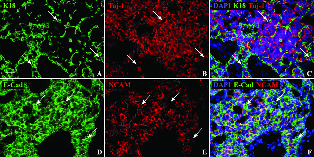
Figure 8.
Sox9(+)/Tuj-1(+) tumor cells do not follow the usual pattern of staining of olfactory epithelial gland cells. A,B,C. The separation of the tumor into two populations is confirmed using K18 and Tuj-1 antibodies. Similar to the two cell populations demonstrated with Sox2 and Sox9 antibodies, immunofluorescent labeling of cell cytoplasm and processes using K18 (arrows), does not co-label with the neuron-specific Tuj-1 antibody. D,E,F. E-Cad has a similar staining pattern as K18 in the epithelium labeling both gland and supporting cells, however within the tumor K18 is restricted to the Sox2(+)/Sox9(−)/Tuj-1(−) population while E-Cad labels the same NCAM(−) non-neuronal population (arrows) as well as the NCAM(+) neuronal population. Scale bar = 25µ.

