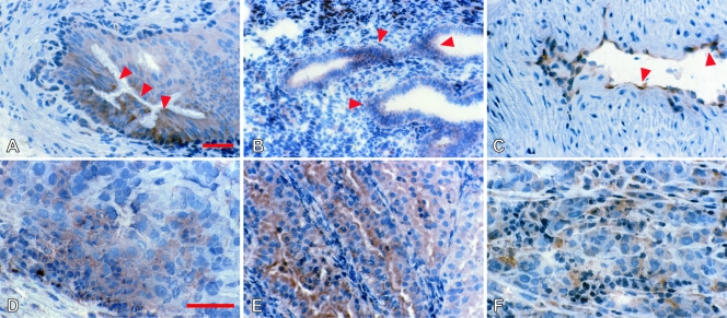
Figure 1.
Immunohistochemical analysis of HLA-E. Normal (A–C) and neoplastic (D–F) nonlymphoid human tissues were stained with MEM-E/02 and nuclear counterstained with Mayer hematoxylin. A ground-glass pattern was detected in the principal but not the basal cells in the epididymis (A, arrows), in endometrial cells (B, arrows), and in the vascular endothelium of the myometrium (C, arrows). In tumors, variable expression was seen in a case of osteosarcoma (D), a well-differentiated endometrial carcinoma (E), and an in transit metastatic melanoma (F). Original magnifications, x160 (A–C, E); x250 (D and F). Scale bars, 100 µm.

