



Abstract
INTRODUCTION
Surgical decision-making in torso trauma is complex. This paper looks at the role of the computed tomography scan in this decision-making process.
PATIENTS AND METHODS
Patients with significant torso trauma (high velocity gunshot wound [HVGSW], blast, stab) admitted to a military role 2 (enhanced) hospital facility during a 7-week period of Operation HERRICK 9 (Afghanistan, October to November 2008) are reported. The management of those patients undergoing a CT scan as part of the decision-making process at the time of admission is discussed.
RESULTS
Twenty eight patients with significant torso trauma were admitted to the facility during the study period; HVGSW (n = 15), blast (n = 9), stab (n = 4). Thirteen patients underwent a CT scan as part of the surgical decision-making process; HVGSW (n = 5), blast (n = 8). Imaging confirmed torso integrity in 12 patients, one of whom subsequently had a laparotomy for vascular control for on-table haemorrhage during lower limb surgery. One patient had a confirmed thoraco-abdominal injury, which was treated conservatively with tube thoracostomy and ‘active observation’.
CONCLUSIONS
A CT scan formed part of the surgical decision-making process in about half of the patients admitted with significant torso trauma, and helped prevent unnecessary laparotomy in this forward military environment. Those patients with a blast injury were more likely to undergo CT scanning than those where the mechanism of injury was a HVGSW.
Keywords: Torso trauma, CT scan, Decision making, Military field hospital
Decision making in patients with penetrating torso trauma is complex. In UK trauma practice, where blunt trauma predominates, computed tomography (CT) is a vital element in diagnosis and definition of injury. It tends to be used to rule in disease in patients who have minimal or reversible physiological compromise, utilising its high specificity.1,2 In the deployed military hospital, CT is a limited resource with only one scanner for often many severely injured patients. While CT may help to define injury in penetrating trauma in some circumstances, there are often other reasons to proceed directly to the operating theatre. One element of surgical triage is an assessment of whether peritoneal penetration has occurred, and whether there is intraperitoneal injury necessitating laparotomy.3
This paper describes the use of CT in clinical decision-making in a series of cases of penetrating torso trauma presenting to a deployed military field hospital.
Patients and Methods
Patients with significant torso trauma (high velocity gunshot wound [HVGSW], blast, stab) admitted to a military role 2 (enhanced) hospital facility during a 7-week period of Operation HERRICK 9 (Afghanistan, October to November 2008) are reported. Patients with haemodynamic instability, anatomical and mechanistic injuries obviously requiring surgery, or those in whom surgery was not considered for other reasons, were excluded from the analysis.
The management of those patients for whom a CT scan was performed as part of the clinical decision-making process at the time of admission is discussed.
Results
Twenty-eight patients with significant torso trauma were admitted to the facility during the study period: HVGSW (n = 15), blast (n = 9), stab (n = 4). Fifteen patients were excluded from the analysis according to the above criteria: HVGSW (n = 10), blast (n = 1), stab (n = 4).
Thirteen patients underwent a CT scan as part of the management decision-making process; HVGSW (n = 5), blast (n = 8). Imaging confirmed torso integrity in 12 patients. One patient subsequently had a laparotomy for vascular control for on-table haemorrhage during lower limb surgery following injury from an explosion.
One patient had a confirmed thoraco-abdominal injury that was treated conservatively with tube thoracostomy and ‘active observation’. This patient was an Afghan male who presented with HVGSWs to the right shoulder and neck. He had three wounds around the shoulder and neck – two wounds in zone 1, and one wound in zone 3. His prehospital vital signs were all within normal limits. He underwent prehospital needle thoracocentesis. His chest X-ray demonstrated a wound track through the right side of the chest and a possible intraperitoneal fragment (Fig. 1). His vital signs remained normal. A CT scan confirmed the intrathoracic injury and demonstrated a fragment below the diaphragm (Fig. 2A–C). In the absence of peritonism, he was managed conservatively with right tube thoracostomy and antibiotics, and made a full recovery.
Figure 1.
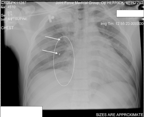
The trauma chest X-ray demonstrates a pneumothorax with debris along the line of the ballistic tract, which extends below the diaphragm (circled with arrows identifying debris).
Figure 2.
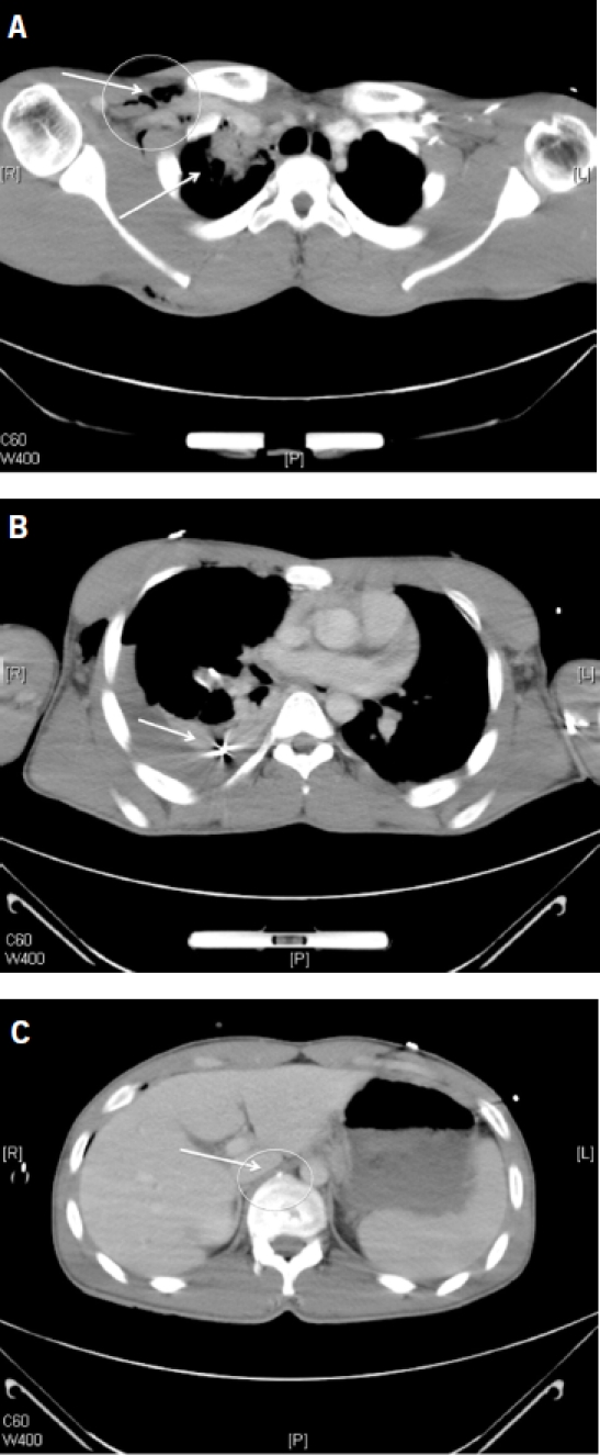
The post-chest drain CT scan demonstrates air in the soft tissues of the right side of the neck (circled) with fragments and contusion at the lung apex (A); haemopneumothorax with fragments inside the chest, see arrow (B); and a small fragment anterior to a vertebral body within the abdomen, circled (C).
Discussion
Diagnostic tests are useful when they aid clinical management by confirming or refuting a diagnosis (ruling in or ruling out disease). Those tests with a high specificity are used to rule in disease (SpIn) and those with a high sensitivity are typically used to rule out disease (SnOut).4 CT has both high specificity and sensitivity for detecting intraperitoneal injury,1,2 but these differ with respect to what the test is looking for – solid organ injury, hollow viscus perforation, the presence of free fluid (usually but not exclusively blood), and other subtle signs of intraperitoneal injury.5
A CT scan formed part of the decision-making process in about half of the patients admitted to our unit with significant torso trauma. It helped prevent unnecessary laparotomy in this forward military environment. Those patients with a blast injury were more likely to undergo CT scanning than those where the mechanism of injury was a HVGSW. Many of these patients had on-going unexplained tachycardia, which could have represented on-going occult haemorrhage.
In the UK, where blunt trauma predominates, CT is most often used as a SpIn; it is used to rule in disease in a population where injury is not obvious. In a deployed military medical facility where penetrating trauma predominates and CT is a limited resource, it is useful in selected patients where the result will affect the management of the patient. In this series, it was effectively used as a SnOut, i.e. used to rule out significant intraperitoneal injury necessitating laparotomy. It was not used in patients who required operative intervention.
This approach may need a significant shift in thinking for clinicians who rely on CT scans on a much larger proportion of trauma patients in their UK practice.
Conclusions
CT is a useful diagnostic tool when used appropriately in a deployed military hospital. In this series, cavity surgery was avoided in patients who may otherwise have undergone coeliotomy. It was effectively used as a SnOut rather than a SpIn.
Acknowledgments
This paper was presented, in part, at the International Surgical Congress of the Association of Surgeons of Great Britain and Ireland, Glasgow, 2009.
References
- 1.Shuman WP. CT of blunt abdominal trauma in adults. Radiology. 1997;205:297–306. doi: 10.1148/radiology.205.2.9356606. [DOI] [PubMed] [Google Scholar]
- 2.ACEP Clinical Policies Committee; Clinical Policies Subcommittee on Acute Blunt Abdominal Trauma. Clinical policy: critical issues in the evaluation of adult patients presenting to the emergency department with acute blunt abdominal trauma. Ann Emerg Med. 2004;43:278–90. doi: 10.1016/j.annemergmed.2003.10.045. [DOI] [PubMed] [Google Scholar]
- 3.Shanmuganathan K, Mirvis SE, Chiu WC, Killeen KL, Scalea TM. Triple-contrast helical CT in penetrating torso trauma: a prospective study to determine peritoneal violation and the need for laparotomy. AJR Am J Roentgenol. 2001;177:1247–56. doi: 10.2214/ajr.177.6.1771247. [DOI] [PubMed] [Google Scholar]
- 4.Sackett DL, Straus SE, Richardson WS, Rosenberg W, Haynes RB. Evidence-Based Medicine. 2nd edn. Edinburgh: Churchill Livingstone; 2000. pp. 249–50. [Google Scholar]
- 5.Brody JM, Leighton DB, Murphy BL, Abbott GF, Vaccaro JP, et al. CT of blunt trauma bowel and mesenteric injury: typical findings and pitfalls in diagnosis. Radiographics. 2000;20:1525–36. doi: 10.1148/radiographics.20.6.g00nv021525. [DOI] [PubMed] [Google Scholar]