Abstract
Background
Open reduction and stabilization of dorsal pelvic ring injuries is accompanied by a high rate of soft tissue complications. Minimally invasive techniques have the potential to decrease soft tissue trauma, but the risk of iatrogenic nerve and vessel damage through the reduced surgical exposure should be considered. We treated these injuries using a transiliac internal fixator (TIFI) in a minimally invasive technique characterized by implantation of a pedicle screw and rod system, bridging the sacroiliac joints and the sacral area.
Questions/purposes
We asked whether (1) we could achieve anatomic restoration with the device, (2) specific complications were associated with this minimally invasive approach (particularly enhanced intraoperative blood loss, soft tissue complications, and iatrogenic neurovascular damage), and (3) function 3 years after trauma was comparable to that of established methods.
Methods
We retrospectively reviewed 67 patients with dorsal pelvic injuries during a 7-year period. We evaluated the (1) reduction by grading the maximal displacement measured with three radiographic views, (2) the complications during the observation period, and (3) the function with a validated questionnaire (Pelvic Outcome Score) in all but five patients at least 3 years after trauma (mean, 37 months; range, 36–42 months).
Results
At last followup we observed a secondary fracture displacement greater than 5 mm in one patient. The intraoperative blood loss was less than 50 mL in all patients. No neurovascular lesions occurred owing to implantation. Four patients had wound infections, one had loosening of a single pedicle screw, and one had an iatrogenic screw malpositioning. Thirty-five of the 62 patients achieved Pelvic Outcome Scores of either a maximum score or 6 of 7 points.
Conclusion
Our observations suggest TIFI is a reasonable alternative to other established fixation devices for injuries of the dorsal pelvic ring with minor risks of major blood loss or iatrogenic neurovascular damage.
Level of Evidence
Level IV, therapeutic study. See Guidelines for Authors for a complete description of levels of evidence.
Introduction
Unstable pelvic ring fractures are severe injuries with a mortality rate of as much as 30%, mainly owing to hemorrhage or accompanying injuries [3, 37]. They usually are attributable to high-energy injuries [27, 38]. The majority of surviving patients consider their quality of life as mediocre or worse [7, 10, 17, 39], and the treatment often is challenging for the surgeon.
One report suggests invasive stabilization is important [13]. However, some surgical approaches for treatment of unstable posterior pelvic ring injuries have disadvantages: (1) anterior plate fixation of the sacroiliac joint places the L5 root at risk during dissection and implant placement, and tremendous blood loss [2, 8]; (2) posterior fixation with transiliac sacral bars that bridge the contralateral sacroiliac joint and cause discomfort in thin patients owing to prominent implants [4]; (3) posterior plate osteosynthesis (open or in a minimally invasive percutaneous technique), intraoperatively, correct bending of the plates is sometimes difficult to achieve and an extended approach for hardware removal might be necessary even in initial percutaneous techniques [19]; and (4) screw fixation of the sacroiliac joint (open or in a percutaneous technique) for which an experienced surgeon and high intraoperative fluoroscopic quality are essential [1, 30, 34]. One mechanical study suggests there are no differences in secondary fracture displacement among these techniques, and the best treatment remains controversial [42].
Surgical exposure with open reduction and fixation can be associated with subsequent wound-healing problems and high infection rates. When using posterior open reduction and internal fixation, the range of wound complications varies among studies from 5% to 25% [15, 18, 32]. Minimally invasive, percutaneous procedures minimize soft tissue injury but the risk of iatrogenic neurovascular lesions is not negligible. Furthermore anatomic reduction does not guarantee patient satisfaction [7]. Long-term problems associated with operatively treated pelvic ring injuries include various complications such as pain, restrictions in activities of daily life, sexual dysfunction, or urinary system complaints [27].
To address the concerns regarding neurovascular injury, we developed a transiliac internal fixator in a minimal incision technique that does not require wide exposure of the fracture side. We minimize the risk of neurovascular injury by inserting only two pedicle screws and connecting them subfascial with a rod. The technique involves bridging the sacroiliac joints and the sacral area instead of a wide surgical exposure of this region. This technique was previously reported in a study of 31 patients [11]. In that study, two patients had wound infections, none experienced iatrogenic neurovascular damage, and overall, patients had high satisfaction with 50% of the patients attaining 6 or 7 of 7 points in the Pelvic Outcome Score. However that study had only a small patient collective with a maximum of 2 years followup, so conclusions for generally recommended use for this device were not possible.
To confirm the findings of the previous study [11], we asked whether (1) we could achieve anatomic restoration with the device, (2) specific complications were associated with this minimally invasive approach (particularly enhanced intraoperative blood loss, soft tissue complications, and iatrogenic neurovascular damage), and (3) function 3 years after trauma were comparable to that of established methods.
Patients and Methods
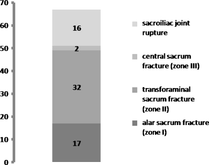
We retrospectively reviewed 67 patients who underwent minimally invasive reduction and fixation of a dorsal pelvic ring injury between January 2000 and December 2007. According to Tile’s classification [37], we classified the injuries as C1 (46 patients), C2 (11 patients), and C3 (10 patients). Of the 67 injuries, 16 were sacroiliac displacements and 51 were sacral fractures. Of the 51 patients with sacral fractures, according to the classification of Denis et al. [6], 17 involved the alae (Zone I), 32 involved the transforamina (Zone II), and two involved the sacral body (Zone III) (Fig. 1). Sixty patients had multiple injuries and seven had isolated pelvic ring injuries accompanied only by minor extremity injuries. Seventeen patients had a complex pelvic ring injury [41]. The predominant injury mechanisms were motor vehicle accidents (30 of 67) followed by falls from a height (26 of 67). The mean patient age at the time of surgery was 36.7 years (range, 16–76 years). Twenty-nine patients were female, and 38 were male (Table 1). The indications for surgery were: (1) sacroiliac displacement and (2) sacral fractures. We considered sacroiliac disruptions with an accompanying osseous lesion of the dorsal ilium and bilateral instabilities as contraindications to surgery. Five of the 67 patients did not complete followup: two died during the early postoperative period (one owing to traumatic brain injury and the other from heart failure), and three moved away or were not available for reevaluation owing to personal reasons. We therefore evaluated 62 patients at a minimum of 36 months (mean, 37 months; range, 36–42 months). No patients were recalled specifically for this study; all data were obtained from medical records and radiographs.
Fig. 1.
Fracture localization of the 67 patients involved in the study is shown.
Table 1.
Patient demographics and injury mechanism
| Variable | Number of patients (N = 67) | Age (years) (mean, 54.3) |
|---|---|---|
| Gender | ||
| Males | 38 | 52.7 |
| Females | 29 | 56.3 |
| Method of injury | ||
| Motor vehicle accident | 30 | |
| Fall from a height | 26 | |
| Motorcycle accident | 6 | |
| Other | 5 | |
We performed initial stabilization according to the Advanced Trauma Life Support program guidelines [35]. Preoperatively we evaluated the severity of the injuries using the polytrauma score (PTS) and the Injury Severity Score (ISS) [14]. The average PTS was 23.8 points (range, 3–60 points) and the average ISS was 20.3 points (range, 9–50 points). Six of the 67 patients had emergency stabilization through pelvic clamp implantation during shock room resuscitation owing to hemodynamic instabilities, despite aggressive fluid and blood replacement. In these three patients we performed the TIFI implantation 3, 8, and 12 days, respectively, after emergency stabilization. Thirty-five patients underwent a symphyseal ventral plate osteosynthesis or implantation of a ventral external fixator to treat ventral pelvic ring injuries before stabilization of the dorsal pelvic ring.
We graded posttraumatic soft tissue wounds using the system of Morel and Lavallée, as reported by Hudson et al., referring to degloving injuries occurring over the region of the greater trochanter region [16]. Seven patients had posttraumatic lesions of the lumbosacral plexis before surgery and six had urogenital lesions; two of these patients, one of whom also had major bleeding from the superior gluteal artery, required interventional embolization.
We determined displacement by grading three radiographic views (AP, 40°-caudad [inlet], 40°-cephaled [outlet]) of the pelvis according to method described by Matta and Tornetta [24]. Displacements were measured to the nearest millimeter on all three views and maximum displacement was recorded. The mean preoperative displacement was 8.7 mm (range, 3–15 mm).
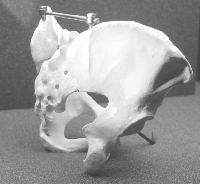
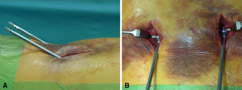
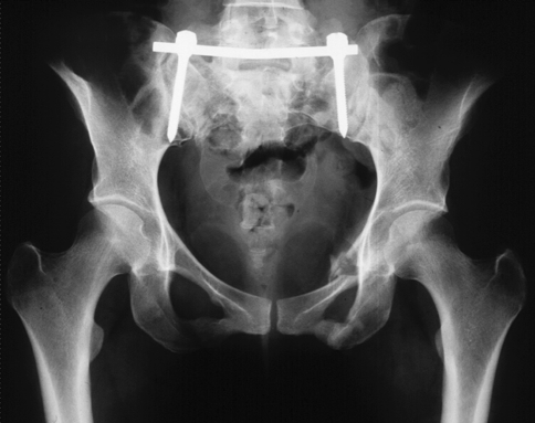
Surgery was scheduled as early as possible to obtain anatomic reduction. We performed surgery within 1 to 12 days after injury; the relatively wide range reflects the time needed for patients to regain hemodynamic and pulmonary stability after the first resuscitation phase. One surgeon (BF), experienced in trauma pelvic surgery, performed all surgeries. Staff placed the patients prone on a radiolucent table. The surgeon marked the posterior superior iliac spine (PSIS) and dorsal iliac crests and used a standard posterior vertical incision of 4 cm, starting 1 cm lateral to the PSIS. The osseous insertion point then was located on the height of the dorsal iliac crest and 1 to 2 cm cranial to the PSIS (Fig. 2). Using a bone awl the surgeon opened the cortical bone and widened the opening parallel to the line of the posterior gluteal line until the instrument reached the opposing cortex. Pedicle screws (7 mm in diameter, Universal Spine System, Synthes, Umkirch, Germany) then were inserted, usually with a length of 50 mm to 60 mm. The same procedure was performed on the contralateral side. In the sagittal plane, a narrow angle (< 30°) was preferred to avoid implant prominence and secondary soft tissue irritation (Fig. 3). The connection bar (6-mm diameter, Universal Spine System, Synthes) then was inserted subfascially and fixed to the pedicle screws. According to the structure of the fracture, distraction or compression was applied using the distraction/compression device from the Universal Spine System (Synthes). Anatomic reduction and screw position then were checked intraoperatively using AP (Fig. 4) and inlet/outlet view radiographs (15). We performed closed reduction manually, except in two patients in whom a Schanz screw [29] was used as a joystick. The average fluoroscopy time was 0.3 minutes (range, 0.1–1.0 minute).
Fig. 2.
The transiliac internal fixator is shown in its correct implantation site bridging the two iliac crests on a hard plastic pelvic model.
Fig. 3A–B.
The operative side of the implanted TIFI is shown before removal of the insertion instrumentation. (A) A side view shows the narrow angle in the sagittal plane that minimizes discomfort. (B) An overview shows the relation between implant size and incision length.
Fig. 4.
An AP view shows the postoperative control of the correctly implanted TIFI.
Patients were restricted to partial weightbearing of 15 kg for the affected limb for 6 to 8 weeks; subcutaneously administered low-molecular-weight heparin was recommended for the entire duration of partial weightbearing as prophylaxis against deep vein thrombosis. On average, patients achieved independent mobilization with limited weightbearing in 12 days (range, 6–18 days, excluding 13 patients with long-time ventilation). Hardware was removed after approximately 11.9 months (range, 0.5–26 months).
Postoperatively, two surgeons (BF, TD) examined all patients at 6, 12, and 52 weeks for signs of wound healing problems or infection. A neurologic examination of the lower extremities was performed in all patients at each followup. When we detected abnormalities we consulted a neurologist. We assessed social reintegration using the German Trauma Society Score [20, 27, 28] (Table 2). This score was developed by the German Pelvis Group (German Chapter of the AO-International and German Trauma Society). Through structured interviews the complaints of the patients regarding social reintegration, malfunction of micturia, sexual malfunction, and pain-free time are assessed as one score on a 7-point scale.
Table 2.
Total Pelvic Outcome Score
| Total Pelvic Outcome Score | Points | Isolated pelvic ring injuries (N = 46) | Pelvic complex injuries Type C (N = 16) |
|---|---|---|---|
| Excellent | 7 | 18 | 1 |
| Good | 6 | 12 | 4 |
| Fair | 4 to 5 | 16 | 9 |
| Poor | 1 to 3 | 0 | 2 |
At 1 year and at the last followup, radiographs were obtained, including AP (Fig. 3), 40°-caudad (inlet), and 40°-cephalad (outlet) pelvic views. Two examiners (BF, TW) graded the achieved reductions by the maximum displacement measured on the three radiographic views of the pelvis according to the technique described Matta and Tornetta [24].
Results
At the last followup of the patients with isolated pelvic trauma, 34 had anatomic reduction and 12 had residual displacement less than 5 mm. For patients with complex trauma, 11 had anatomic reduction, four had residual displacement less than 5 mm, and one had a gap greater than 5 mm owing to implant failure (Table 3).
Table 3.
Maximal displacement in radiographs for Pelvic Outcome Score
| Maximal displacement in radiographs | Points | Isolated pelvic ring injuries (N = 46) | Pelvic complex injuries Type C (N = 16) |
|---|---|---|---|
| Anatomic reduction | 3 | 34 | 11 |
| Displacement of symphysis < 5 mm | |||
| Displacement of os pubis/os ischii fracture < 10 mm | |||
| Posterior displacement < 5 mm | 2 | 12 | 4 |
| Displacement of symphysis < 10 mm | |||
| Displacement of os pubis/os ischii fracture < 15 mm | |||
| Posterior displacement > 5 mm | 1 | 0 | 1 |
| Displacement of symphysis > 10 mm | |||
| Displacement of os pubis/os ischii fracture > 15 mm |
We identified no iatrogenic neurovascular lesions attributable to the implantation. Intraoperative blood loss was less than 50 mL in all patients. Four wound infections occurred during the first 4 weeks postoperatively, all in patients with polytrauma and long-time ventilation. We identified one malpositioned screw without any consequences to the healing process and one secondary displacement attributable to implant failure. Two patients had deep vein thrombosis with secondary pulmonary embolus. Thirteen patients had acute respiratory distress syndrome (ARDS) develop, with a maximum stay in the intensive care unit of 66 days. Twelve patients reported mild local discomfort when in the supine position, in the region of the PSIS that was relieved by standing or walking; the complaints diminished after implant removal. One patient had an asymptomatic screw loosening, which we identified during implant explantation. Five of seven patients with posttraumatic nerve lesions achieved full recovery during the first 3 years. In all six patients with posttraumatic urogenital lesions, sexual dysfunction was persistent and required ongoing urologic treatment.
Twenty-one of 46 patients with pelvic ring injuries without additional pelvic injuries (two of 16 with complex pelvic ring injuries) had no subjective discomfort or restriction during daily activities (Table 4). Fifteen patients (four with complex pelvic ring injuries) had minor restrictions and eight (four with complex pelvic ring injuries) had intermittent pain during mobilization. Two patients experienced permanent pain and incontinence immediately after trauma occurred. Four patients with complex pelvic ring injuries had severe discomfort and restriction with urologic dysfunction, and three had permanent incontinence. Twenty-four of these 46 patients had no restrictions in social life activities, 15 had to make minor adaptations, and seven had major restrictions during work and social life (Table 5). After complex pelvic injury, four patients had no restrictions, seven had minor restrictions, and five had major restrictions during work and social life.
Table 4.
Clinical complaints for Pelvic Outcome Score
| Clinical complaints | Points | Isolated pelvic ring injuries (N = 46) | Pelvic complex injuries Type C (N = 16) |
|---|---|---|---|
| Free of complaints | 4 | 21 | 2 |
| Pain during exercise | 3 | 15 | 4 |
| Minor functional deficits | |||
| Loss of sensitivity | |||
| Pain during minor activities | 2 | 8 | 7 |
| Major functional deficits | |||
| Loss of motor function | |||
| Minor urologic deficits | |||
| Permanent pain | 1 | 2 | 3 |
| Mobilization only with crutches | |||
| Loss of protection sensitivity | |||
| Sexual dysfunction | |||
| Incontinence |
Table 5.
Social reintegration for Pelvic Outcome Score
| Social reintegration | Points | Isolated pelvic ring injuries (N = 46) | Pelvic complex injuries Type C (N = 16) |
|---|---|---|---|
| No changes compared with before trauma | 3 | 24 | 4 |
| Business restriction | 2 | 15 | 7 |
| Occupational redeployment | |||
| Minor restrictions in sport activities or in social life | |||
| At times need external help | |||
| Long-term disability | 1 | 7 | 5 |
| Major restrictions in sport activities or in social life | |||
| Often need external help |
Discussion
Pelvic injuries caused by high-energy lesions have a mortality rate as much as 30%, depending on the associated injuries [9]. Multiple treatment modalities are available with specific advantages and disadvantages, including the experience of the treating surgeon [26]. The stiffness of constructs with various posterior pelvic ring fixation devices is reportedly similar [42]. Several studies suggest anatomic reduction relates to decreased pain symptoms during activities of daily living [26, 31, 33]. Long-term patient satisfaction for patients with operatively treated pelvic injuries needs improvement and patients might experience pain, nonunion, problems with hardware, sexual dysfunction, or major restrictions in activities of daily life [17]. We asked whether (1) we could achieve anatomic restoration with the device, (2) specific complications were associated with this minimally invasive approach (particularly enhanced intraoperative blood loss, soft tissue complications, and iatrogenic neurovascular damage), and (3) function 3 years after trauma was comparable to that of established methods.
Our study has several limitations. First, the lack of a controlled, randomized prospective trial approach does not allow us to directly compare our findings with those with other techniques or to recommend specific guidelines. Second, some authors have emphasized the relevance of an intact ventral pelvic ring regarding the stability of the entire pelvic system [21, 39]. Our cohort included patients with isolated dorsal pelvic ring injuries and combined lesions of the ventral and dorsal structures. However, the number of patients involved in our study is too small to allow meaningful subgroup comparisons. Nonetheless our patient sample is comparable to those in other studies regarding age, gender, trauma mechanism, comorbidity, and injury severity [9, 17, 27]. Similarly, we had too few patients with excessive durations of ventilatory support and compromised immune defense to analyze. Third, reductions were graded by three radiographic views of the pelvis. This might not reflect the maximum amount of displacement and there is debate in the literature regarding the correlation of radiographic displacement and functional outcome [22, 24].
Fourth, the Pelvic Outcome Score is not rigorously validated against another outcome score for patient satisfaction and is more a categorical scoring instrument. This score was first used in a multicenter study with 486 patients in 1996 [28]. Other studies regarding pelvic ring injuries used this tool to investigate complaints of their patients [20, 27, 28]. Lindahl and Hirvensalo [22] compared this score with the scoring system described by Majeed [23], with similar findings. We used this score to compare our findings with those in the literature. However, owing to ceiling effects, it is possible differences between patient subgroups would not be detected. However we believe this categorical classification is reasonable to obtain an overview of patient satisfaction.
One of our 62 patients had persistent displacement greater than 5 mm at last followup. There is controversy in the literature if displacement greater than 5 mm leads to functional impairment. One study suggests an association between residual displacement in the radiographic analysis and pain during activities of daily living [22], whereas another found no such relationship [25]. Additional controversy exists regarding how much displacement is tolerable. In one study, 1-cm residual displacement is tolerated by most patients, whereas remaining displacement greater than 1 cm results in severe pain in as much as 23% of patients [26]. According to Tornetta and Matta [40], reduction is graded as excellent if it is 4 mm or less and good if between 4 mm and 10 mm remaining displacement is achieved. Their suggestion is supported by a study of 64 patients with dorsal pelvic ring injuries in which displacement of 4 mm or greater was linked to increased probability for severe pain and decreased function [5]. Lindahl and Hirvensalo suggested a patient could have a maximum of 5 mm displacement with high functional scores [22]. Another study reported 40 patients and the use of reconstruction plates (30 patients) or percutaneous iliosacral fixation (screws or sacral bars, 10 patients). They found two patients had displacement between 10 and 20 mm whereas the remaining patients had no displacement greater than 10 mm [17]. A displacement of 5 mm or less was achieved in 66 of 101 patients with Type C pelvic ring injuries [22].
In a systematic review of a total of 516 patients with internal fixation of the posterior pelvis [26], a malunion rate of 7% and an implant failure rate of 5% among 455 patients were reported. One of our 67 patients had secondary displacement because of implant failure. In the above-mentioned review, the authors reported a median infection rate of 6% [26], and Lindahl and Hirvensalo reported an infection rate of 5% for 101 patients with Type C injuries undergoing ORIF [22]. Four of our patients had wound infections (7%). These patients were treated with wound irrigation, débridement, and appropriate antibiotics, depending on culture results. All infections occurred during the first 4 weeks postoperatively in patients who required long-term invasive ventilation. The risk of neurologic injury after positioning of sacroiliac screws has been reported between less than 1% and as much as 8% [12, 36], and in a study of 101 patients using either ORIF with plate osteosynthesis or iliosacral screws, one L5 nerve root lesion occurred [22]. We identified no case of neurovascular damage in our 62 patients. There are insufficient data from the literature regarding the time needed for implantation of different devices and intraoperative blood loss. The time for implantation of the TIFI averaged 29 minutes (the longest setting required 48 minutes), and blood loss caused by the approach was no more than 50 mL in each patient. In 40 patients who underwent ORIF for completely unstable pelvic ring fractures (posterior followed by anterior internal fixation), the average volume of intraoperative blood loss was reported as 850 mL [17], and for 103 posterior internal fixations, the mean operative time of 98 minutes and mean total blood loss of 1400 mL were reported [22]. Short duration of wound opening in the operating room and minimized intraoperative blood loss are factors that reduce soft tissue infections and minimize accompanying risks regarding volume therapy and blood transfusion. Therefore, we believe the short operative duration and marginal blood loss are advantages of the TIFI.
In one prospective study including 28 Type C lesions, no or slight pain was present in 50% of patients, moderate pain was present in 43%, and severe pain was present in only 7% [7]. However, half of all patients in this group had to change their profession because of their pelvic ring injuries [7]. Comparable findings were reported by Pohlemann et al. [27], with a followup of 21 patients with Type C injuries. In our study, including patients with pelvic complex trauma, 37% had no or only slight pain during activities, 54% had moderate pain, and 8% had severe pain. In a study of 31 patients with dorsal or ventrodorsal injuries to the pelvic ring, social reintegration according to the pelvic outcome score was complete only in 1/3 of the cases [19] (Table 6). Twenty-eight of 62 of our patients had the same social life activities as before their trauma.
Table 6.
Clinical results of published studies
| Study | Method | Number of patients | Pelvic Outcome Score | ||
|---|---|---|---|---|---|
| Clinical symptoms | Radiologic evaluation of reduction | Social reintegration | |||
| Krappinger et al. [19] | Minimally invasive transiliac plate osteosynthesis | 31 | Very good 34%, | Very good 51%, | Complete 39%, |
| good 39%, | good 29%, | incomplete 43%, | |||
| fair 17%, | fair 16%, | poor 18% | |||
| poor 10% | poor 4% | ||||
| Kabak et al. [17] | Anterior plate osteosynthesis | 40 | Very good 78%, | ||
| good 17%, | |||||
| fair 5%, | |||||
| poor 0% | |||||
| Lindahl and Hirvensalo [22] | Iliosacral screws and/or anterior plate osteosynthesis | 101 | Very good 67%, | Very good 65%, | |
| good 16%, | good 25%, | ||||
| fair 16%, | fair 10%, | ||||
| poor 1% | poor 0% | ||||
| Fuechtmeier et al. [11] | Minimally invasive transiliac internal fixator | 31 | Very good 20%, | Very good 40%, | Complete 40%, |
| good 30%, | good 60%, | incomplete 40%, | |||
| fair 50%, | fair 0%, | poor 20% | |||
| poor 0%, | poor 40% | ||||
| Hao et al. [15] | Minimally invasive posterior plate osteosynthesis | 21 | Very good or | Very good or | |
| good 90%, | good 85%, | ||||
| fair 10%, | fair or | ||||
| poor 0% | poor 15% | ||||
| Current study | Minimally invasive transiliac internal fixator | 67 | Very good 37%, | Very good 73%, | Complete 45%, |
| good 31%, | good 25%, | incomplete 36%, | |||
| fair 24%, | fair 0%, | poor 19% | |||
| poor 8%, | poor 2% | ||||
We assessed displacement, complication rates including intraoperative blood loss, and function in patients treated with a TIFI for an unstable dorsal pelvic ring injury. Long-time displacement in the radiographic analysis, complication rates, and long-term functional outcome were in a similar range as those of other fixation methods. Caution should be exercised in applying our findings to all patients, owing to complications from associated injuries in individual patients. Future multi-institutional, prospective randomized studies may be able to achieve sufficient patient numbers to clarify whether this device is appropriate for all patient subpopulations.
Acknowledgment
We thank Thomas Windisch for assistance with data collection.
Footnotes
Each author certifies that he has no commercial association (eg, consultancy, stock ownership, equity interest, or patent/licensing arrangement) that might pose a conflict of interest in connection with the submitted article.
Each author certifies that his institution has approved or waived approval for the human protocol for this investigation and that all investigations were conducted in conformity with ethical principles of research.
The work was performed at the Department of Trauma Surgery of the University Hospital, Regensburg, Germany.
References
- 1.Altman DT, Jones CB, Routt ML., Jr Superior gluteal artery injury during iliosacral screw placement. J Orthop Trauma. 1999;13:220–227. doi: 10.1097/00005131-199903000-00011. [DOI] [PubMed] [Google Scholar]
- 2.Atlihan D, Tekdemir I, Ates Y, Elhan A. Anatomy of the anterior sacroiliac joint with reference to lumbosacral nerves. Clin Orthop Relat Res. 2000;376:236–241. doi: 10.1097/00003086-200007000-00032. [DOI] [PubMed] [Google Scholar]
- 3.Baylis TB, Norris BL. Pelvic fractures and the general surgeon. Curr Surg. 2004;61:30–35. doi: 10.1016/j.cursur.2003.07.017. [DOI] [PubMed] [Google Scholar]
- 4.Chiu FY, Chuang TY, Lo WH. Treatment of unstable pelvic fractures: use of a transiliac sacral rod for posterior lesions and an external fixator for anterior lesions. J Trauma. 2004;57:141–144. doi: 10.1097/01.TA.0000123040.23231.EB. [DOI] [PubMed] [Google Scholar]
- 5.Cole JD, Blum DA, Ansel LJ. Outcome after fixation of unstable posterior pelvic ring injuries. Clin Orthop Relat Res. 1996;329:160–179. doi: 10.1097/00003086-199608000-00020. [DOI] [PubMed] [Google Scholar]
- 6.Denis F, Davis S, Comfort T. Sacral fractures: an important problem. Retrospective analysis of 236 cases. Clin Orthop Relat Res. 1988;227:67–81. [PubMed] [Google Scholar]
- 7.Draijer F, Egbers HJ, Havemann D. Quality of life after pelvic ring injuries: follow-up results of a prospective study. Arch Orthop Trauma Surg. 1997;116:22–26. doi: 10.1007/BF00434095. [DOI] [PubMed] [Google Scholar]
- 8.Durkin A, Sagi HC, Durham R, Flint L. Contemporary management of pelvic fractures. Am J Surg. 2006;192:211–223. doi: 10.1016/j.amjsurg.2006.05.001. [DOI] [PubMed] [Google Scholar]
- 9.Ertel W, Eid K, Keel M, Trentz O. Therapeutical strategies and outcome of polytraumatized patients with pelvic injuries. Eur J Trauma. 2000;26:278–286. doi: 10.1007/PL00002452. [DOI] [Google Scholar]
- 10.Ferrell M, Bellino M, Olson SA. Pelvic ring injuries. Eur J Trauma. 2005;31:536–542. doi: 10.1007/s00068-005-2129-2. [DOI] [Google Scholar]
- 11.Fuchtmeier B, Maghsudi M, Neumann C, Hente R, Roll C, Nerlich M. The minimally invasive stabilization of the dorsal pelvic ring with the transiliacal internal fixator (TIFI): surgical technique and first clinical findings][in German. Unfallchirurg. 2004;107:1142–1151. doi: 10.1007/s00113-004-0824-9. [DOI] [PubMed] [Google Scholar]
- 12.Giannoudis PV, Tzioupis CC, Pape HC, Roberts CS. Percutaneous fixation of the pelvic ring: an update. J Bone Joint Surg Br. 2007;89:145–154. doi: 10.1302/0301-620X.89B2.18551. [DOI] [PubMed] [Google Scholar]
- 13.Goldstein A, Phillips T, Sclafani SJ, Scalea T, Duncan A, Goldstein J, Panetta T, Shaftan G. Early open reduction and internal fixation of the disrupted pelvic ring. J Trauma. 1986;26:325–333. doi: 10.1097/00005373-198604000-00004. [DOI] [PubMed] [Google Scholar]
- 14.Greenspan L, McLellan BA, Greig H. Abbreviated Injury Scale and Injury Severity Score: a scoring chart. J Trauma. 1985;25:60–64. doi: 10.1097/00005373-198501000-00010. [DOI] [PubMed] [Google Scholar]
- 15.Hao T, Changwei Y, Qiulin Z. Treatment of posterior pelvic ring injuries with minimally invasive percutaneous plate osteosynthesis. Int Orthop. 2009;33:1435–1439. doi: 10.1007/s00264-009-0756-7. [DOI] [PMC free article] [PubMed] [Google Scholar]
- 16.Hudson DA, Knottenbelt JD, Krige JE. Closed degloving injuries: results following conservative surgery. Plast Reconstr Surg. 1992;89:853–855. doi: 10.1097/00006534-199205000-00013. [DOI] [PubMed] [Google Scholar]
- 17.Kabak S, Halici M, Tuncel M, Avsarogullari L, Baktir A, Basturk M. Functional outcome of open reduction and internal fixation for completely unstable pelvic ring fractures (type C): a report of 40 cases. J Orthop Trauma. 2003;17:555–562. doi: 10.1097/00005131-200309000-00003. [DOI] [PubMed] [Google Scholar]
- 18.Kellam JF, McMurtry RY, Paley D, Tile M. The unstable pelvic fracture: operative treatment. Orthop Clin North Am. 1987;18:25–41. [PubMed] [Google Scholar]
- 19.Krappinger D, Larndorfer R, Struve P, Rosenberger R, Arora R, Blauth M. Minimally invasive transiliac plate osteosynthesis for type C injuries of the pelvic ring: a clinical and radiological follow-up. J Orthop Trauma. 2007;21:595–602. doi: 10.1097/BOT.0b013e318158abcf. [DOI] [PubMed] [Google Scholar]
- 20.Kuttner M, Klaiber A, Lorenz T, Fuchtmeier B, Neugebauer R. The pelvic subcutaneous cross-over internal fixator][in German. Unfallchirurg. 2009;112:661–669. doi: 10.1007/s00113-009-1623-0. [DOI] [PubMed] [Google Scholar]
- 21.Leighton RK, Waddell JP, Bray TJ, hapman MW, Simpson L, Martin RB, Sharkey NA. Biomechanical testing of new and old fixation devices for vertical shear fractures of the pelvis. J Orthop Trauma. 1991;5:313–317. doi: 10.1097/00005131-199109000-00010. [DOI] [PubMed] [Google Scholar]
- 22.Lindahl J, Hirvensalo E. Outcome of operatively treated type-C injuries of the pelvic ring. Acta Orthop. 2005;76:667–678. doi: 10.1080/17453670510041754. [DOI] [PubMed] [Google Scholar]
- 23.Majeed SA. Grading the outcome of pelvic fractures. J Bone Joint Surg Br. 1989;71:304–306. doi: 10.1302/0301-620X.71B2.2925751. [DOI] [PubMed] [Google Scholar]
- 24.Matta JM, Tornetta P., 3rd Internal fixation of unstable pelvic ring injuries. Clin Orthop Relat Res. 1996;329:129–140. doi: 10.1097/00003086-199608000-00016. [DOI] [PubMed] [Google Scholar]
- 25.Nepola JV, Trenhaile SW, Miranda MA, Butterfield SL, Fredericks DC, Riemer BL. Vertical shear injuries: is there a relationship between residual displacement and functional outcome? J Trauma. 1999;46:1024–1029. doi: 10.1097/00005373-199906000-00007. [DOI] [PubMed] [Google Scholar]
- 26.Papakostidis C, Kanakaris NK, Kontakis G, Giannoudis PV. Pelvic ring disruptions: treatment modalities and analysis of outcomes. Int Orthop. 2009;33:329–338. doi: 10.1007/s00264-008-0555-6. [DOI] [PMC free article] [PubMed] [Google Scholar]
- 27.Pohlemann T, Gansslen A, Schellwald O, Culemann U, Tscherne H. Outcome after pelvic ring injuries. Injury. 1996;27(suppl 2):B31–B38. [PubMed] [Google Scholar]
- 28.Pohlemann T, Tscherne H, Baumgartel F, Egbers HJ, Euler E, Maurer F, Fell M, Mayr E, Quirini WW, Schlickewei W, Weinberg A. Pelvic fractures: epidemiology, therapy and long-term outcome. Overview of the multicenter study of the Pelvis Study Group][in German] Unfallchirurg. 1996;99:160–167. doi: 10.1007/s001130050049. [DOI] [PubMed] [Google Scholar]
- 29.Reilly MC, Norris BL, Bosse MJ. Pelvic fractures: Sacral fixation. In: Wiss DA, ed. Master Techniques in Orthopaedic Surgery, Fractures. 2nd ed. Philadelphia, PA: Lippincott Williams & Wilkins; 2006.
- 30.Routt ML Jr, Nork SE, Mills WJ. High-energy pelvic ring disruptions. Orthop Clin North Am. 2002;33:59–72, viii. [DOI] [PubMed]
- 31.Routt ML, Jr, Simonian PT, Mills WJ. Iliosacral screw fixation: early complications of the percutaneous technique. J Orthop Trauma. 1997;11:584–589. doi: 10.1097/00005131-199711000-00007. [DOI] [PubMed] [Google Scholar]
- 32.Routt ML, Jr, Simonian PT, Swiontkowski MF. Stabilization of pelvic ring disruptions. Orthop Clin North Am. 1997;28:369–388. doi: 10.1016/S0030-5898(05)70295-9. [DOI] [PubMed] [Google Scholar]
- 33.Saiki K, Hirabayashi S, Horie T, Tsuzuki N, Inokuchi K, Tsutsumi H. Anatomically correct reduction and fixation of a Tile C-1 type unilateral sacroiliac disruption using a rod and pedicle screw system between the S1 vertebra and the ilium: experimental and clinical case report. J Orthop Sci. 2002;7:581–586. doi: 10.1007/s007760200104. [DOI] [PubMed] [Google Scholar]
- 34.Schweitzer D, Zylberberg A, Cordova M, Gonzalez J. Closed reduction and iliosacral percutaneous fixation of unstable pelvic ring fractures. Injury. 2008;39:869–874. doi: 10.1016/j.injury.2008.03.024. [DOI] [PubMed] [Google Scholar]
- 35.Styner JK. The birth of Advanced Trauma Life Support (ATLS) Surgeon. 2006;4:163–165. doi: 10.1016/S1479-666X(06)80087-9. [DOI] [PubMed] [Google Scholar]
- 36.Templeman D, Schmidt A, Freese J, Weisman I. Proximity of iliosacral screws to neurovascular structures after internal fixation. Clin Orthop Relat Res. 1996;329:194–198. doi: 10.1097/00003086-199608000-00023. [DOI] [PubMed] [Google Scholar]
- 37.Tile M. Pelvic ring fractures: should they be fixed? J Bone Joint Surg Br. 1988;70:1–12. doi: 10.1302/0301-620X.70B1.3276697. [DOI] [PubMed] [Google Scholar]
- 38.Tile M. Acute pelvic fractures: I. Causation and classification. J Am Acad Orthop Surg. 1996;4:143–151. doi: 10.5435/00124635-199605000-00004. [DOI] [PubMed] [Google Scholar]
- 39.Tile M. Acute pelvic fractures: II. Principles of management. J Am Acad Orthop Surg. 1996;4:152–161. doi: 10.5435/00124635-199605000-00005. [DOI] [PubMed] [Google Scholar]
- 40.Tornetta P, 3rd, Matta JM. Outcome of operatively treated unstable posterior pelvic ring disruptions. Clin Orthop Relat Res. 1996;329:186–193. doi: 10.1097/00003086-199608000-00022. [DOI] [PubMed] [Google Scholar]
- 41.Tosounidis G, Culemann U, Stengel D, Garcia P, Kurowski R, Holstein JH, Pohlemann T. Complex pelvic trauma in elderly patients][in German. Unfallchirurg. 2010;113:281–286. doi: 10.1007/s00113-010-1764-1. [DOI] [PubMed] [Google Scholar]
- 42.Yinger K, Scalise J, Olson SA, Bay BK, Finkemeier CG. Biomechanical comparison of posterior pelvic ring fixation. J Orthop Trauma. 2003;17:481–487. doi: 10.1097/00005131-200308000-00002. [DOI] [PubMed] [Google Scholar]


