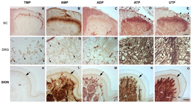
Fig. 1. Nucleotidase enzyme histochemistry.
Nucleotidase histochemical staining of spinal cord, DRG and glabrous paw from mice using TMP, AMP, ADP (3 mM), ATP and UTP (1 mM) as substrates. (A, B) In spinal cord, histochemistry with TMP shows a narrow band of staining restricted to lamina II as widely reported, while AMP staining is broader, suggesting that TMP may be a selective substrate for one of the two known AMPases. (C) Only limited staining is seen with ADP. (D, E) Strong ATPase activity (ATP, UTP) is restricted to the superficial dorsal horn, similar to AMP but with low-intensity diffuse staining in the grey matter that is not evident with AMP. Staining includes axons coursing through the dorsal root and entering the spinal cord (arrowheads). (F, G) In DRG, AMPase activity (TMP, AMP) is evident in small neurons (arrowheads, asterisks show negative cells). (H) As in spinal cord, ADP shows substantial staining only in blood vessels. (I, J), Extensive non-neuronal as well as neuronal staining is evident with ATP and UTP (arrowheads show positive cell, asterisks show negative cells). (K–O) In the foot pad, epidermal staining (arrows) is restricted to keratinocytes of the stratum granulosum for all nucleotides except ADP, which does not produce staining in epidermis. Intense staining is also evident in sweat glands, ducts and blood vessels for all nucleotides except TMP (suggesting that TMP may be a selective substrate for some but not all AMPases). SC, stratum corneum; Epi, epidermis; Der, dermis; SG, sweat gland. Scale bars: A–E, K–O, 100 uM; F–J, 50 uM.

