Abstract
The improvement in survival and local control measures in children with neoplasm in the head and neck region may lead to increased iatrogenic adverse effects of treatment. The aim of this study was to report a new case of the long-term effects of chemoradiotherapy on oral health and dental development in a patient treated for Hodgkin’s disease at an early age. In this case report, a 26-year-old female is presented, who at the age of 5 years received chemotherapy and radiotherapy for Hodgkin’s disease in the neck region. The patient consulted the Department of Oral Medicine because of dental changes and tooth loss despite adequate dental care and oral hygiene. Clinical examination revealed loose teeth and inflamed gingiva of the mandible, x-ray showed premature root resorption, V-shaped and shortened roots and alveolar bone loss. After examination, the patient was referred for extracting the mandibular teeth and then wassent to the prosthetics department. Therefore, in order to decrease dental treatment sequelae in patients who have had cured malignant disease, these cases should have life-long dental care and follow-up.
Keywords: Chemo radiotherapy, Hodgkin Disease, Childhood, Dental Development
INTRODUCTION
Hodgkin’s lymphoma was first described in 1832, but the nature of the pathognomic Reed-Sternberg cells, on which diagnosis of the disease is based has only been cleared in the past few years. Radiotherapy has been employed for treatment of localized disease from the 1940s and combination chemotherapy was presented for anatomically progressive diseases in the 1960s. There has been great improvement in the outcome in the last three decades concluing to the fact that Hodgkin’s lymphoma is mentioned as one of the most curable non-cutaneous malignancies [1]. Combined therapy (chemotherapy and radiotherapy) can lead to the cure of more than 90% of children and adolescents with Hodgkin’s disease, but the intensive treatment may cause early and late complications. Damage of soft tissues, respiratory, cardiovascular, skeletal and endocrine systems, dental development disturbance and second cancers are considered to be late complications of such treatment. These complications may have adverse effects on the patient’s quality of life after discontinuation of treatment [2].
Therefore, chemotherapy and radiotherapy have deleterious effects on dentition in children and adolescents [3]. Hypodontia (partial anodontia), microdontia, altered eruption patterns and root stunting are some of the stated complications [4].
Developmental abnormalities resulting after malignant chemotherapy occur when the patient is treated prior to six years of age [5]. Diminished root surface area due to radiation exposure is the reason for early tooth loss. Combined radio chemotherapy which is commonly used for the treatment of childhood neoplasia causes periodontium effects [6,7].There are just few reports of the complications of the dental system caused by radiotherapy in childhood in the literature. We report a girl who received chemotherapy and radiation of the head and neck area at five years of age and demonstrated developmental disturbances following chemoradiotherapy.
MATERIALS AND METHODS
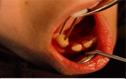
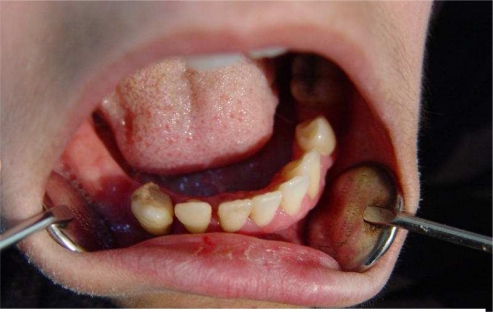
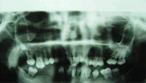
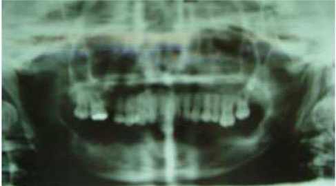
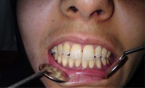
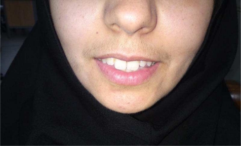
This case report describes a 26-year-old girl, who at the age of 5 years, received chemotherapy and radiotherapy for Hodgkin’s disease in the neck region. The patient consulted the Department of Oral Medicine, School of Dentistry, Tehran University of Medical Sciences because of dental changes and tooth loss despite adequate dental care and oral hygiene. Clinical examination of the oral cavity revealed loose teeth (grade 3) (Fig 1) and in flamed mandibular gingivae, but the maxillary teeth were not mobile and oral hygiene was fairly good (Fig 2). Panoramic view x-ray revealed that the roots of the mandibular teeth showed resorption, had become V-shaped and shortened together with alveolar bone loss and no remarkable changes in the enamel and dentin of the crowns was detected (Fig 3). Medical work-up did not show cervical lymphadenopathy. Considering the patient’s age, her height (149 cm) and weight (50 kg) were lower than normal, while her parents were nearly tall. No problem was found in physical examination. Altogether, she was alert and healthy, did not show any relapse of disease and was not taking any medicine. In the medical history, the patient had stage 1 nodular sclerosis type classical Hodgkin’s lymphoma in the lymph nodes of the neck at the age of 5 years. She was treated by chemotherapy (Bleomicin+Vinblastin+Dacarbazine+Prednisone for 6 periods) plus 2400-cGy Radiation therapy. The patient had radiotherapy for Hodgkin in the neck area to include the cervical nodes. The lower jaw was placed in the radiation field. In family history, her father had died of heart disease about three years ago. Her mother was alive and in complete health. She had two brothers, aged 24 and 15 years, who were alive and healthy. They did not have any problem with their teeth. In social history, she did not have a career and did not smoke. After examination, the patient was referred for extracting mandibular teeth (Fig 4) and was subsequently sent to the prosthetics department in order to restore the missing teeth with a lower jaw of complete denture (Figs 5,6).
Fig 1.
Clinical view of the oral cavity and loosened teeth (grade 3).
Fig 2.
Clinical view of the oral cavity and teeth.
Fig 3.
Panoramic view showing resorption, V-shaping and shortening in the roots and alveolar bone loss in the mandible.
Fig 4.
Prosthetics treatment for the lower jaw.
Fig 5.
Fig 6.
Smile of patient after treatment.
DISCUSSION
Chemotherapy and radiotherapy may have serious effects on developing teeth such as delayed dental development, microdontia, hypoplasia, agenesis and V-shaped and shortened roots [8–10]. In some studies the effects of chemotherapy and radiotherapy on dentition in children has been demonstrated [11]. They evaluated the effects of therapy on dentofacial development in 27 acute lymphoblastic leukemia pediatric patients before 10 years of age who were treated with radiotherapy (RT) alone or chemotherapy together with 1800–2400-cGy cranial radiotherapy. The severity of these abnormalities was greater in children who received RT treatment before 5 years of age. Minicucci observed in 76 children who were treated with high-dose chemoradiotherapy that 82.9% of them showed at least one dental abnormality including tooth agenesis, arrested root development, microdontia and enamel dysplasia [9].
Holtta studied dental development in young children after myeloablative therapy which was either chemotherapy and fractionated total body irradiation (TBI) or chemotherapy alone (non-TBI). In the TBI group, 9 out of 10 patients had very severe root defects in contrast to none in the non-TBI group [8].
Helpin presented a case of rhabdomyosarcoma of the infratemporal and parapharyngeal region.
This patient received therapeutic radiation and exhibited undesirable consequences in dentition and the mandible [12].
Takinam reported hypoplasia of the mandible and teeth in a 4-year-old boy who had cystic hygroma. At the age of 7 months he had been treated with 2400-cGy to the head and neck, followed by surgery. Panoramic view showed hypoplasia of the roots of the canines, molars and permanent teeth [13]. Futhermore, Moller and Carl like Takinam, presented dentomandibular defects after chemo-radiation therapy in boys aged 9 years and 4 years with Hodgkin’s lymphoma. Clinical and radiographic evaluation showed dental abnormalities such as root blunting, mild to severe root shortening, premature closure of the root apices and severe radiation caries with mandibular and maxillary hypoplasia [14,15]. They observed that new megavoltage radiation machines have reduced the chances of bone complications in adults. In this study, we reported a girl who was treated with chemotherapy plus 2400-cGy radiation therapy at the age of 5 for a lesion of Hodgkin’s disease in the neck area. She had disturbances in the root development of the mandibular teeth including short roots, arrested root development, V-shaped root and loose teeth. In contrast, roots of the maxillary teeth were normal and the root apexes were closed.
Our report was similar to the results of other studies on the effects of radiotherapy on dentition and showed the long-term effects of cancer therapy on teeth. Although chemoradiation therapy cures more than 90% of children with lymphoma or leukemia, it often causes dental abnormalities that may affect their quality of life These abnormalities are probably caused by the type, intensity and frequency of treatment and the age of the patients at the time of radiotherapy which might have important consequences on the children’s dental development [2,3].
Finally, the improved outcome of patients with Hodgkin’s disease makes it imperative to consider the long-term side effects of treatment and to ameliorate them as soon as possible. In this way, dental evaluation after diagnosis and frequent follow-ups may help to ensure appropriate preventive measures and minimize dental and periodontal diseases.
Cases similar to this report are rare in the literature and the importance of such cases lies in the fact that dentists and other health care professionals should be aware of the oral and facial manifestations related to treatment of malignances, especially those related to the effects of radiation on bony structures. Another important aspect of this case was inadequate attention to the oral condition of the patient for a long time and consequences of such neglect on her general health status such as the impact on height and weight.
CONCLUSION
Treatment regimes for childhood cancer are known to affect the root development in the long-term survivors but the available data is subjective in nature with very few quantitative data in the literature.
The report of this patient introduces a new case in order to minimize the dental treatment sequel by supportive measures and also to initiate timely dental and prosthetic management.
Unfortunately, for this case dental treatment was not adequate and she could not eat and brush her teeth well because of loose teeth for many years.
It seems that this problem had effected her growth and oral health. Therefore, the health status of children and adolescents cured from Hodgkin’s disease and other childhood cancers should be regularly evaluated.
Acknowledgments
The author appreciates the kind collaboration of Kousar Sodagar.
REFERENCES
- 1.Yung L, Linch D. Hodgkin’s lymphoma. Lancet. 2003 Mar;361(9361):943–51. doi: 10.1016/S0140-6736(03)12777-8. [DOI] [PubMed] [Google Scholar]
- 2.Balwierz W, Moryl-Bujakowska A, Kaczmarek-Kanold M, Wachowiak J, Matysiak M, Sopyło B, et al. The health state evaluation in persons after therapy of Hodgkin’s disease in childhood: report of the Polish Pediatric Leukemia/Lymphoma Study Group. PrzeglLek. 2006;63(1):25–8. [PubMed] [Google Scholar]
- 3.Kaste SC, Hopkins KP, Jones D, Crom D, Greenwald CA, Santana VM. Dental abnormalities in children treated for acute lymphoblasticleukemia. Leukemia. 1997 Jun;11(6):792–6. doi: 10.1038/sj.leu.2400670. [DOI] [PubMed] [Google Scholar]
- 4.Kaste SC, Hopkins K, Jenkins J. Abnormal odontogenesis in children treated with radiation and chemotherapy: imaging findings. AJR Am J Roentgenol. 1994 Jun;162(6):1407–11. doi: 10.2214/ajr.162.6.8192008. [DOI] [PubMed] [Google Scholar]
- 5.Folwaczny M, Hickel R. Impaired dentofacial development after radiotherapy of a non-Hodgkin lymphoma: report of case. ASDC J Dent Child. 2000 Nov-Dec;67(6):428–30. [PubMed] [Google Scholar]
- 6.Herrmann T, Dorr W, Koy S, Lesche A, Lehmann D. Early loss of teeth after treatment for childhood leukemia. StrahlentherOnkol. 2004 Jun;180(6):371–4. doi: 10.1007/s00066-004-1244-z. [DOI] [PubMed] [Google Scholar]
- 7.Cheng CF, Huang WH, Tsai TP, Ko EW, Liao YF. Effects of cancer therapy on dental and maxillofacial development in children: report of case. ASDC J Dent Child. 2000 May-Jun;67(3):218–22. [PubMed] [Google Scholar]
- 8.Hölttä P, Alaluusua S, Saarinen-Pihkala UM, Wolf J, Nyström M, Hovi L. Long-term adverse effects on dentition in children with poor risk neuroblastoma treated with high-dose chemotherapy and autologous stem cell transplantation with or without total body irradiation. Bone Marrow Transplant. 2002 Jan;29(2):121–7. doi: 10.1038/sj.bmt.1703330. [DOI] [PubMed] [Google Scholar]
- 9.Minicucci E, Lopes L, Crocci A. Dental abnormalities in children after chemotherapy treatment for acute lymphoid leukemia. Leuk Res. 2003 Jan;27(1):45–50. doi: 10.1016/s0145-2126(02)00080-2. [DOI] [PubMed] [Google Scholar]
- 10.Avsar A, Elli M, Darko O, Pinarli G. Long-term effects of chemotherapy on caries formation, dental development, and salivary factors in childhood cancer survivors. Oral Surg Oral Med Oral Pathol Oral RadiolEndod. 2007 Dec;104(6):781–9. doi: 10.1016/j.tripleo.2007.02.029. [DOI] [PubMed] [Google Scholar]
- 11.Sonis AL, Tarbell N, Valachovic RW, Gelber R, Schwenn M, Sallan S. Dentofacial development in long-term survivors of acute lymphoblastic leukemia. A comparison of three treatment modalities. Cancer. 1990 Dec;66(12):2645–52. doi: 10.1002/1097-0142(19901215)66:12<2645::aid-cncr2820661230>3.0.co;2-s. [DOI] [PubMed] [Google Scholar]
- 12.Helpin ML, Krejmas NL, Krolls O. Complications following radiation therapy to the head. Oral Surg Oral Med Oral Pathol. 1986 Mar;61(3):209–12. doi: 10.1016/0030-4220(86)90361-0. [DOI] [PubMed] [Google Scholar]
- 13.Takinami S, Kaga M, Yahata H, Kure A, Oguchi H, Yasuda M. Radiation-induced hypoplasia of the teeth and mandible: A case report. Oral Surg Oral Med Oral Pathol. 1994 Sep;78(3):382–4. doi: 10.1016/0030-4220(94)90072-8. [DOI] [PubMed] [Google Scholar]
- 14.Moller P, Perrier M. Dento-maxillofacial sequelae in a child treated for a rhabdomyosarcoma in the head and neck: A case report. Oral Surg Oral Med Oral Pathol Oral RadiolEndod. 1998 Sep;86(3):297–303. doi: 10.1016/s1079-2104(98)90175-5. [DOI] [PubMed] [Google Scholar]
- 15.Carl W, Wood R. Effects of radiation on the developing dentition and supporting bone. J Am Dent Assoc. 1980 Oct;101(4):646–8. doi: 10.14219/jada.archive.1980.0368. [DOI] [PubMed] [Google Scholar]


