Abstract
Odontogenic keratocysts (OKCs) are one of the most frequent features of nevoid basal cell carcinoma syndrome (NBS). It is linked with mutation in the PTCH gene. Partial expression of the gene may result in occurrence of only multiple recurring OKC. Our patient presented with nine cysts with multiple recurrences over a period of 11 years without any other manifestation of the syndrome.
Keywords: Odontogenic Cysts; Basal Cell Nevus Syndrome; Pathology, Surgical, Jaw Cysts
INTRODUCTION
Nevoid basal cell carcinoma syndrome (NBS) is associated with a triad of multiple basal nevi, multiple odontogenic keratocysts (OKCs) and skeletal abnormalities [1]. These triad of symptoms may be associated with other manifestations involving skeletal, craniofacial, neurological, skin, sexual, ophthalmic and cardiac anomalies [2]. Multiple OKCs have been known to occur in non-syndromic cases though it is very rare [3]. These multiple lesions may be the first manifestation of the NBS or otherwise it may be because of the multifocal nature of OKCs [4,5].
We present a case of recurrent multiple OKCs which is not associated with any syndrome. We emphasize here that the present case may be because of partial expression of the PTCH gene and long-term follow-up in such cases is mandatory. After a long follow-up, we could not see any recurrence. So probably the importance of this case is just due to the aberrant nature of OKCs rather than being associated with the syndrome.
CASE REPORT
A 20-year-old female, who is a dental student of our institution, reported to the department of oral and maxillofacial surgery with the chief complaint of discharge from the right maxillary posterior region for two years. The patient gave a history of multiple isolated jaw cysts at the age of nine years, all the cysts were enucleated and histopathologically diagnosed as OKCs. She also gave a history of jaw surgery three years before for OKC in association with maxillary right second molar (17) (Table 1).
Table 1.
Summary of patient’s history for multiple recurring cysts.
| Age | Cyst no. | Site |
Findings |
Recurrence | Follow-up | ||
|---|---|---|---|---|---|---|---|
| Clinical | Radiography | at Surgery | |||||
| 9 years | 3 | Right body of mandible | Painful swelling | Well-defined unilocular | Tooth attached to the outer surface of cyst. Necrotic material within the lumen | No | 9 years |
| Symphysis | Asymptomatic | Well-defined unilocular | Inner surface was smooth | After 9 years, (two separate lesions on either side of midline) | 9 years | ||
| Left ramus of mandible | Asymptomatic | Well-defined unilocular | Thin wall | No | 9 years | ||
| 17 years | 1 | Right maxillary posterior region | Aysmptomatic | Well-defined unilocular | Nothing significant | After 3 years | 3 years |
| 20 years | 3 | Right maxillary posterior region | Discharge (from the posterior maxilla and nasal disc) | Well-defined unilocular | Thin lining | No | 3 years |
| Right parasymphisis | Swelling | Well-defined (minimal cortical plate expansion) | Smooth and regular margin | No | 3 years | ||
| Left parasymphisis | Aysmptomatic | Well-defined unilocular | Nothing significant | No | 3 years | ||
no.= number
Intra-oral examination revealed a mild ill-defined swelling in the right maxillary tuberosity region. There was also a spheroidal swelling measured approximately 1×1 cm in the mandibular left lateral incisor (32) region, mild cortical plate expansion was also seen in relation with the right mandibular lateral incisor (42) to the left mandibular central incisor (31) regions.
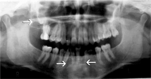
Radiographically, three separate radiolucencies were seen such as a well-defined 2.5×2.5 cm2 radiolucency in the right maxillary tuberosity region, a well-defined radiolucency in the mandibular left central incisor (31) area and a well-defined radiolucency in between the roots of the right deciduous mandibular canine (73) and the right mandibular first pre-molar (44) measured approximately 1×1 cm2 (Fig 1).
Fig 1.
Radiolucencies (arrows) in the first, third and fourth quadrant.
Based on the clinical and radiographic findings, a provisional diagnosis of recurrent OKCs was considered. Chest and skull radiograph findings were insignificant. Blood investigation values were in normal limits. Dermatology consultation did not reveal any cutaneous abnormality including palmar and plantar defects.
Enucleation of all the three lesions was done separately along with curettage. The specimen from the right tuberosity region was approximately 2×2 cm2 in size, creamish white in color and containing a yellowish cheesy material within the cystic space. The other two specimens were small roughly 0.5×0.5 cm2 in size and creamish white in color.
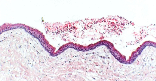
Histopathologically, all the three specimens showed multiple cystic spaces filled with keratin flakes. These cystic spaces were lined by parakeratinized stratified squamous cystic epithelial lining of predominantly even thickness with palisaded basal cells and a corrugated surface (Fig 2).
Fig 2.
Cystic lining showing parakeratinized stratified squamous epithelium of uniform 6–8 cell thickness with surface corrugation.
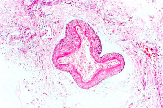
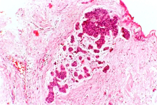
The capsule was fibrous and occasional chronic inflammatory cells were evident. Daughter cyst (Fig 3) and odontogenic epithelial cells arranged in groups were seen in the stroma (Fig 4). The stroma also showed focal areas of calcifications. The final diagnosis of OKC was confirmed. The patient is being followed up regularly and there has been no evidence of recurrence in the last three years after the last treatment.
Fig 3.
Daughter cyst in the cystic capsule.
Fig 4.
Odontogenic epithelial islands in cystic capsule.
DISCUSSION
Multiple OKCs usually occur as a component of syndromes such as NBS, orofacial digital syndrome, Noonan syndrome, Ehler-Danlos syndrome and Simpson-Golabi-Behmel syndrome [3,6–8]. In a study by Brannon [9], 5.1% of 312 cases were associated with NBS and 5.8% were accompanied by multiple keratocysts, but without any other features of the syndrome. However, 8.1% of the total 83 cases were associated with NBS and 7.6% of them showed recurrence; none of these cases with multiple OKCs were non-syndromic in a study on the Iranian population [10].
The present case showed only multiple recurrent OKCs without any other notable deformities such as basal cell carcinoma, skeletal defects, orofacial defects, stunted growth, bleeding diathesis and hyperextensible skin and hypermobile joints.
NBS involves various skeletal, craniofacial, neurological, oro-pharyngeal, cutaneous, sexual, ophthalmic and cardiac anomalies [3]. In our patient, none of these features indicative of NBS was present.
NBS is associated with mutation in the PTCH gene [9q (22.3-q31)]. Mutation within PTCH occurs in sporadic OKCs as well as those associated with NBS. It is suggested that a “two-hit” mechanism may underlie the variable expression of NBS and sporadic OKCs. In NBS, the basal cell carcinomas and keratocysts arise as a consequence of a “first hit” of allelic loss of PTCH within the precursor cell. The development of basal cell carcinoma and keratocysts in the absence of NBS reflects two somatic hits in which there are mutations of PTCH within locally susceptible cells that ultimately result in allelic loss. The absence of all the manifestation of NBS may be due to variability of the PTCH gene expression as mentioned by Auluck et al [3].
The results of the study by Dominguez and Keszler [11] showed, NBS keratocysts were frequently associated with parakeratinization, intramural epithelial remnants and satellite cysts compared to that of solirary keratocysts. In the present case, cysts showed parakeratinisation, daughter cysts and intra-mural epithelial rests. Immunohistochemical expression of cytokeratin 17 and 19 are considered as better indicators of OKCs when it is distinguished from other odontogenic cysts such as dentigerous and radicular cyst [12] and also over expression of PCNA and Ki-67 proves its highly aggressive behavior and recurrence [13]. However, in the present case, diagnosis was not a problem as it showed characteristic histopathological features of OKC and its aggressive nature is a well-known fact. The correlation between the proliferative markers and its recurrence is still debated [14].
Recently, a similar case report of multiple non-syndromic OKC has been reported in a young adult in the maxillary canine and mandibular third molar region [15]. Multiple non-syndromic OKCs have been reported by Auluck et al [3]. However, there are no surgical details, proper follow up and recurrence details for these, as our case showed multiple recurrences in different regions of the jaws for a follow-up period of nearly 14 years. In addition to that, the case was histopathologically diagnosed as a dentigerous cyst once previously [3].
OKCs associated with NBS have higher recurrence rates compared to solitary OKCs. It is believed that the aggressive behavior and high rate of recurrence of OKCs associated with NBS is due to a higher rate of proliferation of the epithelial lining [2]. In the present case, the lesion has recurred in the symphysis and posterior maxillary region. No family history of OKCs was seen here as there were cases of multiple OKCs with familial history [16]. It is said that multiple OKCs seen in the early age may be considered as a first manifestation of NBS which was proposed by a study [4]. However, in the present case, there has been no recurrence in the last three years and no other sign of NBS syndrome is evident even after 14 years, first time the lesion appeared. Thus, this case may be because of the multifocal nature of OKCs rather than the NBS as discussed in a previous article [5].
The patient was followed regularly and after three years of treatment had no symptoms of recurrence of cysts and no other features associated with NBS was seen. It has been reported that multiple OKCs may occur a decade before other symptoms associated with NBS [17]. However, the present case never showed any other symptoms even after 14 years of follow-up after the appearance of these lesions for the first time.
CONCLUSION
This case shows multiple OKC with frequent recurrences without any other notable features which are indicative of Gorlin Goltz Syndrome. As such the occurrence of multiple recurrent OKC’s may be the first and only manifestation of GGS indicating partial expression of the PTCH gene. Thus, it is imperative that patients having multiple OKC’s should be screened for the presence of syndromes. However, multiple OKCs may occur without the syndrome and need not be because of gene defect and probably as a result of the multifocal nature of OKCs. Due to the high rate of recurrences associate with such cases, careful follow-up is mandatory.
Acknowledgments
Prof. Bhasker Rao C, director and Prof. Srinath Thakur, Principal, SDM College of Dental Sciences, Dharwad, Karnataka, India, for their support and encouragement.
REFERENCES
- 1.Gorlin RJ, Goltz RW. Multiple nevoid basal cell epithelioma, jaw cysts and bifid rib. N Engl J Med. 1960 May;262:908–12. doi: 10.1056/NEJM196005052621803. [DOI] [PubMed] [Google Scholar]
- 2.Manfredi M, Vescovi P, Bonanini M, Porter S. Nevoid basal cell carcinoma syndrome: a reiview of the literature. Int. J Oral Maxillofac Surg. 2004 Mar;33(2):117–24. doi: 10.1054/ijom.2003.0435. [DOI] [PubMed] [Google Scholar]
- 3.Auluck A, Suhas S, Pai KM. Multiple odontogenic keratocysts: report of a case. J Can Dent Assoc. 2006 Sep;72(7):651–6. [PubMed] [Google Scholar]
- 4.Lo Muziol L, Nocini P, Bucci P, Pannone G, Consolo U, Procaccini M. Early diagnosis of nevoid basal cell carcinoma syndrome. J Am Dent Assoc. 1999 May;130(5):669–74. doi: 10.14219/jada.archive.1999.0276. [DOI] [PubMed] [Google Scholar]
- 5.Boyne PJ, Hou D, Moretta C, Pritchard T. The multifocal nature of odontogenic keratocysts. J Calif Dent Assoc. 2005 Dec;33(12):961–65. [PubMed] [Google Scholar]
- 6.Lindeboom JA, Kroon FH, de Vires J, van den Akker HP. Multiple recurrent and de-novo odontogenic keratocysts associated with oral-facial-digital syndrome. Oral Surg Oral Med Oral Pathol Oral Radiol Endod. 2003 Apr;95(4):458–62. doi: 10.1067/moe.2003.35. [DOI] [PubMed] [Google Scholar]
- 7.Carr RJ, Green DM. Multiple odontogenic keratocysts in a patient with type II (mitis) Ehler-Danlos syndrome. Br J Oral Maxfacial Surg. 1988 Jun;26(3):205–14. doi: 10.1016/0266-4356(88)90164-7. [DOI] [PubMed] [Google Scholar]
- 8.Connor JM, Evans DA, Goose DH. Multiple odontogenic keratocysts in a case of the Noonan syndrome. Br J Oral Surg. 1982 Sep;20(3):213–6. doi: 10.1016/s0007-117x(82)80041-3. [DOI] [PubMed] [Google Scholar]
- 9.Brannon RB. The odontogenic keratocyst. A clinicopathologic study of 312 cases. Part 1. Clinical features. Oral Surg Oral Med Oral Pathol. 1976 Jul;42(1):54–72. doi: 10.1016/0030-4220(76)90031-1. [DOI] [PubMed] [Google Scholar]
- 10.Habibi A, Saghravanian N, Habibi M, Mellati E. Keratocystic odontogenic tumor: a 10year retrospective study of 83 cases in an Iranian population. J Oral Sci. 2007 Sep;49(3):229–35. doi: 10.2334/josnusd.49.229. [DOI] [PubMed] [Google Scholar]
- 11.Dominguez FV, Keszler A. Comparative study of keratocysts, associated and non-associated with nevoid basal cell carcinoma syndrome. J Oral Pathol. 1988 Jan;17(1):39–42. doi: 10.1111/j.1600-0714.1988.tb01503.x. [DOI] [PubMed] [Google Scholar]
- 12.Stoll C, Stollenwerk C, Riediger D, Mittermayer C, Alfer J. Cytokeratin expression patterns for distinction of odontogenic keratocysts from dentigerous and radicular cysts. J Oral Pathol Med. 2005 Oct;34(9):558–64. doi: 10.1111/j.1600-0714.2005.00352.x. [DOI] [PubMed] [Google Scholar]
- 13.el Murtadi A, Grehan D, Toner M, McCartan BE. Proliferating cell nuclear antigen staining in syndrome and nonsyndrome odontogenic keratocysts. Oral Surg Oral Med Oral Pathol Oral Radiol Endod. 1996 Feb;81(2):217–20. doi: 10.1016/s1079-2104(96)80418-5. [DOI] [PubMed] [Google Scholar]
- 14.Kuroyanagi N, Sakuma H, Miyabe S, Machida J, Kaetsu A, Yokoi M. Prognostic factors for keratocystic odontogenic tumor (odontogenic keratocyst): analysis of clinicopathologic and immunohistochemical findings in cysts treated by enucleation. J Oral Pathol Med. 2009 Apr;38(4):386–92. doi: 10.1111/j.1600-0714.2008.00729.x. [DOI] [PubMed] [Google Scholar]
- 15.Parikh NR. Nonsyndromic multiple odontogenic keratocysts: report of case. J Adv Dental Research. 2010;2(1):71–4. [Google Scholar]
- 16.Yucetas S, Cetiner S, Oygur T. Suspected familial odontogenic keratocysts related to Gorlin-Goltz syndrome. Saudi Med J. 2006 Feb;27(2):250–53. [PubMed] [Google Scholar]
- 17.Blanchard SB. Odontogenic keratocysts: Review of the literature and report of a case. J Periodontol. 1997 Mar;68(3):306–11. doi: 10.1902/jop.1997.68.3.306. [DOI] [PubMed] [Google Scholar]


