Abstract
Objective:
Contradictory results have been reported over microleakage studies of restorative materials and methods. Despite the number of publications on leakage there are few evidences comparing the different microleakage evaluation methods. The purpose of the present study was to compare the clearing technique and longitudinal sectioning in the evaluation of dye penetration along a glass fiber post.
Materials and Methods:
Fifteen single-rooted human teeth were endontically prepared and obturated with gutta percha points and a resin based sealer (AH26). A glass fiber post (Glassix) was cemented into each post space with a dual polymerizing resin cement (Varilink II) and the composite core (Tetric Ceram) was fabricated. Specimens were immersed in Indian ink solution for 72 hours after completion of 1500 cycles of thermal cycling. Then demineralized, cleared and evaluated for the deepest length of dye penetration using a stereomicroscope. Specimens were then cut longitudinally and the length of penetration was measured again by the same instrument. The mean difference of the penetrated length was analyzed by two methods using the paired t test and an analysis of correlation (α = 0.05).
Results:
No significant difference was found in the mean microleakage measured by the two methods (P= 0.07). Significant correlation was found between them (P=0.0001, r= 0.9)
Conclusion:
The clearing technique and longitudinal sectioning showed the same results in microleakage of Glassix post and composite core within the limitation of the present study.
Keywords: Microleakage, Clearing Technique, Longitudinal Sectioning, Glass Fiber Post
INTRODUCTION
To retain the definitive restoration in severely destructed teeth an appropriate post and core system is required [1].
For ultimate success, a sealed unit in the oral environment and resist the masticatory loads [2]. There is a growing number of evidence suggesting that coronal seal along with apical seal is of great importance for prognosis of endodontically treated teeth [3–5]. Coronal leakage at the restoration margin could allow saliva or bacterial endotoxins to penetrate into the post space, resulting in recurrent caries and/or cement disintegration or even failure of endodontic therapy [6–7]. Over the past decades, the sealing ability of different restorative techniques and materials has been investigated using several methods such as bacterial leakage, electrochemical, fluid filtration and penetration of tracers [8–12]. The dye leakage evaluation due to its simplicity and convenience, is the most commonly used technique. [13,14]. Upon immersion of specimens in the dye solution, three methods have been applied to evaluate the degree of leakage, longitudinal sectioning, transverse sectioning and clearing [15,16]. With the single longitudinal technique the entry path of the tracer is difficult to define [17]. In addition, the sectioning process itself alters the spread of the tracer. The single section may not show the deepest part of dye penetration as the pattern of leakage is not uniform around the margins of restorations and endodontic fillings [15,16,18]. Thus, it has been regarded as a less reliable evaluating method. Cross sectioning overcomes shortcoming of the longitudinal slices by providing multiple sections through the canal. It, however, may result in loss of a part of the dentinal tissue and dye due to the problems inherent to the technique such as the saw thickness. Therefore, it is only possible to disclose Therefore, it is only possible to disclose the presence or absence of penetration in each section [19]. The clearing technique, introduced by Okumura in 1927, offers a simple method of non-destructive, continuous and direct assessment of dye penetration in which the maximum depth of dye penetration could be accurately recorded three dimensionally [20]. All the above mentioned methods, however, could not be used for measurement of the volume of tracer ingress [21]. Despite the increasing body of publications on leakage, there are contradictory results between those studies even when the same filling materials have been investigated. It has been suggested that more research should be performed on leakage methodology instead of the materials and techniques [22–23]. Tamse et al [15] studied apical leakage of four dyes with two clearing and cross sectioning methods. Both methods ranked the investigated dyes in the same order. However, the cross sectioning showed a higher range of penetration. Federlin et al [20] used a modified clearing technique in microleakage evaluation of class II composite resin restoration and found no significant difference when the results were compared with the multiple sectioning techniques. However, apical leakage of four endodontic sealers determined by clearing was significantly higher compared with cross sectioning in a study by Martin et al [19]. To the knowledge of the authors, no evidence of comparing the clearing technique and longitudinal sectioning in evaluating coronal microleakage of a post and core system was found. The increasing demand for esthetic and improvement in composite resin technology have introduced prefabricated composite post systems as an alternative to the traditional cast post-and-core [25]. Therefore, the purpose of the present study was to compare microleakge of a glass fiber post by two dye penetration techniques; namely, the longitudinal sectioning and clearing technique. The null hypothesis was there was no difference in the microleakage of the glass fiber post assessed by these two methods.
MATERIALS AND METHODS
Fifteen human anterior single-rooted teeth were selected out of 75 recently extracted human single-rooted teeth. Teeth with a straight root, a mature apex, no caries, no fracture and no extensive wear were included in the study. Teeth were measured within buccolingual and mesiodistal dimensions of 7.9 ±.6 mm and 5.2±.7 mm, respectively. Tissue remnants were cleaned from the tooth surfaces with a hand scaler and stored in 1 % chloramine solution for 48 hours and in normal saline during the study. Crowns of the teeth were sectioned with a diamond disk (Edenta AG Dentalprodukte, Hauptstrausse, Switzerland) at the most coronal point along the cementoenamel junction (CEJ) perpendicular to the long axis of the root.
The root canals with an approximately circular cross section were included in the study. The roots were prepared with K files (MANI Inc, Kiohara Industrial Park, Tochigi, Japan) using a step-back technique to a master file size of 40, a working length of 1 mm less than the canal length, and the canals were flared up to a size of 80.
During instrumentation, a 5.25% hypochlorite solution was used for irrigation. Prepared roots were dried with paper points (GAPA Dent Co, Tianjin City, China) and fitted with a gutta-percha master cone (GAPA Dent Co) that showed “tug-back” at the working length. Each canal was obturated with accessory cones and a eugenol-free sealer (AH26, Lot 0711000699; Dentsply De Trey GmbH, Konstanz Germany) using a lateral condensation technique. The excess material was removed with a heated plugger and sealed with a zinc oxide eugenol-based provisional material (Coltosol, Lot BCL068; Coltene/Whaledent Inc, Apadana Tak, Tehran, Iran).
The apical one-fourth was also sealed externally with a glass ionomer cement (Ionofill, Lot 002033; VOCO; 3M ESPE). All teeth were then stored in normal saline for one week.For preparation of post space, gutta-percha was removed with #2 special reamers of Glassix post, lot 09133, Harald Nordin sa, Chailly/Montreux, Switzerland) to create post spaces 10 mm in length. The canals were cleaned with water and dried with paper points (GAPA Dent Co) and air. Posts were conditioned with saline solution (Monobond S, Vivadent).The canals were etched with a 37% phosphoric acid solution (Total etch; Ivolcar Vivadent, Schaan, Liechtenstein) for 15 s, rinsed and dried with paper cones. A dual cure bonding agent (Excite DSC, Lot 0804150; Vivadent) was applied to the post space in 3–4 layers with a micro brush tip (Microbush International, Waterford, Ireland).
The excess bonding agent was also removed with paper points (GAPA Dent Co). Equal amounts of base and catalyst pastes of a dual cured resin cement (Variolink II, Lot J05817; Ivolcar Vivadent) were mixed and applied to the post and post space with a lentulo spiral (Dentsply-Maillefer). Posts were inserted and retained with finger pressure until initially polymerized. The excess cement was removed and then light polymerized (Coltolux 50; Coltene/ Whaledent, Cuyahoga Falls, Ohio) for 60 seconds, 500mW/cm2, at a distance of 1.0 mm.A hybrid particulated light polymerizing composite resin finishing bur ((#7004, SSWhite Burs, Inc, Lakewood, NJ). A silicone index (Putty Material; Speedex, Coltene/Whaledent Inc, Apadana Tak, Iran) was used to standardize the core preparation. After storage in water at 27 °C room temperature for one week, all specimens were subjected to 1500 cycles of thermocycling between 5°C and 55°C for 60 seconds each and a dwelling time of 30 seconds. Teeth were dried with air, covered with two layers of nail varnish (MY, Kahl & Co, Ghazvin, Iran) 1 mm apical to the core interface and immersed in a 5% solution of Indian ink (Certistain (powder); Merck, Darmstadt, Germany) for 72 hours. Every 2 specimens were immersed in a glass bottle to ensure that all surfaces are sufficiently exposed to the dye. Two teeth that were treated similar to the experimental group were served as positive and negative controls to show the efficiency of nail polisher in preventing penetration of the dye.
Positive control was immersed in dye solution without any varnish coating while all surfaces of negative control were covered by two layers of nail varnish.After dye exposure, the specimens were rinsed under tap water and dried.
The coating was removed using an acetone based varnish remover (Pharmaceutical Lab Ibrhimi, Sharake Sanatyi Abbasabad, Iran). The clearing protocol introduced by Fox et al [3] was followed involving demineralization in 5% nitric acid (E Merck, Darmstadt, Germany) for 72 hours.
The acid solution was replaced daily. Demineralization was considered completed when the texture of the root was rubbery and a 30 gauge dental needle (Jahan syringe, Tehran, Iran) could go through easily. The specimens were then rinsed under running water for 5 minutes and stored in distilled water for 6 hours. The specimens were dehydrated in 75%, 90% and 99% concentrations of ethyl alcohol (E Merck 99%)for 24 hours.
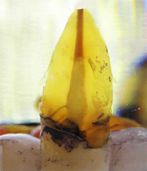
Specimens were then cleared in methyl salicylate liquid (E Merck) (Fig 1). Crystallization of the tooth structure occurred due to bonding of methyl salicylate to the dentinal
Figure 1.
A cleared specimen
The length of the penetrated interfaces was measured in tenth of millimeter at ×10 magnification using a stereomicroscope (Zeiss OPM1; Carl Zeiss, Oberkochen, Germany) by one operator, SCh, with an intrarater reliabilty coefficient of repeated measurements of 0.75.
The deepest length was measured for each specimen. Teeth were thoroughly dried with tissue and embedded in a clear acrylic resin (Pink Tray Resin; Dentsply DeTrey).
They were then sectioned into two halves longitudinally using a low speed saw (IsoMet; Buehler GmbH, Dusseldorf, Germany).
Embedded specimens appeared no longer clear.
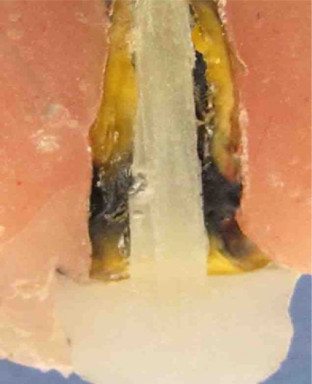
Therefore, the same specimens were examined by longitudinal sectioning (Fig 2).
Figure 2.
A longitudinally sectioned specimen
The observed length of the penetrated interfaces was measured again in tenth of millimeter using a stereomicroscope (Zeiss OPM1; Carl Zeiss).
The mean difference of penetrated length of interfaces observed in twoboth methods were statistically analyzed using paired t test and an analysis of correlation using SPSS ver 11 software (SPSS Inc, Chicago, Ill) at the level of significance of α=0.5.
RESULTS
The microleakage values measured by the two methods are summarized in Table 1. The maximum and minimum lengths of dye penetration in cleared specimens were 8.3 mm and 0.0, respectively. The minimum and maximum values in longitudinal sections were 7.9 mm and 0.0, respectively.
Table I.
Descriptive statistics of microleakage.
| Methods | Number of specimens | Minimum (mm) | Maximum (mm) | Mean | Std deviation |
|---|---|---|---|---|---|
| Clearance | 14 | 0.0 | 8.3 | 3.1 | 2.5 |
| Sectioning | 14 | 0.0 | 7.9 | 3.2 | 2.6 |
In positive controls the dye penetrated along the root canals (11.5 mm). No dye penetrated to the post space in negative controls. Paired t test revealed no significant difference in the mean microleakage of post and cores evaluated by the two methods (P= 0.7) (Table II). A significant correlation was found between the results obtained with the two methods (P=000.1 and r = 0.9). path of penetration was impossible to define.
Table II.
The results of Paired-t test
| Pair | Mean difference±SD | t | 995% confidence interval | df | P value | |
|---|---|---|---|---|---|---|
| lower | upper | |||||
| Clearing-Sectioning | 1.9±3.7 | 1.97 | 3.9 | 6.1 | 14 | .068 |
The longest length of the stained area was then measured in these two specimens.
DISCUSSION
In the present study, the microleakage of Glassix fiber post was evaluated by two methods of dye penetration measuring, longitudinal sectioning and clearing.
The results of thise study showed no difference in the amount of microleakage depicted by the two methods. An acceptable correlation was found between the two methods. Thus the null hypothesis of the research was accepted. Although no significant difference was found between the two methods, each showed potentials and limitations.
For the purpose of statistical analysis only the deepest part of penetration was counted in clearedspecimens. The cleared specimens however, In the present study, microleakage of Glassix fiber post was evaluated by two methods of dye penetration measuring; longitudinal sectioning and clearing.
The results of this study showed no difference in the amount of microleakage depicted by the two methods. An acceptable correlation was found between the two methods. Thus the null hypothesis of the research was accepted.
Although no significant difference was found between the two methods, each showed potentials and limitations. For the purpose of statistical analysis only the deepest part of penetration was counted in cleared specimens. The cleared specimens however, demonstrated a three dimensional view of dye entry and its pathway which is difficult to define in a single longitudinal slice. On the other hand, extensive ingress of dye into the dentinal tubules made it difficult to investigate the microleakage pathway in two of the cleared specimens that was also reported before [20].The method of quantification of a continuous area is also problematic in cleared specimens [20].
Microleakage has been measured as an ordinal score, linear leakage length or percentage of the leakage length to the total length of observed interfaces in the related studies [16]. It was suggested that the missing information occurring in the interval distance of scores might lead to less power in detecting the difference between investigated variables [8]. Hence, linear measurement was adopted in the present study which required high standardization of the specimens. Therefore, teeth with an approximate same size, length and age were included. All the preparations and post length were made identical. In addition, using the same teeth for both methods strengthened the thorough comparison in the present study. No microleakage was observed in the three specimens in the present study which is contradictory to several previous investigations. The method of microleakage measurement and the dye materials used are among the contributing factors. For instance, no tested material showed complete seal using fluid filtration system [5,10].
Regarding the dye material, Indian ink has been used in several studies as tracer. It has been reported that the molecule sizes are in the range of bacteria found in the root canal [11]. Indian ink is also relatively stable during acidic and alcoholic treatments of the clearing or sectioning procedures [15].
However, the molecules grow in size due to agglomeration results in the dentinal tubule blockage and may limit penetration compared with smaller molecule dyes such as methylene blue which was frequently used in other studies [11]. Entrapped air within voids and imperfections along post and core may interfere with the penetration of the tracer [13,15].
Therefore, application of a centrifuge or vacuum has been recommended to overcome this problem [4,12].
It has been indicated that by elimination of voids the dye movement would be facilitated .For the sake of convenience, none of the above mentioned procedures which were used in the present study requires to be investigated in the future. On the other hand, the results of in vitro experiments will be closer to clinical situations if the test design includes cyclic loading to simulate masticatory force [10].
Cyclic loading helps crack propagation in brittle materials or bonded surfaces. In the case of post and cores, the majority of cracks begin from flaws or imperfections found in the bonded interfaces [7].
Less disintegration of the cement layer and less microleakage was expected as test specimens were not exposed to loading in the present study.One of the disadvantages of in vitro microleakage studies is high variation in the data [13,16,21], which was also presented in the present study. Blocked tubules by air or dye itself, variability in using resin cements, differences in the configuration of extracted teeth and the sample size may account for some of the high variation in the results.
The result of the present study was consistent to the previous literature, even if different methods were compared to clearing technique. Clearing did not show significant difference in microleakage measurement compared to fluid filtration, [21] and to cross sectioning, though clearing was less discriminative [15,20].
On the other hand, clearing was determined more precisely than cross sectioning by Martin et al [19]. Nevertheless, clearing may be recommended when it is necessary to visualize the leakage pathway three dimensionally and an easy non-destructive method is preferred and it could be complementary to the more conventional methods of dye penetration [19].
In general, the clinical relevance of in vitro leakage evaluation is an area for further research. Few studies have been investigating the correlation between in vitro microleakage studies, including dye penetration and clinical outcome of the results [4,23].
In those studies, apical dye penetration had no predictive value in the determination of a periapical radiolucency. Nevertheless, the in vitro leakage studies may provide us with an idea of the quality of the filling materials or restorations, but not of the success of those materials or restorations [4].
Since clinical studies are time consuming and expensive and the standardization of the test parameters is difficult, developing a valid in vitro method to determine the sealing capacity of filling materials and restorative systems seems worth the effort.
CONCLUSIONS
Within limitation of the present study there was no significant difference between dye penetration along Glassix post measured by longitudinal sectioning and clearing technique.
Acknowledgments
The present study was supported by grant No 7421 from Vice Chancellor for Research of Tehran University of Medical Sciences.
The authors are grateful to Dr. Nasrin Akhoundi for her statistical consultation, the Reference Laboratory of Dental Research Center of Tehran University of Medical Sciences and Ms. Zohreh Dehghan for her technical assistance in the laboratory work.
REFERENCES
- 1.Morgano SM, Bracketett SE. Foundation restoration in fixed prosthodontics: current knowledge and future needs. J Prosthet Dent. 1999 Dec;82(6):643–57. doi: 10.1016/s0022-3913(99)70005-3. [DOI] [PubMed] [Google Scholar]
- 2.Rogic-Barbic M, Segovic S, Pezelj-Ribaric S, Borcic J, Jukic S, Anic I. Microleakage along Glassix glass fiber posts cemented with three different materials assessed using a fluid transport system. Int Endodon J. 2006 May;39(5):363–7. doi: 10.1111/j.1365-2591.2006.01071.x. [DOI] [PubMed] [Google Scholar]
- 3.Fox K, Gutteridge DL. An in vitro study of coronal microleakage in root-canal-treated teeth restored by the post and core technique. Int Endod J. 1997 Nov;30(6):361–8. doi: 10.1046/j.1365-2591.1997.00093.x. [DOI] [PubMed] [Google Scholar]
- 4.Susini G, Pommel L, About I, Camps J. Lack of correlation between ex vivo apical dye penetration and presence of apical radiolucencies. Oral Surg Oral Med Pathol Oral Radiol Endod. 2006 Sep;102(1):19–23. doi: 10.1016/j.tripleo.2006.03.015. [DOI] [PubMed] [Google Scholar]
- 5.Fogel HM. Microleakage of posts used to restore endodontically treated teeth. J Endodon. 1995 Jul;21(7):376–9. doi: 10.1016/S0099-2399(06)80974-X. [DOI] [PubMed] [Google Scholar]
- 6.Heling I, Gorfil C, Stutzky H, Kopolovic K, Zalkid M, Slutzky-Goldberg I. Endodontic failure caused by inadequate restoration procedures: Review and treatment recommendation. J Prosthet Dent. 2002 Jun;87(6):674–8. doi: 10.1067/mpr.2002.124453. [DOI] [PubMed] [Google Scholar]
- 7.Bolhuis P, de Gee A, Feilzer A. Influence of fatigue loading on four post-and-core systems in maxillary premolars. Quintessence Int. 2004 Sep;35(8):657–67. [PubMed] [Google Scholar]
- 8.Mannocci F, Ferrari M, Watson TF. Microleakage of endodontically treated teeth restored with fiber posts and composite cores after cyclic loading: a confocal microscopic study. J Prosthet Dent. 2001 Mar;85(3):284–91. doi: 10.1067/mpr.2001.113706. [DOI] [PubMed] [Google Scholar]
- 9.Bachicha WS, DiFiore PM, Miller DA, Lautenschlager EP. Microleakage of endodontically treated teeth restored with posts. J Endodon. 1998 Nov;24(11):703–8. doi: 10.1016/S0099-2399(98)80157-X. [DOI] [PubMed] [Google Scholar]
- 10.Reid LC, Kazemi RB, Meiers JC. Effect of fatigue testing on core integrity and post Microleakage of teeth restored with different post systems. J Endod. 2003 Feb;29(2):125–31. doi: 10.1097/00004770-200302000-00010. [DOI] [PubMed] [Google Scholar]
- 11.Crispin BJ, Verissimo MD, Vale SM. Methodologies for assessment of apical and coronal leakage of endodontic filling material: a critical review. J Oral Science. 2006;48(3):93–8. doi: 10.2334/josnusd.48.93. [DOI] [PubMed] [Google Scholar]
- 12.Ruyter IE, Oysaed H. Barthel M, Karagenc B, Gencoqlu N, Ersoy M, Canserver G, Kulekci G. Bacterial leakage versus dye leakage in obturated root canal. J Endod. 2006;32(2):110–3. Conversion in different depths. [Google Scholar]
- 13.Wimonchit S, Timpawat S, Vongsavan N. A comparison of technique for assessment of coronal dye leakage. J Endodon. 2002 Jan;28(1):1–4. doi: 10.1097/00004770-200201000-00001. [DOI] [PubMed] [Google Scholar]
- 14.Pommel L, Jacquot B, Camps J. Lack of correlation among three methods for evaluation of apical leakage. J Endodon. 2001 May;27(5):347–50. doi: 10.1097/00004770-200105000-00010. [DOI] [PubMed] [Google Scholar]
- 15.Tamse A, Katz A, Kablan F. Comparison of apical leakage shown by four different dyes with two evaluating methods. Int Endod J. 1998;31(5):333–7. [PubMed] [Google Scholar]
- 16.Karagenc B, Gencoglu N, Ersoy M, Cansever G, Kulekci G. A comparision of four different microleakage tests for assessment of leakage of root canal filling. Oral Surg Oral Med Oral Pathol Oral Radiol Oral Endod. 2006 Jul;102(1):110–3. doi: 10.1016/j.tripleo.2005.10.044. [DOI] [PubMed] [Google Scholar]
- 17.Youngson CC, Jones JC, Manogue M, Smith IS. In vitro dentinal penetration by tracers used in microleakage studies. Int Endodon J. 1998 Mar;31(1):90–9. doi: 10.1046/j.1365-2591.1998.00132.x. [DOI] [PubMed] [Google Scholar]
- 18.Camps J, Pashley D. Reliability of the dye penetration studies. J Endodon. 2003 Sep;29(9):592–4. doi: 10.1097/00004770-200309000-00012. [DOI] [PubMed] [Google Scholar]
- 19.Martin LC, Lauque FM, Rodriguez GP, Gijon RV, Mondelo JM. A comparative study of apical leakage of endomethasone, top seal, and Roeko seal sealer cements. J Endodon. 2002;28(3):423–6. doi: 10.1097/00004770-200206000-00001. [DOI] [PubMed] [Google Scholar]
- 20.Federlin M, Thonemann B, Hiller K, Fertig Ch, Schmalz G. Microleakage in class II composite resin restoration: application of a clearing protocol. Clin Oral Invest. Jun;6(2):84–91. doi: 10.1007/s00784-002-0156-5. 200. [DOI] [PubMed] [Google Scholar]
- 21.Youngson C, Jones J, Fox K, Smith I, Wood D, Gale M. A fluid filtration and clearing technique to assess microleakage associated with three dentine bonding systems. J Dent. 1999 Mar;27(2):223–33. doi: 10.1016/s0300-5712(98)00048-7. [DOI] [PubMed] [Google Scholar]
- 22.Wu MK, De Gee AJ, Wesselink PR, Moorer WR. Fluid transport and dye penetration along root fillings. Int Endod J. 1994 Jul;27(4):203–8. doi: 10.1111/j.1365-2591.1994.tb00261.x. [DOI] [PubMed] [Google Scholar]
- 23.Oliver CM, Abbott PV. Correlation between clinical success and apical dye penetration. Int Endod J. 2001 Dec;34(8):637–44. doi: 10.1046/j.1365-2591.2001.00442.x. [DOI] [PubMed] [Google Scholar]
- 24.Hedlund S, Johansson NG, Sjogren G. Retention of prefabricated and individually cast root canal posts in vitro. Br Dent J. 2003 Aug;195(3):155–8. doi: 10.1038/sj.bdj.4810405. [DOI] [PubMed] [Google Scholar]


