Abstract
Objective:
Better understanding of the furcation anatomy may serve to decrease the risk of root perforation. The purpose of this study was to measure the thickness of root walls in the danger zone in mandibular first molars.
Materials and Methods:
The roots of 53 extracted human mandibular first molars were sectioned in the horizontal plane 4 mm below the orifice of the mesial and distal root canals. For each cut surface buccal, lingual, mesial, and distal thickness of the root wall was measured. Mean values of the thickness at each location were calculated and compared by ANOVA and t-test.
Results:
The results showed that the mean thickness in the distal portion of the mesial root was smaller in comparison to all other portions of the roots (P<0.05) and this difference was statistically significant except for the mesial portion of the distal root (P=0.463). The mean thickness of radicular dentin at the distal aspect of mesial roots was 1.2 millimeter.
Conclusion:
Our study suggests that knowledge of the root dentin thickness in the danger zone is essential for preventing endodontic mishaps leading to failure.
Keywords: Endodontics, Dentin, Instrumentation, Molar, Mandible
INTRODUCTION
A thorough knowledge of the root canal anatomy is essential for successful endodontic therapy [1]. The thickness of root canal walls is an important factor since any false assumptions about it may lead to problems such as strip perforation.
Strip perforations [2,3] and vertical root fractures [4] are possible outcomes of excessive removal of radicular dentin especially in zones that have been termed danger zones. Perforation of the root during post space preparation [5–7] and pulpal injury during rotary odontoplasty, a procedure often used in conjunction with guided tissue regeneration for correction of root surface contour [8,9] are the other dangers of inattention to root canal and furcation anatomy.
Stripping is a lateral perforation caused by over instrumentation through a thin wall (danger zone) in the root [10,11]. In the danger zone there is less tooth structure compared with more peripheral portion (safety zone) of the root dentin. To minimize the risk of stripe perforations in the roots with figure-eight cross section and thin walls, such as mandibular incisors, the mesial root of mandibular molars and the mesiobuccal roots of maxillary molars, anticurvature filing should be employed [12–15].
A stripping perforation into the furcation generally results into failure if obturating material extrudes into the periodontium. Prevention is the key, because these types of perforations are very difficult to treat [16].
The new sentence is: Abou-Rass first described the anticurvature filing method to maintain the integrity of canal walls at their thin portion and reduces the possibility of root perforation or stripping [14].
Kessler et al reported that the danger zone is located 4 to 6 mm below the canal chamber orifice [11]. There is little information in literature concerning the thickness of radicular dentin.
Hence the aim of this study was to obtain precise measurements of the thickness of radicular dentin 4 mm below the orifice of canals of the first mandibular molars.
MATERIALS AND METHODS
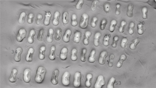
A total of 53 mandibular first molars extracted due to caries or periodontal disease were collected. Teeth that were endodontically treated and those with root caries were excluded from this study. Age, gender and the systemic condition of the patients were unknown. Teeth were stored in a 2% sodium hypochlorite solution. The teeth were then cleaned with an ultrasonic instrument (Greatcare, China) to remove calculus and remnant of the periodontal ligament. The teeth were dried and submitted for sectioning and measuring. The teeth were then sliced off 4 mm below the orifice of the mesial and distal root canals using a diamond disc (Tizcavan, Iran) with a thickness of 0.2 mm under water spray (Fig 1).
Fig 1.
The sections of mandibular first molars 4 mm below the orifice of the root.
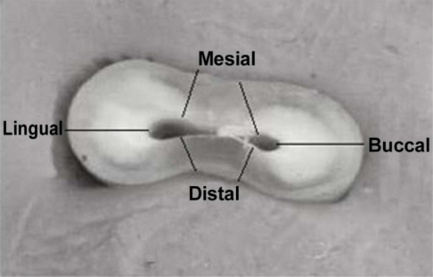
A photograph was taken of each sample surface by digital camera (Nikon S550, Japan, Maximum resolution 3648×2736) and transmitted to a computer. The thickness of dentin in different aspects of roots (including mesial, distal, buccal and lingual areas) were measured with Photoshop software (Adobe system incorporated, US patent, Ver 4.0) at 6× magnification. Measurements of the dentin thickness in different root wall were taken from the external limit of the root canal to the surface of the root. In each wall the thinnest area between the canal wall and external root surface was recorded. In roots with two canals only the smallest diameter was recorded (Fig 2). Data were subjected to paired t-test and ANOVA with a significance level of 0.05.
Fig 2.
Measurement of dentin thickness in two-canal roots.
RESULTS
The average and standard deviation of the values obtained for each group of roots are listed in Tables 1 and 2. The minimum value of the remaining thickness was in the distal wall of the mesial root (0.4 mm) and the maximum value was in the buccal wall of the distal root (3.6 mm).
Table 1.
Mean dentin thickness and standard deviation (SD) in mesial roots at different aspects.
| Aspects |
Thickness (mm) |
|||
|---|---|---|---|---|
| Maximum | Minimum | Mean | SD | |
| Mesial | 2.6 | 0.8 | 1.96 | 0.3 |
| Distal | 1.8 | 0.4 | 1.2 | 0.3 |
| Buccal | 2.7 | 1.6 | 2.17 | 0.2 |
| Lingual | 2.9 | 1.7 | 2.2 | 0.3 |
Table 2.
Mean dentin thickness and standard deviation (SD) in distal roots at different aspects.
| Aspects |
Thickness (mm) |
|||
|---|---|---|---|---|
| Maximum | Minimum | Mean | SD | |
| Mesial | 1.9 | 0.7 | 1.3 | 0.3 |
| Distal | 2.9 | 1.0 | 1.98 | 0.3 |
| Buccal | 3.6 | 1.5 | 2.33 | 0.5 |
| Lingual | 3.2 | 1.8 | 2.38 | 0.4 |
The mean thickness in the distal portion of the mesial root was smaller in comparison to all other portions of roots (P<0.05) and this difference was statistically significant except for the mesial portion of the distal root (P=0.463).
DISCUSSION
The variation of root dentin thickness in different areas supports the notion that it is very important for practitioners to increase their knowledge in regard to root canal anatomy.
Raiden et al [7] reported that the remaining dentin thickness was greater in radiographs than what was actually present and should not therefore be considered to be a reliable method for measuring the thickness of the tooth wall [3]. This procedure also has the limitation of presenting only a 2-dimensional image of a 3-dimensional object and permits the evaluation of the proximal walls only. In contrast with radiographic evaluation, the study of internal anatomy provides true assumptions about the dentin thickness, the number of canals and canal morphology of the tooth.
Our study showed that the thickness of dentin at the area of furcation was less than other surfaces in most of the specimens.
The minimal radicular dentin thickness was 0.4 mm at the distal aspect of the mesial root. In regard to mesial roots, the minimum average of dentin thickness was 1.2 mm at the distal aspect. On the other hand, the mean value of radicular dentin in distal roots was 1.3 mm at the mesial aspect.
Kessler et al [11] reported a mean value of 1.119 mm (SD=0.273) for the danger zone. Furthermore, Lim and Stock [15] studied the risks of perforation in mandibular molars and found danger zone with an average size of 1.05 mm (SD=0.33) in the mesiobuccal and 1.05 mm (SD=0.24) in the mesiolingual canal, with a mean size of 1.05 mm (SD=0.28). Bryant [10] reported that the mean size of the danger zone for 200 canals used was 0.79 mm. Studies by Filho et al [3] and Montgomery [12] reported a mean value of 0.789 mm (SD=0.182) and 0.976 mm (SD=0.24) for the danger zone, respectively.
All of these studies show the minimal thickness of dentin in the danger zone and emphasis on the need for assessment of the root wall thickness in this area before starting canal preparation. Sinai [16] observed that strip perforation in the cervical third of the root canal lead to inflammatory reactions and subsequent breakdown of the supporting structures.
Knowledge of the remaining dentin thickness in the root, especially in the distal aspect of the mesial roots would minimize if not eliminate the occurrence of strip perforations.
CONCLUSION
According to the lower thickness of root dentin near the furcation area in the mandibular first molars, the clinicians must be careful in accurate choice of the best technique of instrumentation and flaring of mandibular first molars to achieve an ideal rate of endodontic success.
Acknowledgments
The authors wish to thanks M.s Hakimian and M.s Azizizan for their assistance in the editing of this paper.
This work was done at the Shahid Sadoughi University of Medical Sciences, Yazd, Iran.
REFERENCES
- 1.Deutsch AS, Musikant BL. Morphological measurements of anatomic landmarks in human maxillary and mandibular molar pulp chambers. J Endod. 2004 Jun;30(6):388–90. doi: 10.1097/00004770-200406000-00003. [DOI] [PubMed] [Google Scholar]
- 2.Sterrett JD, Pelletier H, Russell CM. Tooth thickness at the furcation entrance of lower molars. J Clin Periodontol. 1996 Jul;23(7):621–7. doi: 10.1111/j.1600-051x.1996.tb00585.x. [DOI] [PubMed] [Google Scholar]
- 3.Filho PFG, Letra A, Menezes R, Carmo AMRD. Danger zone in mandibular molars before instrumentation: an in vitro study. J Appl Oral Sci. 2003 Aug;11(4):324–6. doi: 10.1590/s1678-77572003000400009. [DOI] [PubMed] [Google Scholar]
- 4.Testori T, Badino M, Castagnola M. Vertical root fractures in endodontically treated teeth: a clinical survey of 36 cases. J Endod. 1993 Feb;19(2):87–91. doi: 10.1016/S0099-2399(06)81202-1. [DOI] [PubMed] [Google Scholar]
- 5.Pilo R, Shapenco E, Lewinstein I. Residual dentin thickness in bifurcated maxillary first premolars after root canal and post space preparation with parallel-sided drills. J Prosthet Dent. 2008 Apr;99(4):267–73. doi: 10.1016/S0022-3913(08)60059-1. [DOI] [PubMed] [Google Scholar]
- 6.Katz A, Wasenstein-Kohn S, Tamse A, Zuckerman O. Residual dentin thickness in bifurcated maxillary premolars after root canal and dowel space preparation. J Endod. 2006 Mar;32(3):202–5. doi: 10.1016/j.joen.2005.10.048. [DOI] [PubMed] [Google Scholar]
- 7.Raiden G, Koss S, Costa L, Hernandez JL. Radiographic measurement of residual root thickness in premolars with post preparation. J Endod. 2001 Apr;27(4):296–8. doi: 10.1097/00004770-200104000-00017. [DOI] [PubMed] [Google Scholar]
- 8.Stamfelj I, Vidmar G, Cvetko E, Gaspersic D. Cementum thickness in multirooted human molars: a histometric study by light microscopy. Ann Anat. 2008;190(2):129–39. doi: 10.1016/j.aanat.2007.10.006. [DOI] [PubMed] [Google Scholar]
- 9.Bellucci C, Perrini N. A study on the thickness of radicular dentine and cementum in anterior and premolar teeth. Int Endod J. 2002 Jul;35(7):594–606. doi: 10.1046/j.1365-2591.2002.00537.x. [DOI] [PubMed] [Google Scholar]
- 10.Bryant ST, Dummer PM, Pitoni C, Bourba M, Moghal S. Shaping ability of 0.04 and 0.06 taper profile rotary nickel-titanium instruments in stimulated root canals. Int Endod J. 1999 May;32(3):155–64. doi: 10.1046/j.1365-2591.1999.00256.x. [DOI] [PubMed] [Google Scholar]
- 11.Kessler JR, Peters DD, Lorton L. Comparison of the relative risk of molar root perforations using various endodontic instrumentation techniques. J Endod. 1983 Oct;9(10):439–47. doi: 10.1016/S0099-2399(83)80260-X. [DOI] [PubMed] [Google Scholar]
- 12.Montgomery S. Root canal wall thickness of mandibular molar after biomechanical preparation. J Endod. 1985 May;1(2):257–63. doi: 10.1016/S0099-2399(85)80181-3. [DOI] [PubMed] [Google Scholar]
- 13.Berutti E, Fedon G. Thickness of cementum / dentin in mesial roots of mandibular first molars. J Endod. 1992 Nov;18(11):545–8. doi: 10.1016/S0099-2399(06)81211-2. [DOI] [PubMed] [Google Scholar]
- 14.Abou-Rass M, Frank AL, Glick DH. The anticurvature filling method to prepare the curved root canal. J Am Dent Assoc. 1980 Nov;101(5):792–4. doi: 10.14219/jada.archive.1980.0427. [DOI] [PubMed] [Google Scholar]
- 15.Lim SS, Stock CJ. The risk of perforation in the curved canal: anticurvature filling compared with step back technique. Int Endod J. 1987 Jan;20(1):33–9. doi: 10.1111/j.1365-2591.1987.tb00586.x. [DOI] [PubMed] [Google Scholar]
- 16.Sinai IH. Endodontic perforations: their prognosis and treatment. J Am Dent Assoc. 1977 Jul;95(1):90–5. doi: 10.14219/jada.archive.1977.0531. [DOI] [PubMed] [Google Scholar]


