Abstract
This study aimed to investigate the influence of different peritoneal dialysis regimens on blood pressure control, the diurnal pattern of blood pressure and left ventricular hypertrophy in patients on peritoneal dialysis. Forty-four patients undergoing peritoneal dialysis were enrolled into the study. Patients were treated with different regimens of peritoneal dialysis: 26 patients on continuous ambulatory peritoneal dialysis (CAPD) and 18 patients on automated peritoneal dialysis (APD). All patients performed 24-hour ambulatory blood pressure monitoring (ABPM) and echocardiography. Echocardiography was performed for measurement of cardiac parameters and calculation of left ventricular mass index (LVMI). There were no significant differences in average of systolic and diastolic blood pressure during 24-hour, daytime, and nighttime between CAPD and APD groups. There were no significant differences in diurnal variation of blood pressure, systolic and diastolic blood pressure load, and LVMI between CAPD and APD groups. LVMI was associated with 24 hour systolic blood pressure load (r = 0.311, P < 0.05) and daytime systolic blood pressure load (r = 0.360, P < 0.05). In conclusion, this study found that there is no difference in blood pressure control, diurnal variation of blood pressure and left ventricular hypertrophy between CAPD and APD patients. The different peritoneal dialysis regimens might not influence blood pressure control and diurnal variation of blood pressure in patients on peritoneal dialysis.
Keywords: blood pressure monitoring, ambulatory; continuous ambulatory peritoneal dialysis (CAPD); automated peritoneal dialysis (APD); left ventricular mass index
Introduction
Automated peritoneal dialysis (APD) is increasingly used in patients with end-stage renal disease. There are multiple reasons for this, including factors related to peritoneal membrane transport characteristics and dialysis adequacy, lifestyle, and the increasing age1).
APD has been reported to have several advantages over continuous ambulatory peritoneal dialysis (CAPD) including lower incidence of peritonitis, better small solute clearances, reduced incidences of hernias, and better dialysis acceptability for workers2-4). However, APD has potential disadvantages such as a more rapid loss of residual renal function, less sodium removal and more peritoneal protein loss5-10). The short dwell times and less sodium and fluid removal of APD compared with CAPD seem to be associated with changes in hemodynamics and blood pressure control7-9).
Cardiovascular disease is the most common cause of morbidity and mortality in patients with end-stage renal disease (ESRD)11). Hypertension in dialysis patients is an important risk factor for development of left ventricular hypertrophy (LVH) and for increased cardiovascular mortality. The main reason for hypertension in dialysis patients is volume overload12,13). Information on the compared ability of CAPD and APD to control blood pressure levels and prevent cardiovascular complications is very limited.
The aim of this study is to investigate the influence of different peritoneal dialysis regimens on blood pressure control, the diurnal pattern of blood pressure and left ventricular hypertrophy in patients on peritoneal dialysis.
Subjects and Method
A total of 44 stable patients undergoing peritoneal dialysis at Chungbuk National University Hospital for more than 6 months were enrolled in this study. Patients were treated by different regimens of peritoneal dialysis: 26 patients on CAPD and 18 patients on APD. Patients' preference was the main reason to start APD. All patients performed 24-hour ambulatory blood pressure monitoring (ABPM) and echocardiography.
The ABPM device (90207 ABP Monitor; Spacelabs Healthcare Inc., Issaquah, Washington, USA) was employed for measurement of blood pressure using an oscillometric technique. The monitor was automatically programmed to inflate every 30 minutes from 7 AM until 11 PM (daytime). From 11 PM to 7 AM (nighttime), the device inflated every 60 minutes. Mean 24-hour, daytime, and nighttime systolic BP and diastolic BP were derived from 24-hour ABPM as the average of measurements taken over the respective time period. ABPM recordings were downloaded, evaluated, and analyzed in a blinded manner at Spacelabs Healthcare. During the monitoring period, patients were instructed to attend to their usual activities, diet, and medications. Mean arterial pressure (MAP) was calculated as diastolic pressure plus one third of the difference between systolic and diastolic pressure. Diurnal blood pressure variation was characterized by an index calculated as the ratio between the blood pressure difference of daytime and nighttime and the daytime blood pressure value, multiplied by 100. Non-dipper was defined as a reduction in nighttime systolic blood pressure or diastolic blood pressure less than 10% of the daytime blood pressure. Systolic blood pressure load and diastolic blood pressure load were defined as the percentage of readings higher than 135/85 mmHg for daytime blood pressure, and 125/75 mmHg for nighttime blood pressure. A blood pressure load greater than 30% was considered significant14).
Two-dimensional and M-mode echocardiography was performed using an iE 33 echocardiography system (Phillips Medical Systems, Eindhoven, The Nethelands). Measurements of end-systolic and end-diastolic left ventricular internal dimensions, end-diastolic interventricular septal thickness, end-diastolic posterior wall thickness, aortic root dimension, and left atrial dimension were made according to the American Society of Echocardiography guidelines15). Criterias for LVH were left ventricular mass index (LVMI) 125 g/m2 for males and 100 g/m2 for females.
Left ventricular mass (LVM) was calculated by the formula derived by Devereux and Reichek16);
LVM (g) = 1.04 × [(LVIDd + IVSd + LVPWd)3 - (LVIDs)3] - 13.6
where LVIDd is left ventricular end diastolic internal dimension, IVSd is interventricular septal thickness measured at end-diastole and LVPWd is posterior wall thickness.
LVMI was standardized by dividing it to body surface area, which was calculated using DuBois formula17).
The following data were collected for each patient: age, gender, body mass index, body weight, primary cause of ESRD, duration of hypertension and peritoneal dialysis, use of erythropoietin, hemoglobin, hematocrit, serum blood urea nitrogen, creatinine, albumin, calcium, and phosphate. Data of peritoneal dialysis therapy were collected, including peritoneal dialysis adequacy (weekly Kt/V and weekly creatinine clearance) and peritoneal equilibration test (PET). Routine PET was performed according to the method of Twardowski18). Weekly Kt/V was calculated as 7 × (24-hour urea clearance/total body water), where total body water was estimated according to the formula of Watson et al.19).
Weekly creatinine clearance was calculated as 7 × (24-hour creatinine clearance/1.73 m2 body surface area), where body surface was estimated by the method of DuBois17).
In this study, CAPD patients were treated with 4 exchanges daily using 1,500-2,000 mL peritoneal dialysis solution containing 1.5%, 2.5%, or 4.25% dextrose for ultrafiltration as appropriate. For patients on APD, the cycler was programmed to deliver 4-5 cycles of 1,500-2,000 mL peritoneal dialysis solution during the night. A final exchange of 1,000-1,500 mL was introduced and allowed to remain in the peritoneal cavity until the next cyclic exchange at night.
SPSS 12.0 for windows software was used for all statistical analysis (SPSS Inc, Chicago, IL, USA). Values are expressed as mean ± SD. The comparisons between groups were assessed by unpaired t-test or Wilcox singed-rank test. Chi-squre test was used for 2 × 2 contingency tables when appropriate for non-numeric data. Correlation between numerical parameters were analysed with Spearman's correlation test. A P value of less than 0.05 was considered statistically significant.
Result
1. Patient characteristics
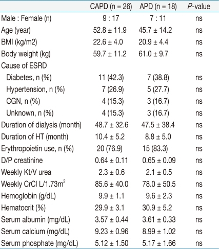
This study group consisted of 44 patients on peritoneal dialysis (16 male, 28 female, mean age: 49.9 ± 13.2 years), including 26 patients on CAPD (9 male, 17 female, mean age: 52.8 ± 11.9 years) and 18 patients on APD (7 male, 11 female, mean age: 45.7 ± 14.2 years). The characteristics of the 44 patients are shown in Table 1.
Table 1.
Clinical Characteristics by CAPD and APD
CAPD, continuous ambulatory peritoneal dialysis; APD, automated peritoneal dialysis; BMI, body mass index; ESRD, end-stage renal disease; CGN, chronic gromerulonephritis; HT, hypertension; CrCl, creatinine clearance; ns, not significant.
Mean time on PD was 48.7 ± 32.6 months in CAPD and 47.5 ± 38.4 months in APD group. Primary causes of ESRD were diabetic nephropathy (n = 18, 40.1%), hypertension (n = 12, 27.3%), chronic glomerulonephritis (n = 7, 15.9%), and unknown etiology (n = 7, 15.9%). There was no significant difference in gender, age, BMI, body weight, primary diagnosis for ESRD, duration of dialysis and hypertension, use of erythropoietin, dialysate/plasma (D/P) creatinine ratio, weekly Kt/V, weekly creatinine clearance, hemoglobin, hematocrit serum albumin, calcium, and phosphate. Only 2 patients of 18 APD had high PET characteristics.
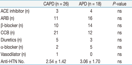
Thirty-six (81.8%) of the 44 PD patients were treated with antihypertensive agents, including angiotensin converting enzyme inhibitors, angiotensin receptor blocker, beta-blockers, calcium channel blockers, alpha-blockers, diuretics or combinations of such medications. There was no significant difference in total number of antihypertensives between the 2 groups (Table 2).
Table 2.
Comparison of Use of Anti-Hypertensive Agent between CAPD and APD
CAPD, continuous ambulatory peritoneal dialysis; APD, automated peritoneal dialysis; ACE inhibitor, angiotensin converting enzyme inhibitor; ARB, angiotensin receptor blocker; CCB, calcium channel blocker; Anti-HTN No., total number of antihypertensive agent; ns, not significant.
2. Comparison of blood pressure control between CAPD and APD
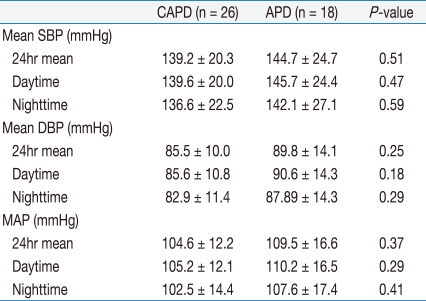
There were no significant differences in ambulatory mean systolic and diastolic blood pressure, daytime systolic and diastolic blood pressure, and nighttime systolic and diastolic blood pressure between the 2 groups (Table 3). Ambulatory systolic and diastolic blood pressure of 24-hour, daytime, and nighttime in the CAPD group were lower than those of the APD group, despite not reaching statistically significant levels.
Table 3.
Comparison of Blood Pressure Control between CAPD and APD
CAPD, continuous ambulatory peritoneal dialysis; APD, automated peritoneal dialysis; SBP, systolic blood pressure; DBP, diastolic blood pressure; MAP, mean arterial pressure.
On analysis for diurnal variation of blood pressure, there was no difference in the non-dipper hypertension rate between 2 groups. Twenty-three of 26 (88.5%) CAPD patients were non-dipper, whereas 15 of 18 (83.3%) APD patients were non-dipper. On comparing diurnal indices of systolic and diastolic blood pressure between two groups, no difference was found in both CAPD and APD groups (Table 4).
Table 4.
Comparison of Diurnal Blood Pressure Variations and Proportions of Non-dipper Hypertension between CAPD and APD
Diurnal index of BP calculated as the ratio between the blood pressure difference of daytime and nighttime and the daytime blood pressure value, multiplied by 100. CAPD, continuous ambulatory peritoneal dialysis; APD, automated peritoneal dialysis; SBP, systolic blood pressure; DBP, diastolic blood pressure; HT, hypertension; ns, not significant; BP, blood pressure.
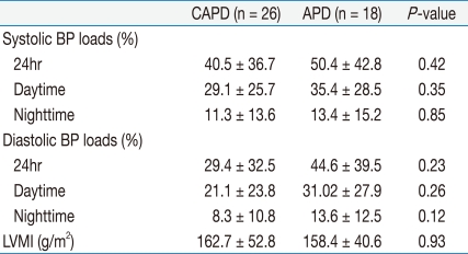
There were no significant differences in systolic and diastolic blood pressure load and LVMI between CAPD and APD groups (Table 5).
Table 5.
Comparison of Blood Pressure Loads and Left Ventricular Hypetrophy between CAPD and APD
CAPD, continuous ambulatory peritoneal dialysis; APD, automated peritoneal dialysis; BP, blood pressure; LVMI, left ventricular mass index.
There were no significant correlations between LVMI and 24-hour mean systolic and diastolic blood pressure, daytime systolic and diastolic blood pressure, and nighttime systolic and diastolic blood pressures.
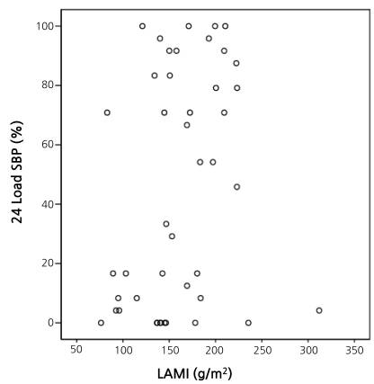
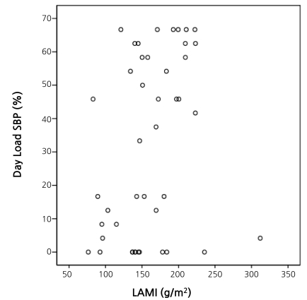
LVMI was correlated with 24-hour systolic BP load (r = 0.311, P < 0.05) and daytime systolic BP load (r = 0.360, P < 0.05) (Fig. 1 and 2).
Fig. 1.
Correlation between LVMI and 24-Hour Systolic Blood Pressure Load (r = 0.311, P < 0.05). LVMI, left ventricular mass index; 24hr Load SBP, 24 hour systolic blood pressure load.
Fig. 2.
Correlation between LVMI and Daytime Systolic Blood Pressure Load (r = 0.36, P < 0.05). LVMI, left ventricular mass index; Day Load SBP, daytime systolic blood pressure load.
Discussion
There is an ongoing controversy in the literature as to whether APD is associated with less effective control of blood pressure as compared with CAPD. Wang et al.20) found similar levels of blood pressure in patients on CAPD and APD, but a higher ventricular mass in patients on APD, which they attributed to potential diurnal hypervolemia in this therapy. Ortega et al.21) detected higher systolic blood pressure levels in APD than in CAPD, but were unable to find a correlation between sodium removal and blood pressure levels.
Our study demonstrates that there were no significant differences in blood pressure control, diurnal variation of blood pressure, and left ventricular mass between CAPD and APD.
Left ventricular hypertrophy is a strong predictor of myocardial infarction, heart failure, sudden death, and stroke in patients with essential hypertension and in patients with ESRD22-26). There are reports of a high incidence of LVH in CAPD patients and that LVH seemed to be strongly associated with a significantly high cardiovascular morbidity and mortality in CAPD patients27). It has reported that each 10 mmHg rise in average mean arterial blood pressure was associated with a 48% increased likelihood of having both concentric LVH and left ventricular dilation on second echocardiogram for patients with ESRD28). However, in this study, we have not observed significant difference in LVMI between CAPD and APD groups. Ambulatory systolic and diastolic blood pressure at 24-hour, daytime and nighttime was not associated with LVMI.
It has been reported that ambulatory nighttime systolic blood pressure load > 30% had an independent association with LVH20). Similarly, we found in our study that LVMI in patients on peritoneal dialysis correlated with 24-hour systolic blood pressure load and daytime systolic blood pressure load.
Diurnal blood pressure rhythm is known to be abnormal in chronic renal failure, dialysis and renal transplantation patients. It has been reported that non-dipper patients have increased risk of target organ damage and mortality in patients with hypertension29-31). Recently, reduced blood pressure diurnal variability has been reported as a risk factor for left ventricular dilatation in hemodialysis patents. Although a number of factors such as autonomic dysfunction, increase in plasma volume, heart failure and sleep disorders have been implicated for abnormal circadian rhythm of BP in dialysis patients, the main reason of it was unclear. Subjectively assessed sleep disturbance during overnight BP monitoring increases the nocturnal BP level and potentially attenuates the correlation with hypertension-related cardiac damage, even though habituation to overnight BP monitoring occurs32).
It has been suggested that APD may be less effective than CAPD for sodium and fluid removal, due to its semi-intermittent nature and frequently lower capacity for ultrafiltration, and also to the short dwell schedule that characterizes the night session in APD, which may result in significant sodium sieving and less efficient sodium removal32). In addition, the alarms or noises of the APD machine may cause sleep disturbance in patients on APD. However, we found no difference in diurnal variation of blood pressure for APD compared with CAPD .
Recently, patients are increasingly selecting APD for lifestyle reasons rather than CAPD because they have high transport membrane characteristics, resulting in an increasing proportion of APD patients having low transport membrane characteristics. In this study, there was no difference in D/P creatinine and only 2 patients had high transporter membrane characteristics in the APD group.
In this study, 81.8% of the peritoneal dialysis patients were receiving antihypertensive agents, and these medications themselves may affect blood pressure control and circadian blood pressure rhythm.
There are limitations to this study. The size of our study population was not large enough to compare the difference between CAPD and APD group. In addition, we did not evaluate the sodium removal and volume status between CAPD and APD groups. Further study is needed to investigate whether the effect of the sodium and fluid removal according to CAPD regimen can influence the blood pressure control.
It has been suggested that the use of a glucose polymeric solution, icodextrin 7.5%, is able to improve volume status in peritoneal dialysis patients due to an improved and prolonged ultrafiltration, especially during long dwells33-35). Further study is needed to study the influence of icodextrin for long dwells in APD on volume status and blood pressure control.
In conclusion, we found no difference in blood pressure control, diurnal variation of blood pressure, and left ventricular hypertrophy for APD compared with CAPD. The different peritoneal dialysis regimens might not influence blood pressure control and diurnal variation of blood pressure in patients on peritoneal dialysis.
Footnotes
The paper was supported by a grant from Chungbuk National University (2010).
References
- 1.Wilson J, Nissenson AR. Determinants in APD selection. Semin Dial. 2002;15:388–392. doi: 10.1046/j.1525-139x.2002.00097.x. [DOI] [PubMed] [Google Scholar]
- 2.Brunkhorst R, Wrenger E, Krautzig S, Ehlerding G, Mahiout A, Koch KM. Clinical experience with home automated peritoneal dialysis. Kidney Int Suppl. 1994;48:S25–S30. [PubMed] [Google Scholar]
- 3.Holley JL, Bernardini J, Piraino B. Continuous cycling peritoneal dialysis is associated with lower rates of catheter infections than continuous ambulatory peritoneal dialysis. Am J Kidney Dis. 1990;16:133–136. doi: 10.1016/s0272-6386(12)80567-1. [DOI] [PubMed] [Google Scholar]
- 4.Rodriguez AM, Diaz NV, Cubillo LP, et al. Automated peritoneal dialysis: a Spanish multicentre study. Nephrol Dial Transplant. 1998;13:2335–2340. doi: 10.1093/ndt/13.9.2335. [DOI] [PubMed] [Google Scholar]
- 5.Hiroshige K, Yuu K, Soejima M, Takasugi M, Kuroiwa A. Rapid decline of residual renal function in patients on automated peritoneal dialysis. Perit Dial Int. 1996;16:307–315. [PubMed] [Google Scholar]
- 6.Hufnagel G, Michel C, Queffeulou G, Skhiri H, Damieri H, Mignon F. The influence of automated peritoneal dialysis on the decrease in residual renal function. Nephrol Dial Transplant. 1999;14:1224–1228. doi: 10.1093/ndt/14.5.1224. [DOI] [PubMed] [Google Scholar]
- 7.Rodriguez-Carmona A, Fontan MP. Sodium removal in patients undergoing CAPD and automated peritoneal dialysis. Perit Dial Int. 2002;22:705–713. [PubMed] [Google Scholar]
- 8.Ortega O, Gallar P, Carreno A, et al. Peritoneal sodium mass removal in continuous ambulatory peritoneal dialysis and automated peritoneal dialysis: influence on blood pressure control. Am J Nephrol. 2001;21:189–193. doi: 10.1159/000046246. [DOI] [PubMed] [Google Scholar]
- 9.Rodriguez-Carmona A, Perez-Fontan M, Garca-Naveiro R, Villaverde P, Peteiro J. Compared time profiles of ultrafiltration, sodium removal, and renal function in incident CAPD and automated peritoneal dialysis patients. Am J Kidney Dis. 2004;44:132–145. doi: 10.1053/j.ajkd.2004.03.035. [DOI] [PubMed] [Google Scholar]
- 10.Westra WM, Kopple JD, Krediet RT, Appell M, Mehrotra R. Dietary protein requirements and dialysate protein losses in chronic peritoneal dialysis patients. Perit Dial Int. 2007;27:192–195. [PubMed] [Google Scholar]
- 11.Erturk S, Ertug AE, Ates K, et al. Relationship of ambulatory blood pressure monitoring data to echocardiographic findings in haemodialysis patients. Nephrol Dial Transplant. 1996;11:2050–2054. doi: 10.1093/oxfordjournals.ndt.a027095. [DOI] [PubMed] [Google Scholar]
- 12.Katzarski KS, Charra B, Luik AJ, et al. Fluid state and blood pressure control in patients treated with long and short haemodialysis. Nephrol Dial Transplant. 1999;14:369–375. doi: 10.1093/ndt/14.2.369. [DOI] [PubMed] [Google Scholar]
- 13.Gunal AI, Duman S, Ozkahya M, et al. Strict volume control normalizes hypertension in peritoneal dialysis patients. Am J Kidney Dis. 2001;37:588–593. [PubMed] [Google Scholar]
- 14.Phillips RA, Diamond JA. Ambulatory blood pressure monitoring and echocardiography--noninvasive techniques for evaluation of the hypertensive patient. Prog Cardiovasc Dis. 1999;41:397–440. doi: 10.1016/s0033-0620(99)70019-8. [DOI] [PubMed] [Google Scholar]
- 15.Sahn DJ, DeMaria A, Kisslo J, Weyman A. Recommendations regarding quantitation in M-mode echocardiography: results of a survey of echocardiographic measurements. Circulation. 1978;58:1072–1083. doi: 10.1161/01.cir.58.6.1072. [DOI] [PubMed] [Google Scholar]
- 16.Devereux RB, Reichek N. Echocardiographic determination of left ventricular mass in man. Anatomic validation of the method. Circulation. 1977;55:613–618. doi: 10.1161/01.cir.55.4.613. [DOI] [PubMed] [Google Scholar]
- 17.Dubois D, Dubois EF. A formula to estimate the approximate surface area if height and weight be known. Arch Intern Med. 1916;17:863–871. [Google Scholar]
- 18.Watson PE, Watson ID, Batt RD. Total body water volumes for adult males and females estimated from simple anthropometric measurements. Am J Clin Nutr. 1980;33:27–39. doi: 10.1093/ajcn/33.1.27. [DOI] [PubMed] [Google Scholar]
- 19.Twardowski ZJ. Clinical value of standardized equilibration tests in CAPD patients. Blood Purif. 1989;7:95–108. doi: 10.1159/000169582. [DOI] [PubMed] [Google Scholar]
- 20.Wang MC, Tseng CC, Tsai WC, Huang JJ. Blood pressure and left ventricular hypertrophy in patients on different peritoneal dialysis regimens. Perit Dial Int. 2001;21:36–42. [PubMed] [Google Scholar]
- 21.Ortega O, Gallar P, Carreno A, et al. Peritoneal sodium mass removal in continuous ambulatory peritoneal dialysis and automated peritoneal dialysis: influence on blood pressure control. Am J Nephrol. 2001;21:189–193. doi: 10.1159/000046246. [DOI] [PubMed] [Google Scholar]
- 22.Casale PN, Devereux RB, Milner M, et al. Value of echocardiographic measurement of left ventricular mass in predicting cardiovascular morbid events in hypertensive men. Ann Intern Med. 1986;105:173–178. doi: 10.7326/0003-4819-105-2-173. [DOI] [PubMed] [Google Scholar]
- 23.Frohlich ED. Left ventricular hypertrophy as a risk factor. Cardiol Clin. 1986;4:137–144. [PubMed] [Google Scholar]
- 24.Anderson KP. Sudden death, hypertension, and hypertrophy. J Cardiovasc Pharmacol. 1984;6(Suppl 3):S498–S503. [PubMed] [Google Scholar]
- 25.McLenachan JM, Henderson E, Morris KI, Dargie HJ. Ventricular arrhythmias in patients with hypertensive left ventricular hypertrophy. N Engl J Med. 1987;317:787–792. doi: 10.1056/NEJM198709243171302. [DOI] [PubMed] [Google Scholar]
- 26.Levy D, Garrison RJ, Savage DD, Kannel WB, Castelli WP. Prognostic implications of echocardiographically determined left ventricular mass in the Framingham Heart Study. N Engl J Med. 1990;322:1561–1566. doi: 10.1056/NEJM199005313222203. [DOI] [PubMed] [Google Scholar]
- 27.Silaruks S, Sirivongs D, Chunlertrith D. Left ventricular hypertrophy and clinical outcome in CAPD patients. Perit Dial Int. 2000;20:461–466. [PubMed] [Google Scholar]
- 28.Chung JH, Yun NR, Ahn CY, Lee WS, Kim HL. Relationship between serum N-terminal pro-brain natriuretic peptide level and left ventricular dysfunction and extracellular water in continuous ambulatory peritoneal dialysis patients. Electrolyte Blood Press. 2008;6:15–21. doi: 10.5049/EBP.2008.6.1.15. [DOI] [PMC free article] [PubMed] [Google Scholar]
- 29.Sturrock ND, George E, Pound N, Stevenson J, Peck GM, Sowter H. Non-dipping circadian blood pressure and renal impairment are associated with increased mortality in diabetes mellitus. Diabet Med. 2000;17:360–364. doi: 10.1046/j.1464-5491.2000.00284.x. [DOI] [PubMed] [Google Scholar]
- 30.White WB. How well does ambulatory blood pressure predict target-organ disease and clinical outcome in patients with hypertension? Blood Press Monit. 1999;4(Suppl 2):S17–S21. [PubMed] [Google Scholar]
- 31.Covic A, Goldsmith DJ, Covic M. Reduced blood pressure diurnal variability as a risk factor for progressive left ventricular dilatation in hemodialysis patients. Am J Kidney Dis. 2000;35:617–623. doi: 10.1016/s0272-6386(00)70007-2. [DOI] [PubMed] [Google Scholar]
- 32.Verdecchia P, Angeli F, Borgioni C, Gattobigio R, Reboldi G. Ambulatory blood pressure and cardiovascular outcome in relation to perceived sleep deprivation. Hypertension. 2007;49:777–783. doi: 10.1161/01.HYP.0000258215.26755.20. [DOI] [PubMed] [Google Scholar]
- 33.Struijk DG, Krediet RT. Sodium balance in automated peritoneal dialysis. Perit Dial Int. 2000;20(Suppl 2):S101–S105. [PubMed] [Google Scholar]
- 34.Posthuma N, ter Wee PM, Donker AJ, Oe PL, Peers EM, Verbrugh HA The Dextrin in APD in Amsterdam (DIANA) Group. Assessment of the effectiveness, safety, and biocompatibility of icodextrin in automated peritoneal dialysis. Perit Dial Int. 2000;20(Suppl 2):S106–S113. [PubMed] [Google Scholar]
- 35.Johnson DW, Arndt M, O'Shea A, Watt R, Hamilton J, Vincent K. Icodextrin as salvage therapy in peritoneal dialysis patients with refractory fluid overload. BMC Nephrol. 2001;2:2. doi: 10.1186/1471-2369-2-2. [DOI] [PMC free article] [PubMed] [Google Scholar]


