Abstract
Francisella tularensis has been associated with naturally occurring waterborne outbreaks and is also of interest as a potential biological weapon. Recovery of this pathogen from water using cultural methods is challenging due to the organism's fastidious growth requirements and interference by indigenous bacteria. A 15-min acid treatment procedure prior to culture on a selective agar was evaluated for recovery of F. tularensis from seeded water samples. Mean levels of reduction of virulent strains of F. tularensis subsp. holarctica and subsp. tularensis were less than 20% following acid treatment. The attenuated live vaccine strain (LVS) was less resistant to acid exposure. The acid treatment procedure coupled with plating on cystine heart agar with rabbit blood and antibiotics (CHARBab) allowed the isolation of F. tularensis seeded into five natural water samples.
TEXT
Tularemia is an infectious zoonotic disease caused by the facultative, intracellular, Gram-negative bacterium Francisella tularensis. F. tularensis subsp. holarctica (type B), found throughout the Northern Hemisphere, but particularly in Eurasia, causes severe disease but does not generally result in death. F. tularensis subsp. tularensis (type A), found predominantly in North America, is the more virulent biotype, and without antibiotic treatment has a mortality rate of 5 to 15% (5). Humans can become infected from bites of infected arthropod vectors, from handling of sick or dead animals, from inhalation of particles from contaminated soil or vegetation, and by consumption of contaminated food and water. The organism may survive for a year in water or mud (4). Reports of naturally occurring waterborne outbreaks (7, 10) have been limited primarily to contamination by the holarctica subspecies.
Owing to the organism's infectivity, ease of transmission, and potential use as a bioweapon, virulent strains of F. tularensis are listed as Class A Select Agents by the U.S. Centers for Disease Control and Prevention. Both the United States and the former Soviet Union maintained stockpiles of the weaponized agent prior to 1970 (5). In the event of an intentional release or a natural outbreak, rapid recovery and identification of the agent would expedite characterization and control.
Members of this species are slow-growing, fastidious organisms, which makes isolation by cultural methods very challenging. Many bacteria indigenous to water are likely to have a competitive advantage under the culture conditions required for growth of Francisella spp. Rapidly growing background organisms easily mask the presence of the target colonies on agar media. These indigenous bacteria can also deplete nutrients or produce bacteriocins which can negatively impact the growth of F. tularensis. Reducing the number of these competing microorganisms in water samples is an important consideration for enhancing pathogen recovery. Low-pH treatment of water samples has previously been used to reduce nontarget organisms to aid in the detection of Legionella spp. from water (2). The use of selective agar antibiotic containing medium has been proposed for the isolation of F. tularensis from environmental samples (13). In this study we evaluated a standard acid treatment procedure developed for the recovery of Legionella spp. (15). Coupled with the use of a commercially available selective culture medium (cystine heart agar with rabbit blood and antibiotics [CHARBab]) to enhance the recovery of seeded cultures of F. tularensis from water samples.
Four virulent strains of F. tularensis subsp. holarctica type B and three virulent strains of Francisella tularensis subsp. tularensis type A1, originally collected from various geographical locations in the United States, were used in the study. The avirulent F. tularensis subsp. holarctica live vaccine strain (LVS) was also included (Table 1). All strains were obtained from Laura Rose (U.S. Centers for Disease Control and Prevention, Atlanta, GA). Experiments with virulent strains were conducted under biosafety level 3 conditions at the University of Cincinnati College of Medicine, and all protocols were approved by the university institutional biological safety committee.
Table 1.
F. tularensis strains used in the study
| Subspecies and type designation | Isolate/origin |
|---|---|
| F. tularensis subsp. tularensis (type A1) | F2246/Maryland |
| F. tularensis subsp. tularensis (type A1) | H3563/Oklahoma |
| F. tularensis subsp. tularensis (type A1) | Schu4/Ohio |
| F. tularensis subsp. holarctica (type B) | IN99-1009/Indiana |
| F. tularensis subsp. holarctica (type B) | KY99-3387/Kentucky |
| F. tularensis subsp. holarctica (type B) | NY98/New York |
| F. tularensis subsp. holarctica (type B) | OR96-0246/Oregon |
| F. tularensis subsp. holarctica (type B, attenuated) | LVS |
Frozen stock cultures were stored in brain heart infusion broth (Becton Dickinson, Sparks, MD) with 15% (vol/vol) glycerol at −80°C. Separate frozen isolates of each strain were used for each set of experiments. Isolates were inoculated into 25 ml of tryptic soy broth (Becton Dickinson, Sparks, MD) containing 2% (vol/vol) Iso-Vitalex (Becton Dickinson, Sparks, MD) and incubated at 37°C for 48 h. Cultures were concentrated, resuspended in sterile chlorine-free tap water, and washed twice by centrifugation (3,000 × g for 10 min at 4°C) prior to the inoculation of water samples.
Samples were analyzed using either enriched chocolate agar consisting of GC agar base, bovine hemoglobin with GCHI enrichment (Remel, Lenexa, KS), or cystine heart agar with rabbit blood and antibiotics (CHARBab) (Remel, Lenexa, KS). The composition of CHARBab agar is based upon a modification of the formulation originally proposed by Rhamy (14) and was supplemented with 1 × 10⁶ U liter−1 penicillin and 1 × 10⁶ U liter−1 polymyxin B. All samples were analyzed by the spread plate procedure using a maximum inoculum of 0.1 ml per plate and incubated at 37°C for 6 days.
Colony confirmation was based upon World Health Organization (WHO) diagnostic protocols (16). Presumptive greenish-white, 2- to 4-mm colonies on CHARBab agar were confirmed by subculturing 10% of the target colonies back to CHARBab agar and examining for characteristic greenish-white colonies with a butyrous consistency after incubation at 37°C for 48 h. All presumptive positive colonies were further verified by the slide agglutination assay using hyperimmune rabbit-anti F. tularensis antibody (Becton Dickinson, Sparks, MD).
The acid treatment procedure was conducted as previously described (15). Briefly, 2 ml of sterile KCl-HCl acid (18 parts of 0.2 M KCl plus 1 part of 0.2 M HCl, pH 2.0) was added to 2 ml of the sample, and the resultant solution was mixed and incubated at room temperature for 15 min. After the 15-min exposure time, 2 ml of 0.1 N KOH neutralizer was added to each tube. The pH values for all samples were determined to insure appropriate acidification and neutralization. A negative control (no acid treatment) was prepared by adding 2 ml of sample to 4 ml of sterile water.
To determine the effect of the low-pH acid treatment in pure culture, each of the seven F. tularensis strains was cultured, washed, and resuspended in sterile dechlorinated tap water prior to experimentation as described in reference 15, with final concentrations in the range of 5 to 6 log10 CFU ml−1. These samples were analyzed using both enriched chocolate agar and CHARBab agar. Five environmental water sources, i.e., two creeks, a pond, a river, and a well, were included in the study. One-hundred-milliliter volumes of each sample were seeded with three F. tularensis subsp. holarctica strains, KY99-3387, NY98, and LVS, resulting in final concentrations of 2.0 ± 0.1 log10 CFU ml−1. Strains of F. tularensis belonging to the subspecies holarctica were chosen to seed natural water samples primarily due to the association of this subspecies with natural waterborne outbreaks. Preliminary experiments using acid exposure times of 5, 15, and 30 min (data not shown) indicated that the elimination of background organisms without major decreases in the number of Francisella organisms was best achieved using the 15-min exposure time listed by Bopp et al. (2). Each strain was seeded individually into each water sample and analyzed before and after acid treatment using CHARBab agar. Experiments were conducted in triplicate for each water type. Statistical comparisons were based upon analysis of variance (ANOVA) testing of the log-transformed data.
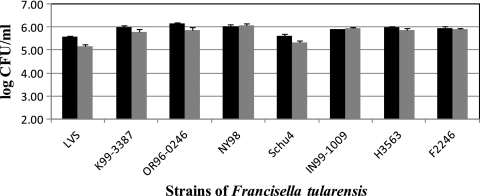
For the sterile tap water sample and the five natural water samples, the mean pH level prior to acidification was 8.0 (range, 7.6 to 8.2). The acid treatment procedure lowered the pH of the samples to a range of 2.1 to 2.6. The results of the acid treatment for the F. tularensis strains seeded into sterile chlorine-free tap water are shown in Fig. 1. Average log reductions after acid treatment for F. tularensis subsp. tularensis, F. tularensis subsp. holarctica, and the LVS were 0.15 (19.2%), 0.11 (15.7%), and 0.39 (61%), respectively. There was no statistical difference in acid resistance between strains of F. tularensis subsp. holarctica and F. tularensis subsp. tularensis (P = 0.829). There was a significant difference between the LVS and the other holarctica strains (P < 0.001) and tularensis subspecies strains (P = 0.001). There was no significant difference in titers of the LVS, F. tularensis subsp. tularensis, and F. tularensis subsp. holarctica (P = 0.8439, P = 0.275, and P = 0.2838), respectively, for any of the subspecies when plated on either enriched chocolate agar or CHARBab agar (data not shown), thus indicating that the use of the selective medium did not decrease recovery.
Fig. 1.
Effect of acid treatment on F. tularensis strains seeded into sterile chlorine-free tap water. Shown are untreated samples (black) and acid-treated samples (gray). Error bars indicate standard deviations for each replicate.
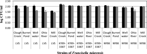
All three target strains were recovered and quantified from the seeded natural water samples. The results of the acid treatment procedure for the seeded natural water samples are shown in Fig. 2. The CHARBab medium reduced the number of background indigenous organisms in the non-acid-treated samples to ≤1.5 log10 CFU ml−1. All presumptive target colonies submitted for verification were confirmed as F. tularensis by using the WHO verification procedure (16). Average log reductions across all water types after acid treatment for F. tularensis subsp. holarctica strains KY99-3387, NY98, and LVS were 0.01 (7%), 0.09 (17%), and 0.29 (49%), respectively.
Fig. 2.
The effect of acid treatment on F. tularensis subsp. holarctica strains recovered from five seeded natural water samples. Shown are untreated samples (black) and acid-treated samples (gray). Error bars indicate standard deviations for each replicate.
To further evaluate recovery in samples containing higher background levels of indigenous bacteria, two additional water samples were collected from the Clough Creek source following two separate rainfall events and seeded with the least acid-resistant strain (F. tularensis subsp. holarctica LVS). A 100-ml portion of each sample was autoclaved to serve as a sterile control to determine the initial titer of the seeded inoculum. The first water sample was seeded with LVS to achieve a final concentration of 2.0 ± 0.1 log10 CFU ml−1. After the 6-day incubation, the background nontarget colony level on CHARBab agar which did not receive the acid treatment was 2.9 log10 CFU ml−1. This level of background organisms precluded isolation of the LVS strain in the non-acid-treated sample. The background organisms from the acid-treated sample were reduced to 0.9 log10 CFU ml−1; this reduction allowed the detection of the target LVS colonies (1.8 log10 CFU ml−1). In the second sample, the background level of nontarget colonies on CHARBab agar without acid treatment was 3.8 log10 CFU ml−1. This sample was seeded with a higher LVS level of 4.4 ± 0.1 log10 CFU ml−1. Even at this higher level of seeded inoculum, we were unable to isolate LVS from the non-acid-treated sample. Following acid treatment, the background organisms on CHARBab agar were reduced to 2.0 ± 0.2 log10 CFU ml−1, and the target strain was detected and quantified at a level of 4.1 ± 0.2 log10 CFU ml−1.
The combination of acid treatment followed by plating on the selective agar medium allowed the recovery of F. tularensis from water and effectively reduced indigenous background organisms. Differences in acid resistance were observed among strains and subgroups of F. tularensis. The overall mean reduction (<20%) was less than the reduction (>50%) that has been previously reported for the procedure when used for detecting Legionella spp. in water (3, 11). The avirulent F. tularensis subsp. holarctica LVS was consistently less resistant to acid treatment than the virulent strains. A similar finding regarding the LVS was recently reported where the attenuated strain was less resistant to chlorination than were virulent strains (12). While the selective CHARBab agar alone was effective in reducing background colonies in most samples, the acid treatment procedure was required for detecting target colonies in samples with high background levels of indigenous bacteria.
Recent developments in the use of ultrafiltration procedures (6, 9) allow the sampling of large volumes of water for the presence of bacterial pathogens. While these concentration methods are amenable for recovering small numbers of target organisms, they also concentrate the indigenous water microbes. The procedure presented in this study may be beneficial in the analysis of these concentrated water samples, which would contain high levels of background organisms.
The ability to isolate and characterize viable strains of F. tularensis from water is an important consideration and would greatly augment molecular assays by providing isolates for further characterization. This information would be useful in the area of microbial forensics and could assist in differentiating between naturally occurring outbreaks and intentional releases associated with a bioterrorist attack (8). The recovery of viable isolates could aid in epidemiological investigations by providing information on strain pathogenicity and antimicrobial resistance patterns. Such procedures would also be helpful in ecological studies designed to study the distribution, transmission, and persistence of members of this genus (1). An area for future research regarding the use of this method should include attempting to accurately replicate natural conditions to which the F. tularensis cells could be exposed prior to testing. To date, attempts at direct isolation of F. tularensis from water have been very limited and confined primarily to analysis by animal inoculation. The results of this study indicate that the acid treatment procedure, coupled with the use of a selective antibiotic-containing agar medium, can be utilized for the isolation and quantification of this organism from water.
Acknowledgments
The opinions expressed in this paper are those of the authors and do not necessarily reflect the official positions and policies of the U.S. Environmental Protection Agency (EPA). Any mention of products or trade names does not constitute recommendation for use by the U.S. EPA.
We thank our colleagues at the U.S. Centers for Disease Control and Prevention for providing the Francisella tularensis strains (Laura Rose) and for subtyping the isolates (Jeannine Petersen). We also thank Mano Sivaganesan, U.S. Environmental Protection Agency, for statistical support.
Footnotes
Published ahead of print on 29 July 2011.
REFERENCES
- 1. Barns S. M., Grow C. C., Okinaka R. T., Keim P., Kuske C. R. 2005. Detection of diverse new Francisella-like bacteria in environmental samples. Appl. Environ. Microbiol. 71:5494–5500 [DOI] [PMC free article] [PubMed] [Google Scholar]
- 2. Bopp C. A., Sumner J. W., Morris G. K., Wells J. G. 1981. Isolation of Legionella spp. from environmental water samples by low-pH treatment and use of a selective medium. J. Clin. Microbiol. 13:714–719 [DOI] [PMC free article] [PubMed] [Google Scholar]
- 3. Boulanger C. A., Edelstein P. H. 1995. Precision and accuracy of recovery of Legionella pneumophila from seeded tap water by filtration and centrifugation. Appl. Environ. Microbiol. 61:1805–1809 [DOI] [PMC free article] [PubMed] [Google Scholar]
- 4. Broekhuijsen M., et al. 2003. Genome-wide DNA microarray analysis of Francisella tularensis strains demonstrates extensive genetic conservation within the species but identifies regions that are unique to the highly virulent F. tularensis subsp. tularensis. J. Clin. Microbiol. 41:2924–2931 [DOI] [PMC free article] [PubMed] [Google Scholar]
- 5. Dennis D. T., et al. 2001. Tularemia as a biological weapon, medical and public health management. JAMA 285:2763–2773 [DOI] [PubMed] [Google Scholar]
- 6. Francy D. S., et al. 2009. Comparison of traditional and molecular analytical methods for detecting biological agents in raw and drinking water following ultrafiltration. J. Appl. Microbiol. 107:1479–1491 [DOI] [PubMed] [Google Scholar]
- 7. Gelman A. C. 1961. The ecology of tularemia, p. 89–108In May J. M.(ed.), Studies in disease ecology. Hafner Publishing Co., New York, NY [Google Scholar]
- 8. Grunow R., Finke E.-J. 2002. A procedure for differentiating between the intentional release of biological warfare agents and natural outbreaks of disease: its use in analyzing the tularemia outbreak in Kosovo in 1999-2000. Clin. Microbiol. Infect. 8:510–521 [DOI] [PubMed] [Google Scholar]
- 9. Hill V. R., et al. 2007. Multistate evaluation of an ultrafiltration-based procedure for simultaneous recovery of enteric microbes in 100-liter tap water samples. Appl. Environ. Microbiol. 73:4218–4225 (Erratum, 73:6327.) [DOI] [PMC free article] [PubMed] [Google Scholar]
- 10. Karpoff S. P., Antononoff N. I. 1936. The spread of tularemia through water as a new factor in its epidemiology. J. Bacteriol. 32:243–258 [DOI] [PMC free article] [PubMed] [Google Scholar]
- 11. Leoni E., Legnani P. P. 2001. Comparison of selective procedures for isolation and enumeration of Legionella species from hot water systems. J. Appl. Microbiol. 90:27–33 [DOI] [PubMed] [Google Scholar]
- 12. O'Connell H. A., Rose L. J., Shams A. M., Arduino M. J., Rice E. W. 2011. Chlorine disinfection of Francisella tularensis. Lett. Appl. Microbiol. 52:84–86 [DOI] [PubMed] [Google Scholar]
- 13. Petersen J. M., et al. 2009. Direct isolation of Francisella spp. from environmental samples. Lett. Appl. Microbiol. 48:663–667 [DOI] [PubMed] [Google Scholar]
- 14. Rhamy B. W. 1933. A new and simplified medium for Pasteurella tularensis and other delicate organisms. Am. J. Clin. Pathol. 3:121–124 [Google Scholar]
- 15. U.S. Department of Health and Human Services 1992. Procedures for the recovery of Legionella from the environment, p. 5, 12 Centers for Disease Control and Prevention, Atlanta, GA [Google Scholar]
- 16. World Health Organization 2007. WHO guidelines on tularemia, p. 61–66 WHO Press, Geneva, Switzerland [Google Scholar]


