Abstract
H5N1 highly pathogenic avian influenza viruses (HPAIV) of clade 2.2 spread from Southeast Asia to Europe. Intriguingly, in contrast to all common avian strains specifying glutamic acid at position 627 of the PB2 protein (PB2-627E), they carry a lysine at this position (PB2-627K), which is normally found only in human strains. To analyze the impact of this mutation on the host range of HPAIV H5N1, we altered PB2-627K to PB2-627E in the European isolate A/Swan/Germany/R65/2006 (R65). In contrast to the parental R65, multicycle growth and polymerase activity of the resulting mutant R65-PB2K627E were considerably impaired in mammalian but not in avian cells. Correspondingly, the 50% lethal dose (LD50) in mice was increased by three orders of magnitude, whereas virulence in chicken remained unchanged, resulting in 100% lethality, as was found for the parental R65. Strikingly, R65-PB2K627E reverted to PB2-627K after only one passage in mice but did not revert in chickens. To investigate whether additional R65 genes influence reversion, we passaged R65-PB2K627E reassortants containing genes from A/Hong Kong/156/97 (H5N1) (carrying PB2-627E), in avian and mammalian cells. Reversion to PB2-627K in mammalian cells required the presence of the R65 nucleoprotein (NP). This finding corresponds to results of others that during replication of avian strains in mammalian cells, PB2-627K restores an impaired PB2-NP association. Since this mutation is apparently not detrimental for virus prevalence in birds, it has not been eliminated. However, the prompt reversion to PB2-627K in MDCK cells and mice suggests that the clade 2.2 H5N1 HPAIV may have had a history of intermediate mammalian hosts.
INTRODUCTION
Highly pathogenic avian influenza viruses (HPAIV) cause outbreaks in poultry with high mortality leading to enormous economic losses and shortages in food supply. Since HPAIV outbreaks have largely been regionally limited in the past, the recent panzootic of HPAIV H5N1 is exceptional. In 1997, the first 18 human cases of HPAIV H5N1 infection have been identified in Hong Kong, resulting in six deaths (7, 55) which demonstrated a prominent zoonotic potential of these viruses. Humans continue to become infected with HPAIV H5N1 with case/fatality rates exceeding 50% (22), prompting the concern that an H5N1 HPAIV could initiate a devastating pandemic in which a virulent strain with novel antigenic properties would sweep through an immunologically naive human population.
In late 2003, H5N1 HPAIV emerged in Southeast Asia again and have spread since then in an unprecedented manner to more than 60 countries on three continents paralleled by rapid virus evolution and further divergence into clades and subclades (63, 64). Clade 2.2 H5N1 viruses had first been identified in May 2005 in dead bar-headed geese (Anser indicus) at the Qinghai Lake in China (6, 31, 67) followed by inter- and transcontinental transmission to Mongolia, the Middle East, Africa, and Europe (5, 10, 44, 48, 60). These strains induced lethal disease in several mammalian species, including tigers (Pantherus tigris) and leopards (Pantherus pardus) at zoos in Thailand (25, 58), domestic cats (Felis catus) in Cambodia, Iraq, Indonesia, and Germany (9, 26, 61), masked palm civets (Paguma larvata) in Vietnam (42), a dog (Canis familiaris) in Thailand (3, 49), and a stone marten (Martes foina) in Germany (26, 61).
The recently isolated Qinghai Lake strains and their predecessors, the clade 2.2 viruses, have been unusual in that they carry a lysine residue at position 627 of the PB2 polymerase subunit (PB2-627K) (4, 30, 63) which is present in human isolates like seasonal strains (54), the 1918 pandemic viruses (57), one H7N7 isolate (12), and some H5N1 isolates (15, 20, 24) except for the novel pandemic 2009 H1N1 strains (65). In contrast, the avian strains carry a glutamic acid residue (E) at this position (PB2-627E), making PB2-627K a strong mammalian host range marker (54). In different HPAIV, the presence of PB2-627K leads to high levels of virulence in experimentally infected mice, guinea pigs, and ferrets (15, 19, 20, 32, 43, 45, 53) and has led to fatal outcomes of several zoonotic infections in humans (8, 12). In early 2006, these clade 2.2 viruses eventually emerged in Western Europe (14, 35, 44, 61, 63). A particularly large outbreak in swans (Cygnus sp.) occurred on the German island of Rügen, accompanied with high mortality (50, 61). Represented by the prototypic isolate A/Swan/Germany/R65/06 (H5N1) (R65) (61), this virus transmitted to 20 different free-living bird species, including mute (Cygnus olor) and whooper swans (Cygnus cygnus), Canada geese (Anser canadensis), and tufted ducks (Aythya fuligula) (17), to a black swan in a zoo (Cygnus atratus) (50), and to poultry (chicken, turkeys) (23). In addition, infection of three stray cats and one stone marten (26, 61) demonstrated the ability of these viruses to infect mammalian species as well. Phylogenetic analysis classified R65 as belonging to subclade 2.2.2 (Northern type of Qinghai-like-viruses from 2006) (50) carrying PB2-627K.
To analyze the importance of PB2-627K for the broad host range of H5N1 HPAIV involving several avian and mammalian species, we generated a mutant virus from R65 which carries the avian-specific PB2-627E. Remarkably, during initial rescue experiments in mammalian cells, this mutant reverted to PB2-627K promptly. This observation led us to study the reversion in avian and mammalian cells and in chickens and mice. Furthermore, we elucidated which R65 genes play a role in the reversion to PB2-627K in mammalian cells.
MATERIALS AND METHODS
Cells and viruses.
Madin-Darby canine kidney cells (MDCK), human embryonic kidney cells (HEK-293T), Vero cells, and human basal lung epithelial cells (A549) were maintained in minimal essential medium supplemented with 10% fetal calf serum (FCS). Chicken fibroblast (DF-1) and quail fibroblast (QT-6) cells were grown in Iscove's modified Dulbecco's medium containing 10% FCS. Virus strains A/Swan/Germany/R65/06 (H5N1) (R65) and A/Hong Kong/156/97 (H5N1) (Hk156) were propagated in embryonated chicken eggs prior to cloning of viral genes for reverse genetics (2, 18, 51). All experiments with R65 or Hk156 were performed under biosafety level 3+ conditions.
Recombinant viruses.
The generation of recombinant R65 has been described (2, 18, 51) (R65). The amino acid substitution PB2-K627E was introduced into R65 PB2 by site-directed mutagenesis using the Quikchange protocol (primer sequences available upon request). Recombinant viruses except R65 were rescued by cotransfecting the eight R65-specific plasmids together with expression plasmids pCAGGS-PR8-PB2, pCAGGS-PR8-PB1, pCAGGS-PR8-PA, and pCAGGS-PR8-NP (a kind gift of P. Palese) into HEK-293T cells. Virus stocks were propagated in MDCK cells (R65) or embryonated chicken eggs (R65-PB2K627E). After cotransfection into HEK-293T cells, the R65-PB2K627E supernatant (500 μl) was subjected to one subsequent passage in DF1 cells (T25 flask, 6.5 ml). This master stock was used to propagate the working stock in embryonated chicken eggs (inoculum, 200 μl; dilution, 1:10−5). Presence of the introduced mutations and absence of unwanted alterations were verified by sequencing. All eight segments of Hk156 were cloned into plasmid pHWSccdB as described previously (51). R65/Hk156 reassortant viruses were rescued after transfection of HEK-293T cells and subsequent infection of DF-1 cells with HEK-293T supernatants 2 days posttransfection. Virus titers were determined by plaque assay as described previously (52).
Growth kinetics.
Vero, A549, DF-1, and QT-6 cells were inoculated at a multiplicity of infection of 10−3 for 1 h at 37°C, washed once with phosphate-buffered saline (PBS), once with citrate-buffered saline (CBS), and again twice with PBS, and then incubated in the appropriate medium supplemented with 0.2% bovine serum albumin (BSA) at 37°C. Virus titers at 0, 8, 24, 48, and 72 h postinfection (p.i.) were determined by plaque assay on MDCK cells after one freeze-thaw cycle (three independent infection experiments).
Luciferase assay.
We cotransfected the pHWSccdB plasmid constructs encoding the polymerase proteins PB2, PB1, PA, and NP together with the luciferase reporter plasmids pPolI-Luc-RT in HEK-293T cells (16) and pPRC-PolI-Luc-RT (kindly provided by Daniel Mayer) in DF-1 cells using Lipofectamine 2000 (Invitrogen). For normalization of transfection efficiency, we included the plasmid pIE-Renilla-Luc (a kind gift from G. Keil). The appropriate medium containing 0.2% bovine serum albumin (BSA) was replaced 6 h posttransfection. Expression of firefly and renilla luciferases was measured after cell lysis 24 h posttransfection according to the manufacturer's protocol (Dual Glow luciferase assay; Promega), using a Sirius Luminometer (Berthold, Pforzheim, Germany). Each luciferase value represents the average of results of five independent experiments.
Ethics statement.
The animal experiments with chickens and mice were evaluated by the responsible ethics committee of the State Office for Agriculture, Food Safety and Fishery in Mecklenburg-Western Pomerania (LALFF M-V) and gained governmental approval.
Chicken experiments.
Eight-week-old White Leghorn specific-pathogen-free chickens were infected oculonasally with virus. Each bird was observed daily for clinical signs and classified as healthy (0), ill (1) (exhibiting one of the following: respiratory symptoms, depression, diarrhea, cyanosis, edema, or central nervous system [CNS] symptoms), severely ill (2) (severe or more than one of the aforementioned symptoms), or dead (3) according to OIE guidelines (1). Brains, lungs, pancreas, and spleens from infected birds were homogenized using a TissueLyzer (Qiagen), and DF-1 cells were inoculated with homogenates. Supernatants were sequenced after extraction of RNA (RNeasy minikit; Qiagen) and one-step reverse transcriptase PCR (RT-PCR) (Qiagen) (primer sequences available on request).
Mouse experiments.
Four-week-old female BALB/c mice (Charles River, Sulzfeld, Germany) were housed in an ISOcage (Tecniplast). For determination of the 50% lethal dose (LD50), they were anesthetized by inhalation of isoflurane and subsequently intranasally inoculated with 50 μl of virus serially diluted in PBS or mock-inoculated with an equal volume of PBS. The animals were observed daily for body weight, clinical symptoms, or death over a period of 14 days. The LD50 was calculated by the method of Reed and Muench (40). Virus titers from lungs, hearts, and brains were determined by plaque assay from entire organ homogenates.
RESULTS
Generation of recombinant viruses.
Recombinant parental virus A/Swan/Germany/R65/06 (R65) was generated as described previously (2). R65 PB2-627K was replaced by R65 PB2-627E using site-directed mutagenesis to obtain the mutant virus R65-PB2K627E by cotransfecting the mutated PB2 and the other seven R65 gene segments. Intriguingly, the first rescue attempts using the standard set of eight plasmids (51) were not successful, but additional cotransfection of plasmids pCAGGS-PR8-PB2, pCAGGS-PR8-PB1, pCAGGS-PR8-PA, and pCAGGS-PR8-NP expressing the PB2, PB1, PA, and NP proteins, respectively, from strain A/Puerto Rico/8/34 (PR8) enabled rescue of the mutant virus R65-PB2K627E. Full-length sequencing of the PB2, PB1, PA, and NP genes of R65-PB2K627E demonstrated the absence of recombination with the PR8 gene fragments (data not shown). Remarkably, subsequent infection of MDCK cells with R65-PB2K627E resulted in rapid reversion of PB2-627E to PB2-627K (data not shown). The same reversion was observed in another rescue experiment in which a modified HA with a monobasic instead of the polybasic cleavage site (18) was used for virus rescue. Reproducibly, replication of R65-PB2K627E in MDCK cells after several independent rescue experiments led to rapid reversion to PB2-627K. However, no reversion of R65-PB2K627E occurred during propagation in the avian cell line DF-1 or in embryonated hen eggs (data not shown).
Growth kinetics on avian and mammalian cells.
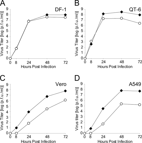
To investigate multicycle growth properties of R65 and R65-PB2K627E depending on the origin of cells, we infected avian DF-1 and QT-6 as well as mammalian Vero and A549 cells. Both viruses replicated to high titers in DF-1 and QT-6 cells with no or only minor differences, respectively (Fig. 1 A and B). However, in Vero and A549 cells, R65-PB2K627E replicated with delay and reached final titers two to three orders of magnitude lower than that of the parental R65 (Fig. 1C and D). These data demonstrate that substitution of PB2-627K by PB2-627E resulted in considerably impaired replication in mammalian but not in avian cells.
Fig. 1.
Viral replication on avian and mammalian cells. (A) DF-1, (B) QT-6, (C) Vero, and (D) A549 cells were inoculated with R65 (black diamonds) or R65-PB2K627E (white circles) at a multiplicity of infection of 10−3. Viral titers in the supernatant were determined by plaque assay at the indicated time points. Values are averages of results from three independent experiments.
Differences in polymerase activities.
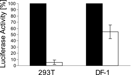
To analyze whether the impaired replication of R65-PB2K627E in mammalian cells resulted from a decrease in polymerase activity, we cotransfected plasmids expressing wild-type PB2-627K or mutant PB2-627E with PB1, PA, and NP expression plasmids, including the luciferase reporter plasmid pPolI-Luc-RT or pPRC-PolI-Luc-RT into mammalian 293T or avian DF-1 cells, respectively. Compared to PB2-627K, the mutant plasmid PB2-627E resulted in a 95% decrease in luciferase activity in 293T cells, whereas in DF-1 cells the reporter induction was only reduced by half (Fig. 2). Therefore, the substitution PB2-K627E in R65 led to a considerable reduction of polymerase activity in mammalian cells and an only moderate decline in avian cells, suggesting that PB2-627K elevates the polymerase activity of HPAIV irrespective of the host species but most strikingly in mammalian cells.
Fig. 2.
Polymerase activities. Luciferase reporter activities in lysates of human 293T or avian DF-1 cells were determined after cotransfection of PB2, PB1, PA, and NP plasmids and a firefly luciferase minigene plasmid containing either an avian or human promoter (pPolI-Luc-RT or pPRC-PolI-Luc-RT, respectively) (five independent experiments). R65 PB2-627K (black bars) was set to 100% in comparison to the mutated PB2K627E (white bars).
Virulence in chickens.
Since the polymerase activity of R65-PB2K627E was also decreased in avian cells, we investigated the virulence of this mutant in chickens. To this end, 10 8-week-old White Leghorn chickens per group were inoculated oculonasally with 104 PFU of either R65 or R65-PB2K627E and monitored for progression of disease as described previously (1). The two viruses caused the same clinical symptoms like depression, ruffled feathers, and diarrhea after 2 days postinfection (d.p.i.) and caused death in all infected animals within 3 d.p.i. (Fig. 3) resulting in identical clinical scores of 2.6. Macroscopic observation of organs revealed the same severe alterations, such as necrosis of comb and wattles, conjunctivitis, pancreatitis, and pneumonia. Hence, the substitution PB2-K627E did not alter the virulence of R65 in chickens.
Fig. 3.
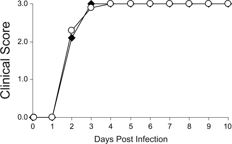
Virulence in chickens. Ten 8-week-old chickens were infected oculonasally with 104 PFU of R65 (black diamonds) or R65-PB2K627E (white circles) and scored daily for clinical symptoms. The birds were observed for 10 days for clinical signs and scored as healthy (0), ill (1), severely ill (2), or dead (3); the daily clinical score was calculated from the sum of individual clinical scores from all birds divided by the number of animals per group.
Virulence and replication efficiency in mice.
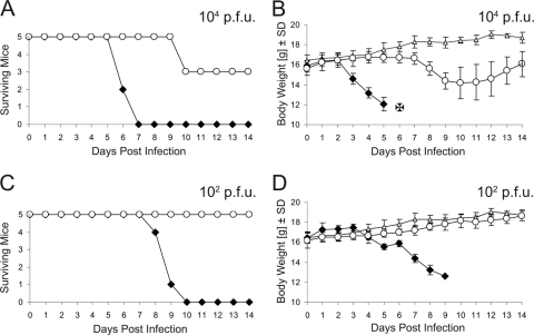
To study the impact of the PB2-K627E substitution in a mammalian host, we inoculated groups of five mice each intranasally with virus at doses ranging from 104 to 10 PFU. Overall, with increasing virus doses, the disease progressed faster. R65-infected mice developed symptoms like anorexia (body weight loss), ruffled fur, hunched posture, shivering, apathy, conjunctivitis, and neurological failure (spinning when holding it at the tail, paralysis) from day 3 to day 9 and died between days 6 and 10 p.i. From five mice infected with 10 PFU, only one animal survived, indicating an LD50 of R65 below 10 PFU. All R65-PB2K627E-infected mice which had received the highest doses of 106 and 105 PFU died within 4 and 9 days, respectively. In contrast, infection with 104 PFU led to death in only two of five mice and 103 PFU killed one of five mice, whereas all animals survived the infection with 102 or 10 PFU without noticeable weight loss, indicating an LD50 of R65-PB2K627E of approximately 104 PFU (Fig. 4, Table 1). Taken together, the results show that the mutation PB2-K627E considerably increased the LD50 in mice.
Fig. 4.
Virulence in mice. Survival (animal numbers) and body weight (average and standard deviation) of 4-week-old female BALB/c mice inoculated intranasally with PBS (white triangles), R65 (black diamonds), or R65-PB2K627E (white circles) at doses of 104 PFU (A and B) or 102 PFU (C and D). Animals with weight losses of 25% or more were euthanized (B).
Table 1.
Virulence in mice: organ titers and LD50
| Virus | Average virus titer per organ (log10 PFU) ± SD (no. of virus-positive organs/total no. of organs) |
LD50 (PFU) | ||
|---|---|---|---|---|
| Lung | Heart | Brain | ||
| R65 | 6.6 ± 0.1 (3/3) | 2.6 ± 2.6 (2/3) | 2.7 ± 0.3 (2/3) | <10 |
| R65-PB2K627E | 4.6 ± 0.4 (3/3) | 3.0 (1/3) | 0 (0/3) | >104 |
Next, we investigated whether the decreased virulence of R65-PB2K627E in mice is paralleled by reduced replication efficiency in organs. We infected three mice intranasally with 104 PFU of either R65 or R65-PB2K627E and removed lungs, hearts, and brains at day 3 p.i. In contrast to R65-infected mice, the virus titers in the lungs of the animals infected by R65-PB2K627E were reduced by two orders of magnitude and no virus was detected in the brains (Table 1), indicating impaired replication of R65-PB2K627E. Overall, the decreased viral loads in lungs and the absence of virus in brains correspond to the considerably reduced virulence of R65-PB2K627E in mice.
Reversion of PB2-K627E in mice but not in chickens.
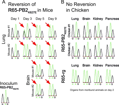
To follow the stability of the PB2-K627E mutation in a mammalian host, we infected mice with 104 or 105 PFU of R65-PB2K627E and removed lungs and brains from two animals each on days 1, 3, and 5 p.i. Whole RNA was extracted for RT-PCR to determine the PB2 sequences. In addition, we included the organs from two animals from the previous experiment which had died on day 9 p.i. Remarkably, the presence of revertant virus was detected in the virus population already on day 3 p.i. in lungs (Fig. 5A), at a time when no virus had been isolated from the brains (Table 1). Revertant virus became predominant in lungs and brains at days 5 and 9 p.i. (Fig. 5A). To reveal whether reversion occurs also in an avian host, we examined likewise the lungs, brains, kidneys, and pancreas from chickens infected with R65-PB2K627E (two animals) or R65 (one animal). None of the PB2 sequences from RT-PCR amplicons indicated the presence of any revertants (Fig. 5B). Taken together, these data demonstrate that reversion of PB2-627E to PB2-627K occurred in mice but not in chickens. Thus, selection for the PB2-627K virulence marker in R65 is enforced only during replication in a mammalian host.
Fig. 5.
Reversion of R65-PB2K627E in mice but not in chickens. (A) Mouse passages of R65-PB2K627E. Lungs and brains were taken on days 1, 3, 5, or 9 from mice infected intranasally with 105 (1, 5, or 9 days p. i.) or 104 (3 days p. i.) PFU virus. From organ homogenates, viral RNA was isolated, and the PB2 segment was amplified by RT-PCR and sequenced to monitor the presence of PB2-627E. Red arrows on electropherograms indicate emergence of revertants carrying PB2-G27K. (B) Chicken passage of R65-PB2K627E. Brains, lungs, pancreas, and spleens from two R65-PB2K627E-infected and one R65-infected animal (104 PFU oculonasally) were homogenized, and the virus was propagated in avian DF-1 cells. From supernatants, viral RNA was isolated, and the PB2 segment was amplified by RT-PCR and sequenced to monitor the presence of PB2-627E.
NP of R65 is required for reversion of R65-PB2K627E in mammalian cells.
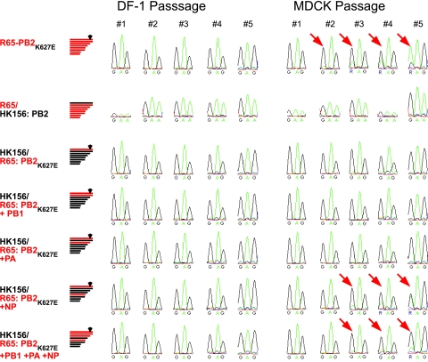
After having demonstrated reversion of PB2-627E to PB2-627K in mammalian cells and in mice, we addressed the question whether the genetic background of R65 determines reversion. To this end, we generated reassortants of R65-PB2K627E by replacing one or more R65 gene segments with those from HPAIV A/Hong Kong/156/97 (H5N1) (Hk156), a strain that carries PB2-627E. The resulting reassortants were designated according to their R65 gene segments: Hk156/R65:PB2K627E, Hk156/R65:PB2K627E+PB1, Hk156/R65:PB2K627E+PA, Hk156/R65:PB2K627E+NP, and Hk156/R65:PB2K627E+PB1+PA+NP. To investigate whether a PB2-627E from a different virus strain would also revert to PB2-627K in the R65 background, we generated the reassortant R65/Hk156:PB2, composed of the Hk156 PB2 gene encoding PB2-627E and the remaining R65 genes. All reassortant viruses and the homologous mutant R65-PB2K627E were passaged five times in DF-1 or MDCK cells. After RT-PCR from isolated supernatant RNA, we sequenced the PB2 gene region encompassing amino acid position 627. In DF-1 cells, R65-PB2K627E remained stable, whereas in MDCK cells, revertants emerged at the second passage and became predominant during subsequent passages (Fig. 6). In contrast, no reversion was observed in reassortant R65/Hk156:PB2 and its reciprocal counterpart Hk156/R65:PB2K627E, indicating that R65 PB2 requires other R65 proteins to select for PB2-627K in mammalian cells. Finally, revertants were found only after infection with the reassortants Hk156/R65:PB2K627E+NP and Hk156/R65:PB2K627E+PB1+PA+NP, which carry the R65 NP gene (Fig. 6). However, they did not become predominant, in contrast to the revertant of the homologous R65-PB2K627E. Thus, the presence of the R65 NP protein is the minimum prerequisite for reversion of R65-PB2K627E in mammalian cells.
Fig. 6.
R65 NP enforces reversion of R65-PB2K627E in mammalian cells. Reassortant viruses containing the gene segments of R65-PB2K627E (red; mutation PB2-627E represented by a diamond) and Hk156 (black) were passaged in avian DF-1 or mammalian MDCK cells. From infectious supernatants, viral RNA was isolated, and the PB2 segment was amplified by RT-PCR and sequenced to monitor the presence of PB2-627E. Red arrows on electropherograms indicate emergence of revertants carrying PB2-627K.
DISCUSSION
HPAIV, and in particular those of the H5N1 subtype, have a wide host range and cause zoonotic infections in humans (22, 23, 36, 63). Whereas in the past, HPAIV outbreaks were short and localized events, since 2005 they are unprecedented in their duration and transcontinental spread from Southeast Asia to Europe and Africa. Some markers in HPAIV facilitating zoonotic transmission to humans have been identified, including amino acid residues like PB2-701N and NP-319K (8). Whereas those changes were found mainly in various avian-like mammalian strains and zoonotic HPAIV (13), the only marker discriminating human from avian strains (54) is PB2-627K. All common avian strains and the novel pandemic H1N1 strains carry PB2-627E. However, on several occasions, PB2-627K was recognized in H5N1 and H7N7 HPAIV to result in enhanced replication efficiency, leading to high pathogenicity in mammals (15, 19, 20, 32, 43, 45, 53), including humans (8, 12), but those transmissions to mammals were considered dead-end events. Remarkably, the 2005-2006 clade 2.2 strains retained PB2-627K during their transcontinental journey (63). This observation led us to analyze the importance of this mutation for the broad host range of those viruses. For that, we generated a mutant of the Western European clade 2.2.2 strain R65 in which PB2-627K was replaced by E: R65-PB2K627E. First attempts to generate and maintain this mutant in mammalian cells led to immediate reversion to the parental virus. This observation could be reproduced in three independent experiments in different facilities (FLI Insel Riems and Department of Virology, University of Freiburg). Since the detection limit of mutants by Sanger sequencing of PCR products lies above 10% (29), notable amounts of revertants have accumulated within the viral population. However, R65-PB2E627K was stably maintained in avian cells and embryonated chicken eggs, indicating an effect of avian versus mammalian hosts on the stability of this mutation. Therefore, we further studied the PB2-K627E mutation in avian and mammalian cells and in chicken and mice. Subsequently, we investigated whether other R65 genes determine reversion to PB2-627K in mammalian cells.
We found that the PB2-627E substitution in the R65 genetic background resulted in (i) decreased replication in mammalian but not avian cells, (ii) severely decreased polymerase activity in mammalian cells but only slightly reduced polymerase activity in avian cells, and (iii) considerably decreased virulence accompanied with reduced replication efficiency in mice, but it did not affect virulence in chickens. Above all, whereas PB2-627E remained stable in avian cells and in chickens, it reverted to PB2-627K in mammalian cells and mice. Passaging of R65-PB2K627E reassortants carrying Hk156 gene segments in MDCK cells revealed that the presence of R65-PB2 and NP was crucial for reversion.
The PB2 K627E mutation led in the mammalian cells to a severely reduced activity of reconstituted RNP of 5%, corresponding to a notably decreased viral replication: viral growth decreased by two magnitudes, and LD50 in mice increased by three magnitudes. However, the 50% reduction of RNP activity in avian cells is not accompanied by measurable decreased viral replication efficiency indicated by equivalent virus propagation in DF1 cells and virulence in chickens. This lower impact of PB2-627K on virus replication in avian cells may be attributed to the absence of a dominant inhibitory activity, whereas in mammalian cells, this mutation enables optimized replication in the presence of a postulated human cofactor (33).
The polymerase activity of avian viruses was shown to be reduced in mammalian cells due to inefficient association of PB2 with NP, a defect which can be repaired by PB2-627K (34, 37, 39). Furthermore, a yet unknown cellular factor, X, was supposed to stabilize the PB2-NP interaction, thereby enhancing viral replication (28). This postulated PB2-X interaction may be impaired in human cells if PB2 originates from an avian strain and carries the avian signature PB2-627E. On the PB2 surface, PB2-627K resides in a patch of basic amino acid residues which is thought to mediate interaction with cellular host factors. Disruption of this basic patch by PB2-627E in avian strains might interfere with those interactions (56). Our finding that both the R65 PB2 and NP are necessary for positive selection for PB2-627K in mammalian cells indicates that compensation of the impaired PB2-NP interaction by PB2-627K is essential for the high replication efficiency in clade 2.2 HPAIV in mammals. It appears that enhanced polymerase activity of HPAIV might compensate insufficient adaptation to the mammalian host. In mouse lung (at 37°C), the virus replication in the presence of PB2-627E is impaired by several magnitudes but is still detectable (reference 21 and our data); however, in the nasal turbinates (at 33°C), virus was undetectable (21). Therefore, these observations support the notion that the reversion to PB2-627K occurred in the lungs first, allowing the virus then to outreach the upper respiratory tract.
Many recent H5N1 isolates of clade 2.2 and genotype Z (5, 19, 25, 38), as well as the Dutch H7N7 HPAIV from 2003 which led to the death of a veterinarian (12), carry PB2-627K, suggesting that this mutation arises early after transmission from birds to mammals, including humans. Correspondingly, we observed a rapid reversion of PB2-K627E to K within one mouse passage corresponding to the results of others (32). PB2-627K is known to increase the replication efficiency in mice but not to affect the cell tropism (45). Furthermore, in our experiments, virus became detectable in the brain only on day 5 p.i., whereas the appearance of revertants in the lung was observed already on day 3 p.i. Therefore, CNS infection may be a consequence of the selection of revertants during primary infection in the lung.
Since their emergence in 2005 at Qinghai Lake, clade 2.2 viruses were detected in many wild bird species and domestic poultry in Europe, Africa, and Asia (11, 46, 61). They were also isolated from wild pikas at the Qinghai Lake area (66). Free-ranging wild pikas (Ochotona curzoniae), which serve as food source for raptorial birds and carnivorous mammals (47), are a mammalian host infected by H5N1 HPAIV in the natural environment of Qinghai Lake (66). Pikas might be infected at the common weed-foraging sites of wild birds such as bar-headed geese and then transmit the viruses to predators spreading them to poultry farms. Furthermore, because of successful transmission of clade 2.2 H5N1 viruses from poultry to felids and between felids, it had been suggested that felids may serve as intermediate hosts and further transmit the virus to humans (62). Accordingly, cats were shown to be infected both by horizontal transmission and by consumption of virus-infected birds resulting in severe pneumonia accompanied with extensive virus spread to brain, liver, kidney, and heart (41) and excretion via the respiratory, urinary, or rectal tracts (27, 59). Thus, cats or other felids may serve as intermediate hosts for avian influenza viruses during their adaptation to mammals.
In summary, we conclude that clade 2.2 H5N1 HPAIV may have acquired the PB2-627K mutation rapidly facilitated by a unique constellation of PB2 and NP and that this mutation might compensate for insufficient adaptation to mammalian hosts. However, in avian hosts, the PB2-627K mutation has been maintained with no apparent disadvantages and, therefore, it has not been eliminated. The prompt reversion of R65-PB2K627E to PB2-627K in a mammalian host suggests that the clade 2.2 H5N1 HPAIV may have had a history of mammalian intermediate hosts like felids. Overall, the broad host range due to the acquisition and maintenance of PB2-627K might have supported the unprecedented duration and panzootic spread of the clade 2.2 HPAIV.
ACKNOWLEDGMENTS
We thank A. Brandenburg, C. Meinke, M. Schmidt, M. Gräber, M. Grawe, and the animal care workers for their excellent technical assistance. Furthermore, we are very grateful to A. Osterhaus for providing the A/Hong Kong/156/97 (H5N1) virus, P. Palese for the expression plasmids pCAGGS-PR8-PB2, pCAGGS-PR8-PB1, pCAGGS-PR8-PA, and pCAGGS-PR8-NP, G. Keil for the renilla expression plasmid pIE-Renilla-Luc, and D. Mayer for providing the plasmid pPRC-PolI-Luc-RT.
This study was supported by the European Commission (FP6-2005-SSP-5B INFLUENZA [Flupol]), the Forschungssofortprogramm Influenza of the German government (FSI 2.44), and the Bundesministerium für Bildung und Forschung (BMBF, FluResearchNet).
Footnotes
Published ahead of print on 17 August 2011.
REFERENCES
- 1. Alexander D. J. 2008. Avian influenza, chapter 2.3.4, p. 465–481In Vallat B. (ed.), Manual of diagnostic tests and vaccines for terrestrial animals, 6th ed. OIE, Paris, France [Google Scholar]
- 2. Bogs J., et al. 2010. Highly pathogenic H5N1 influenza viruses carry virulence determinants beyond the polybasic hemagglutinin cleavage site. PLoS One 5:e11826. [DOI] [PMC free article] [PubMed] [Google Scholar]
- 3. Butler D. 2006. Thai dogs carry bird-flu virus, but will they spread it? Nature 439:773. [DOI] [PubMed] [Google Scholar]
- 4. Chen H., et al. 2006. Properties and dissemination of H5N1 viruses isolated during an influenza outbreak in migratory waterfowl in western China. J. Virol. 80:5976–5983 [DOI] [PMC free article] [PubMed] [Google Scholar]
- 5. Chen H., et al. 2006. Establishment of multiple sublineages of H5N1 influenza virus in Asia: implications for pandemic control. Proc. Natl. Acad. Sci. U. S. A. 103:2845–2850 [DOI] [PMC free article] [PubMed] [Google Scholar]
- 6. Chen H., et al. 2005. Avian flu: H5N1 virus outbreak in migratory waterfowl. Nature 436:191–192 [DOI] [PubMed] [Google Scholar]
- 7. Claas E. C., et al. 1998. Human influenza A H5N1 virus related to a highly pathogenic avian influenza virus. Lancet 351:472–477 [DOI] [PubMed] [Google Scholar]
- 8. de Jong M. D., et al. 2006. Fatal outcome of human influenza A (H5N1) is associated with high viral load and hypercytokinemia. Nat. Med. 12:1203–1207 [DOI] [PMC free article] [PubMed] [Google Scholar]
- 9. Desvaux S., et al. 2009. Highly pathogenic avian influenza virus (H5N1) outbreak in captive wild birds and cats, Cambodia. Emerg. Infect. Dis. 15:475–478 [DOI] [PMC free article] [PubMed] [Google Scholar]
- 10. Ducatez M. F., et al. 2007. Molecular and antigenic evolution and geographical spread of H5N1 highly pathogenic avian influenza viruses in western Africa. J. Gen. Virol. 88:2297–2306 [DOI] [PubMed] [Google Scholar]
- 11. Ducatez M. F., Webster R. G., Webby R. J. 2008. Animal influenza epidemiology. Vaccine 26(Suppl. 4):D67–D69 [DOI] [PMC free article] [PubMed] [Google Scholar]
- 12. Fouchier R. A., et al. 2004. Avian influenza A virus (H7N7) associated with human conjunctivitis and a fatal case of acute respiratory distress syndrome. Proc. Natl. Acad. Sci. U. S. A. 101:1356–1361 [DOI] [PMC free article] [PubMed] [Google Scholar]
- 13. Gabriel G., et al. 2005. The viral polymerase mediates adaptation of an avian influenza virus to a mammalian host. Proc. Natl. Acad. Sci. U. S. A. 102:18590–18595 [DOI] [PMC free article] [PubMed] [Google Scholar]
- 14. Gall-Recule G. L., et al. 2008. Double introduction of highly pathogenic H5N1 avian influenza virus into France in early 2006. Avian Pathol. 37:15–23 [DOI] [PubMed] [Google Scholar]
- 15. Gao P., et al. 1999. Biological heterogeneity, including systemic replication in mice, of H5N1 influenza A virus isolates from humans in Hong Kong. J. Virol. 73:3184–3189 [DOI] [PMC free article] [PubMed] [Google Scholar]
- 16. Ghanem A., et al. 2007. Peptide-mediated interference with influenza A virus polymerase. J. Virol. 81:7801–7804 [DOI] [PMC free article] [PubMed] [Google Scholar]
- 17. Globig A., et al. 2009. Epidemiological and ornithological aspects of outbreaks of highly pathogenic avian influenza virus H5N1 of Asian lineage in wild birds in Germany, 2006 and 2007. Transbound. Emerg. Dis. 56:57–72 [DOI] [PubMed] [Google Scholar]
- 18. Gohrbandt S., et al. 2011. Amino acids adjacent to the haemagglutinin cleavage site are relevant for virulence of avian influenza viruses of subtype H5. J. Gen. Virol. 92:51–59 [DOI] [PubMed] [Google Scholar]
- 19. Govorkova E. A., et al. 2005. Lethality to ferrets of H5N1 influenza viruses isolated from humans and poultry in 2004. J. Virol. 79:2191–2198 [DOI] [PMC free article] [PubMed] [Google Scholar]
- 20. Hatta M., Gao P., Halfmann P., Kawaoka Y. 2001. Molecular basis for high virulence of Hong Kong H5N1 influenza A viruses. Science 293:1840–1842 [DOI] [PubMed] [Google Scholar]
- 21. Hatta M., et al. 2007. Growth of H5N1 influenza A viruses in the upper respiratory tracts of mice. PLoS Pathog. 3:1374–1379 [DOI] [PMC free article] [PubMed] [Google Scholar]
- 22. Horimoto T., Kawaoka Y. 2001. Pandemic threat posed by avian influenza A viruses. Clin. Microbiol. Rev. 14:129–149 [DOI] [PMC free article] [PubMed] [Google Scholar]
- 23. Kalthoff D., Globig A., Beer M. 2010. (Highly pathogenic) avian influenza as a zoonotic agent. Vet. Microbiol. 140:237–245 [DOI] [PubMed] [Google Scholar]
- 24. Katz J. M., et al. 2000. Molecular correlates of influenza A H5N1 virus pathogenesis in mice. J. Virol. 74:10807–10810 [DOI] [PMC free article] [PubMed] [Google Scholar]
- 25. Keawcharoen J., et al. 2004. Avian influenza H5N1 in tigers and leopards. Emerg. Infect. Dis. 10:2189–2191 [DOI] [PMC free article] [PubMed] [Google Scholar]
- 26. Klopfleisch R., et al. 2007. Encephalitis in a stone marten (Martes foina) after natural infection with highly pathogenic avian influenza virus subtype H5N1. J. Comp. Pathol. 137:155–159 [DOI] [PubMed] [Google Scholar]
- 27. Kuiken T., et al. 2004. Avian H5N1 influenza in cats. Science 306:241. [DOI] [PubMed] [Google Scholar]
- 28. Labadie K., Dos Santos Afonso E., Rameix-Welti M. A., van der Werf S., Naffakh N. 2007. Host-range determinants on the PB2 protein of influenza A viruses control the interaction between the viral polymerase and nucleoprotein in human cells. Virology 362:271–282 [DOI] [PubMed] [Google Scholar]
- 29. Li J., et al. 2008. Replacing PCR with COLD-PCR enriches variant DNA sequences and redefines the sensitivity of genetic testing. Nat. Med. 14:579–584 [DOI] [PubMed] [Google Scholar]
- 30. Li Y., et al. 2010. Persistent circulation of highly pathogenic influenza H5N1 virus in Lake Qinghai area of China. Avian Dis. 54:821–829 [DOI] [PubMed] [Google Scholar]
- 31. Liu J., et al. 2005. Highly pathogenic H5N1 influenza virus infection in migratory birds. Science 309:1206. [DOI] [PubMed] [Google Scholar]
- 32. Mase M., et al. 2006. Recent H5N1 avian influenza A virus increases rapidly in virulence to mice after a single passage in mice. J. Gen. Virol. 87:3655–3659 [DOI] [PubMed] [Google Scholar]
- 33. Moncorge O., Mura M., Barclay W. S. 2010. Evidence for avian and human host cell factors that affect the activity of influenza virus polymerase. J. Virol. 84:9978–9986 [DOI] [PMC free article] [PubMed] [Google Scholar]
- 34. Naffakh N., Massin P., Escriou N., Crescenzo-Chaigne B., van der Werf S. 2000. Genetic analysis of the compatibility between polymerase proteins from human and avian strains of influenza A viruses. J. Gen. Virol. 81:1283–1291 [DOI] [PubMed] [Google Scholar]
- 35. Nagy A., et al. 2007. Highly pathogenic avian influenza virus subtype H5N1 in Mute swans in the Czech Republic. Vet. Microbiol. 120:9–16 [DOI] [PubMed] [Google Scholar]
- 36. Peiris J. S., de Jong M. D., Guan Y. 2007. Avian influenza virus (H5N1): a threat to human health. Clin. Microbiol. Rev. 20:243–267 [DOI] [PMC free article] [PubMed] [Google Scholar]
- 37. Poole E., Elton D., Medcalf L., Digard P. 2004. Functional domains of the influenza A virus PB2 protein: identification of NP- and PB1-binding sites. Virology 321:120–133 [DOI] [PubMed] [Google Scholar]
- 38. Puthavathana P., et al. 2005. Molecular characterization of the complete genome of human influenza H5N1 virus isolates from Thailand. J. Gen. Virol. 86:423–433 [DOI] [PubMed] [Google Scholar]
- 39. Rameix-Welti M. A., Tomoiu A., Dos Santos Afonso E., van der Werf S., Naffakh N. 2009. Avian influenza A virus polymerase association with nucleoprotein, but not polymerase assembly, is impaired in human cells during the course of infection. J. Virol. 83:1320–1331 [DOI] [PMC free article] [PubMed] [Google Scholar]
- 40. Reed L. H., Muench H. 1938. A simple method of estimating fifty percent endpoints. Am. J. Hyg. (Lond.) 27:493–497 [Google Scholar]
- 41. Rimmelzwaan G. F., et al. 2006. Influenza A virus (H5N1) infection in cats causes systemic disease with potential novel routes of virus spread within and between hosts. Am. J. Pathol. 168:176–183, quiz 364 [DOI] [PMC free article] [PubMed] [Google Scholar]
- 42. Roberton S. I., et al. 2006. Avian influenza H5N1 in viverrids: implications for wildlife health and conservation. Proc. Biol. Sci. 273:1729–1732 [DOI] [PMC free article] [PubMed] [Google Scholar]
- 43. Salomon R., et al. 2006. The polymerase complex genes contribute to the high virulence of the human H5N1 influenza virus isolate A/Vietnam/1203/04. J. Exp. Med. 203:689–697 [DOI] [PMC free article] [PubMed] [Google Scholar]
- 44. Salzberg S. L., et al. 2007. Genome analysis linking recent European and African influenza (H5N1) viruses. Emerg. Infect. Dis. 13:713–718 [DOI] [PMC free article] [PubMed] [Google Scholar]
- 45. Shinya K., et al. 2004. PB2 amino acid at position 627 affects replicative efficiency, but not cell tropism, of Hong Kong H5N1 influenza A viruses in mice. Virology 320:258–266 [DOI] [PubMed] [Google Scholar]
- 46. Siengsanan J., et al. 2009. Comparison of outbreaks of H5N1 highly pathogenic avian influenza in wild birds and poultry in Thailand. J. Wildl. Dis. 45:740–747 [DOI] [PubMed] [Google Scholar]
- 47. Smith A. T., Formozov N. A., Hoffmann R. S. 1990. The pikas, p. 69.In Chapman J. A., Flux J. E. C.(ed.), Rabbits, hares and pikas: status survey and conservation action plan. IUCN, Gland, Switzerland [Google Scholar]
- 48. Smith G. J., et al. 2006. Evolution and adaptation of H5N1 influenza virus in avian and human hosts in Indonesia and Vietnam. Virology 350:258–268 [DOI] [PubMed] [Google Scholar]
- 49. Songserm T., Amonsin A., Jam-on R., et al. 2006. Fatal avian influenza A H5N1 in a dog. Emerg. Infect. Dis. 12:1744–1747 [DOI] [PMC free article] [PubMed] [Google Scholar]
- 50. Starick E., et al. 2008. Phylogenetic analyses of highly pathogenic avian influenza virus isolates from Germany in 2006 and 2007 suggest at least three separate introductions of H5N1 virus. Vet. Microbiol. 128:243–252 [DOI] [PubMed] [Google Scholar]
- 51. Stech J., et al. 2008. Rapid and reliable universal cloning of influenza A virus genes by target-primed plasmid amplification. Nucleic Acids Res. 36:e139. [DOI] [PMC free article] [PubMed] [Google Scholar]
- 52. Stech J., Xiong X., Scholtissek C., Webster R. G. 1999. Independence of evolutionary and mutational rates after transmission of avian influenza viruses to swine. J. Virol. 73:1878–1884 [DOI] [PMC free article] [PubMed] [Google Scholar]
- 53. Steel J., Lowen A. C., Mubareka S., Palese P. 2009. Transmission of influenza virus in a mammalian host is increased by PB2 amino acids 627K or 627E/701N. PLoS Pathog. 5:e1000252. [DOI] [PMC free article] [PubMed] [Google Scholar]
- 54. Subbarao E. K., London W., Murphy B. R. 1993. A single amino acid in the PB2 gene of influenza A virus is a determinant of host range. J. Virol. 67:1761–1764 [DOI] [PMC free article] [PubMed] [Google Scholar]
- 55. Subbarao K., et al. 1998. Characterization of an avian influenza A (H5N1) virus isolated from a child with a fatal respiratory illness. Science 279:393–396 [DOI] [PubMed] [Google Scholar]
- 56. Tarendeau F., et al. 2008. Host determinant residue lysine 627 lies on the surface of a discrete, folded domain of influenza virus polymerase PB2 subunit. PLoS Pathog. 4:e1000136. [DOI] [PMC free article] [PubMed] [Google Scholar]
- 57. Taubenberger J. K., et al. 2005. Characterization of the 1918 influenza virus polymerase genes. Nature 437:889–893 [DOI] [PubMed] [Google Scholar]
- 58. Thanawongnuwech R., et al. 2005. Probable tiger-to-tiger transmission of avian influenza H5N1. Emerg. Infect. Dis. 11:699–701 [DOI] [PMC free article] [PubMed] [Google Scholar]
- 59. Vahlenkamp T. W., Teifke J. P., Harder T. C., Beer M., Mettenleiter T. C. 2010. Systemic influenza virus H5N1 infection in cats after gastrointestinal exposure. Influenza Other Respi Viruses. 4:379–386 [DOI] [PMC free article] [PubMed] [Google Scholar]
- 60. Wallace R. G., Hodac H., Lathrop R. H., Fitch W. M. 2007. A statistical phylogeography of influenza A H5N1. Proc. Natl. Acad. Sci. U. S. A. 104:4473–4478 [DOI] [PMC free article] [PubMed] [Google Scholar]
- 61. Weber S., et al. 2007. Molecular analysis of highly pathogenic avian influenza virus of subtype H5N1 isolated from wild birds and mammals in northern Germany. J. Gen. Virol. 88:554–558 [DOI] [PubMed] [Google Scholar]
- 62. Webster R. G., Govorkova E. A. 2006. H5N1 influenza—continuing evolution and spread. N. Engl. J. Med. 355:2174–2177 [DOI] [PubMed] [Google Scholar]
- 63. Webster R. G., Guan Y., Peiris M., Chen H. 2006. H5N1 influenza continues to circulate and change. Microbe 1:559–565 [Google Scholar]
- 64. WHO/OIE/FAO/H5N1 Evolution Working Group 2008. Toward a unified nomenclature system for highly pathogenic avian influenza virus (H5N1). Emerg. Infect. Dis. 14:e1. [DOI] [PMC free article] [PubMed] [Google Scholar]
- 65. Yamada S., et al. 2010. Biological and structural characterization of a host-adapting amino acid in influenza virus. PLoS Pathog. 6 [DOI] [PMC free article] [PubMed] [Google Scholar]
- 66. Zhou J., et al. 2009. Characterization of the H5N1 highly pathogenic avian influenza virus derived from wild pikas in China. J. Virol. 83:8957–8964 [DOI] [PMC free article] [PubMed] [Google Scholar]
- 67. Zhou J. Y., et al. 2006. Characterization of a highly pathogenic H5N1 influenza virus derived from bar-headed geese in China. J. Gen. Virol. 87:1823–1833 [DOI] [PubMed] [Google Scholar]


