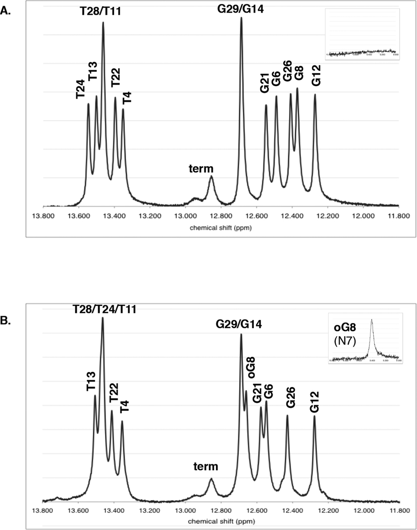
Figure 5. Imino proton region of 1D 1H NMR spectra.
(A) 1–2 parent duplex and (B) 1oxo-2 lesion duplex spectra are shown at 8°C and pH 7.5. An additional small, broad proton resonance can be seen around 9.4 ppm in the 8oxoG lesion duplex spectrum in the absence of catalyst, corresponding to the N7 imino proton of the 8oxoG lesion itself (inset). Since guanine does not normally have an N7 imino proton, this peak is missing from the parent duplex. “Term” refers to the terminal G16 and G30 protons with chemical shifts around 12.9 ppm.

