Abstract
AMP-activated protein kinase (AMPK) is a sensor and regulator of cellular energy metabolism potentially implicated in a broad range of conditions, including obesity and Alzheimer’s disease. Its role in the control of key metabolic enzymes makes this kinase a central player in glucose and lipid homeostasis. Recently, by screening a library of synthetic small molecules selected for their structural similarity with the natural polyphenol resveratrol, we identified RSVA314 and RSVA405 as potent indirect activators of AMPK (half-maximal effective concentration [EC50] = 1 μmol/L in cell-based assays). Here we show that RSVA314 and RSVA405 can significantly activate AMPK and inhibit acetyl-CoA carboxylase (ACC), one target of AMPK and a key regulator of fatty acid biogenesis, in nondifferentiated and proliferating 3T3-L1 adipocytes. We found that RSVA314 and RSVA405 treatments inhibited 3T3-L1 adipocyte differentiation by interfering with mitotic clonal expansion during preadipocyte proliferation (half-maximal inhibitory concentration [IC50] = 0.5 μmol/L). RSVA314 and RSVA405 prevented the adipogenesis-dependent transcriptional changes of multiple gene products involved in the adipogenic process, including peroxisome proliferator–activated receptor (PPAR)-γ, CCAAT/enhancer-binding protein α (C/EBPα), fatty acid synthase, fatty acid binding protein 4 (aP2), RANTES or resistin. Furthermore, orally administered RSVA405 at 20 and 100 mg/kg/d significantly reduced the body weight gain of mice fed a high-fat diet. This work shows that the novel small-molecule activators of AMPK (RSVA314 and RSVA405) are potent inhibitors of adipogenesis and thus may have therapeutic potential against obesity.
INTRODUCTION
AMP-activated protein kinase (AMPK) is a central regulator of cellular and whole-body energy homeostasis. AMPK is a heterotrimeric Ser/Thr protein kinase activated by different upstream kinases via a specific Thr phosphorylation within the activation loop of the catalytic subunit of the protein (1). LKB1 and Ca2+/calmodulin-dependent protein kinase kinase-β (CaMKKβ) are the main AMPK-phosphorylating and -activating kinases (2,3). AMPK regulates cellular energy levels by balancing nutrient availability and energy demand via its control of several proteins involved in glucose and lipid metabolism, including the regulator of fatty acid biosynthesis acetyl-CoA carboxylase (ACC). AMPK is mainly activated by intracellular increases of the AMP:ATP ratio via the recruitment of LKB1, a situation that can occur during cellular stress or low nutrient availability. Once activated, AMPK promotes ATP production while inhibiting ATP consumption, via, for instance, the facilitation of fatty acid oxidation and blockade of fatty acid biosynthesis. AMPK is therefore a promising target for the treatment of metabolic disorders, such as type 2 diabetes and obesity (4,5).
AMPK was proposed to be involved in adipogenesis, the process of adipocyte differentiation that leads to excessive adipose tissue formation in obesity (6). Adipocyte differentiation is controlled by several transcription factors and adipogenic factors, including the CCAAT/enhancer binding proteins (C/EBPs), the peroxisome proliferator–activated receptors (PPARs), fatty acid synthase (FAS) or fatty acid binding protein 4 (aP2) (7,8). Adipogenesis leads to the production of specific hormones or cytokines by the adipose tissue, such as RANTES and resistin, that participate in the adipogenic process. Adipogenesis is initiated by cell cycle reentry of growth-arrested preadipocytes, a mechanism known as mitotic clonal expansion (MCE). The role of AMPK in adipogenesis is not entirely clear, but several AMPK-activating molecules were found to have antiadipogenic effects via MCE inhibition and downregulation of adipogenic transcriptional pathways (9–12).
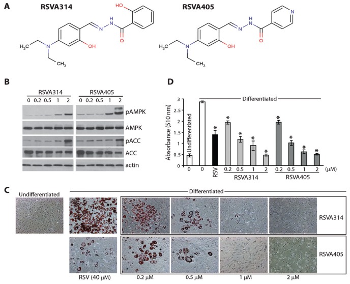
We and others recently revealed that pharmacological activation of AMPK using resveratrol could be achieved in vitro in different cell lines and in vivo in mice via a mechanism implicating CaMKKβ (13) and LKB1 (14). Resveratrol is a natural polyphenol associated with protective effects against the deregulation of energy homeostasis observed in mouse models for type 2 diabetes, and this via a mechanism implicating AMPK (15,16). A large body of literature also supports the antiadipogenic effect of resveratrol in in vitro models (17). Recently, we showed that activation of AMPK by resveratrol results in an increase in the intracellular clearance of the amyloidogenic peptide Aβ, the core component of the amyloid plaques and main culprit in Alzheimer’s disease pathogenesis (13,18,19). In a subsequent study, by screening a library of synthetic small molecules selected for their structural similarity with resveratrol and for their improved antiamyloidogenic properties, we identified the structurally related series of resveratrol analogs (RSVAs), the RSVA series (20). Two of the most potent compounds, RSVA314 and RSVA405 (Figure 1A), were further characterized and were found to share with resveratrol the same mechanism of action by facilitating AMPK activation, and this with a potency nearly 40 times higher than resveratrol (half-maximal effective concentration [EC50] ~ 1 μmol/L) (20).
Figure 1.
Effect of RSVA314 and RSVA405 treatment on 3T3-L1 adipocyte differentiation. (A) Structure of RSVA314 and RSVA405 drawn using Marvin applet. (B) 3T3-L1 preadipocytes were treated for 24 h with the indicated concentrations of RSVAs. Phospho-AMPK, total AMPK, phospho-ACC and total ACC protein levels were assessed by Western blotting. (C, D) 3T3-L1 preadipocytes were treated during differentiation with the indicated concentrations of RSVAs or resveratrol (RSV). Undifferentiated 3T3-L1 cells on day 0 were used as control (undifferentiated). On day 8, cells were stained with Oil Red O and photographed (C). Lipid content in these cells was quantified using isopropanol extraction and absorbance measurement at 510 nm (D). Results are shown as means ± SD of three independent experiments. *P < 0.05 (Student t test).
Here, we evaluated the antiadipogenic potential of RSVA314 and RSVA405. We found that RSVA314 and RSVA405 can significantly activate AMPK and consequently inhibit ACC in 3T3-L1 preadipocytes. Further, RSVA314 and RSVA405 treatments inhibited 3T3-L1 adipocyte differentiation by interfering with MCE during preadipocyte proliferation (half-maximal inhibitory concentration [IC50] ~ 0.5 μmol/L) without affecting cell viability of proliferating preadipocytes. RSVA314 and RSVA405 also interfered with the adipogenesis-dependent transcriptional changes of PPARγ, C/EBPα, FAS, aP2, RANTES and resistin, at concentrations consistent with the effect of these compounds on adipocyte differentiation. Importantly, we also found that orally administered RSVA405 at 20 or 100 mg/kg/day significantly reduced the body weight gain of mice fed a high-fat diet (HFD). Thus, the novel small-molecule activators of AMPK (RSVA314 and RSVA405) are inhibitors of adipogenesis in vitro in cultured cells and in vivo in mice and thus may have therapeutic potential in metabolic diseases associated with excessive adipose tissue formation. This work supports the notion that pharmacological activation of AMPK in preadipocytes is beneficial against obesity.
MATERIALS AND METHODS
Materials
Resveratrol was purchased from Sigma-Aldrich (St. Louis, MO, USA). RSVA314 and RSVA405 (see Figure 1A) were from Chembridge (Hit2Lead compounds 5194489 and 5113025, respectively; San Diego, CA, USA; www.hit2lead.com) and were dissolved in DMSO. Antibodies directed against phospho-AMPKα (Thr172), AMPK, phospho-ACC (Ser79), ACC, PPARγ, FAS and aP2 were from Cell Signaling Technology (Danvers, MA, USA. Anti-actin antibody was from BD Transduction Laboratories (San Jose, CA, USA).
Growth and Differentiation of 3T3-L1 Cells
3T3-L1 preadipocytes were obtained from American Type Culture Collection (Manassas, VA, USA). 3T3-L1 cells were cultured in Dulbecco’s modified Eagle’s medium (DMEM) containing 10% fetal bovine serum. For differentiation, cells were plated in 12-well plates. Two days after confluency (day 0), growth-arrested 3T3-L1 preadipocytes were incubated in differentiation medium containing 5 μg/mL insulin (Sigma-Aldrich), 1 μmol/L dexamethasone (Sigma-Aldrich) and 0.5 mmol/L isobutyl-methylxanthine (Calbiochem, EMD Chemicals, Gibbstown, NJ, USA). On day 2, the medium was changed to DMEM containing 5 μg/mL insulin. Cells were then cultured in normal medium from day 4 to day 8. On day 8, fully differentiated cells were harvested or stained with Oil Red O dye (Sigma-Aldrich).
Oil Red O Staining
Lipid accumulation in differentiated 3T3-L1 cells was assessed by staining of neutral fats and cholesterol esters using Oil Red O dye. Briefly, cells were fixed in paraformaldehyde 4% solution and stained with 0.2% Oil Red O in isopropanol for 50 min. Lipid staining was quantified using isopropanol extraction and absorbance measurement at 510 nm.
Western Blot Analysis
Cells were washed with phosphate-buffered saline and solubilized in ice-cold HEPES (N-2-hydroxyethylpiperazine-N′-2-ethanesulfonic acid) buffer (25 mmol/L HEPES, pH 7.4; 150 mmol/L NaCl; 1 × complete protease inhibitor mixture, Roche Applied Science, Indianapolis, IN, USA) containing 1% sodium dodecyl sulfate. A total of 10–20 μg of cell extracts was resolved by sodium dodecyl sulfate– polyacrylamide gel electrophoresis and transferred to nitrocellulose membrane. A standard enhanced chemiluminescence detection procedure using the antibodies listed above was then performed.
Cell Proliferation and Cell Viability Assays
Cell proliferation was determined using 3-(4,5-dimethylthiazol-2-yl)-2,5-diphenyltetrazolium bromide (MTT) assays according to the manufacturer’s instructions (Promega, Madison, WI, USA). Briefly, cells were seeded on 24-well plates and incubated with each compound for the indicated time periods. Supernatant was discarded and cells were incubated with 200 μL MTT solution (2 mg/mL MTT in DMEM) for 2 h. Absorbance was measured at 570 nm using a reference wavelength of 630 nm. Cell viability was determined using SYTOX Green nucleic acid stain (1 μmol/L; Invitrogen, San Diego, CA, USA), a nucleic acid dye that only targets cells with compromised plasma membranes. Viable cells were stained with Hoechst 33342 (5 μg/mL; Invitrogen). Cells were incubated with SYTOX Green and Hoechst 33342 for 30 min at 37°C and photographed on a fluorescent microscope.
RNA Isolation and Mouse Diabetes Polymerase Chain Reaction Array
DNA-free total cellular RNA was extracted with an RNeasy kit (Qiagen, Valencia, CA, USA) according to the manufacturer’s instructions. RNA from 3T3-L1 cells differentiated for 2 d in the absence or presence of RSVAs (2 μmol/L) was isolated from three independent experiments and separately analyzed on the array. The mouse diabetes RT2 Profiler™ PCR Array (SABiosciences, Frederick, MD, USA) was used to investigate the expression profile of genes relevant to the onset, development and progression of diabetes. The experiment was performed in triplicate following the manufacturer’s instructions. All reagents for real-time polymerase chain reaction (PCR) were purchased from SABiosciences. The plates were run on a Roche LightCycler 480 real-time PCR machine. Gene expression levels were determined by using the data analyzer template provided by the manufacturer (http://www.sabiosciences.com/pcrarraydataanalysis.php) by using glyceraldehyde-3-phosphate dehydrogenase (GAPDH), β-actin, heat shock protein 90kDa alpha (cytosolic), class B member 1 (HSP90AB1), hypoxanthine quinine phosphoribosyl transferase 1 (HPRT1) and glucuronidase, beta (GUSB) as a reference. The nondifferentiated condition was set to 1.
In Vivo Studies
All animal experiments were conducted according to protocols approved by the Feinstein Institute for Medical Research Institutional Animal Care and Use Committee. Four-wk-old male C57BL/6 mice (Taconic, Hudson, NY, USA) were randomly assigned to four groups of five mice each. Mice were fed a basal purified diet with 12% energy from fat (58G7, TestDiet [Richmond, IN, USA]; standard diet group) or a basal purified diet containing 60% energy from fat (58G9, Test-Diet; HFD) or an HFD supplemented with RSVA405 (20 or 100 mg/kg/d, HFD + RSVA405) for 11 wks. We calculated that the mice ingest on average 2.5 g food daily, a result in line with previous studies with this mouse strain (21). On the basis of this calculation and on the average weight of these mice during the experiment (~30 g), diets supplemented with 0.02% (20 mg/kg/d) or 0.1% (100 mg/kg/d) of RSVA405 were prepared by directly mixing to homogeneity RSVA405 powder into the HFD. Body weight and food intake were monitored weekly throughout the study. Blood glucose and plasma cholesterol and triglyceride concentrations were measured at the end of the treatment period of 11 wks by using a portable blood test system (Cardiochek; Polymer Technology Systems, Indianapolis, IN, USA).
RESULTS
The Analogs of Resveratrol, RSVA314 and RSVA405 Inhibit Adipocyte Differentiation
Treatments with 1–2 μmol/L RSVA314 or RSVA405 (Figure 1A) resulted in a significant increase in the phosphorylation of AMPK and its substrate ACC in 3T3-L1 preadipocytes, showing that the two compounds activated AMPK in these cells (Figure 1B), as we previously observed in HEK293 fibroblasts and SH-SY5Y neuroblastoma cells (20). We then examined the antiadipogenic potential of RSVA314 and RSVA405 by assessing their effect on adipocyte differentiation and compared it to the effect of resveratrol. Cultures of 3T3-L1 preadipocytes were exposed to the RSVA compounds at different concentrations during the differentiation process. Oil Red O staining revealed that treatments with RSVA314 or RSVA405, at concentrations as low as 200 nmol/L, significantly inhibited the accumulation of lipid droplets in a dose-dependent manner in 3T3-L1 adipocytes (Figure 1C). As reported before (22), we observed that resveratrol was only effective around 40 μmol/L (Figure 1C). Oil Red O staining was confirmed by quantitative analyses of neutral lipid content (Figure 1D). We estimated that both RSVA314 and RSVA405 inhibited lipid accumulation in 3T3-L1 adipocytes with an apparent IC50 around 0.5 μmol/L (Figure 1D). These results show that the two RSVA compounds are potent inhibitors of adipocyte differentiation.
RSVA314 and RSVA405 Inhibited MCE during Adipocyte Proliferation without Inducing Cell Toxicity
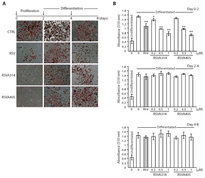
To investigate the mechanism of action of the RSVA compounds on adipogenesis, 3T3-L1 cells were treated at three different phases during the differentiation process: the proliferation phase (days 0–2), the differentiation phase (days 2–4) and the terminal differentiation phase (days 4–8). The effect of the RSVAs on adipocyte differentiation was assessed on day 8 by staining for lipid accumulation with Oil Red O. Treatment with RSVA314 or RSVA405 only during the proliferation phase (day 0–2) resulted in a nearly complete inhibition of lipid accumulation, whereas the two compounds had no noticeable effect when the cells were treated only at later phases during differentiation (day 2–4) or terminal differentiation (day 4–8) (Figures 2A, B). We found that both RSVA314 and RSVA405, when applied during the proliferation phase, inhibited lipid accumulation in 3T3-L1 cells with an apparent IC50 between 0.5 and 1 μmol/L (Figure 2B). These data show that the RSVA compounds specifically acted during the proliferation phase but were unable to reduce the lipid content in already differentiated adipocytes. Using MTT assays and cell counts, we next asked whether the RSVA compounds affect preadipocyte MCE during the proliferation phase. MTT assays revealed that treatments of undifferentiated 3T3-L1 cells with increasing concentrations of RSVA314 or RSVA405 in proliferation medium for 2 d significantly lowered cell proliferation in a dose-dependent manner compared with nontreated control cells (Figure 3A). Cell counts confirmed the reduction in cell numbers during the proliferation phase by RSVA314 or RSVA405, with a nearly complete inhibition of cell expansion at concentrations between 0.5 and 1 μmol/L for both compounds (Figures 3B, C). Cell viability assays in proliferating preadipocytes revealed no noticeable toxicity associated with the treatments with RSVA314 or RSVA405 at concentrations up to 2 μmol/L (Figures 3B, D). Thus, RSVA314 and RSVA405 inhibited adipocyte MCE during the proliferation phase without interfering with cell viability.
Figure 2.
Effect on adipogenesis of RSVA314 and RSVA405 treatment during the different phases of adipocyte differentiation. (A) Differentiating 3T3-L1 cells were treated with resveratrol (RSV; 40 μmol/L), RSVA314 (1 μmol/L), RSVA405 (1 μmol/L) or vehicle (DMSO; CTRL) during the different phases of adipocyte differentiation (see arrows; day 0–2, day 2–4 or day 4–8). Cells were stained with Oil Red O on day 8 and photographed. (B) Lipid content was quantified in cells treated as in A with 40 μmol/L RSV or with the indicated concentrations of RSVA314 or RSVA405. Undifferentiated 3T3-L1 cells on day 0 were used as control (undifferentiated). Results are shown as means ± SD of three independent experiments. *P < 0.05, **P < 0.001 (Student t test).
Figure 3.
Effect of RSVA314 and RSVA405 treatment on cell proliferation and cell viability. (A–D) 3T3-L1 cells were treated with the indicated concentrations of RSVA314 or RSVA405 during the proliferation phase (day 0–2). Undifferentiated 3T3-L1 cells on day 0 were used as control (day 0). Cell proliferation was determined using MTT assays (A). Cells were costained with SYTOX Green and Hoechst (B). Cell numbers were determined by counting Hoechst-positive nuclei (C). Cell viability was assessed by counting SYTOX Green–positive (upper panel, D) and SYTOX Green–negative cells (lower panel, D). Results in A, C and D are shown as means ± SD of three independent experiments. *P < 0.005, **P < 0.01, ***P < 0.001 (Student t test). CTRL, control.
RSVA314 and RSVA405 Activated AMPK during the Proliferation Phase
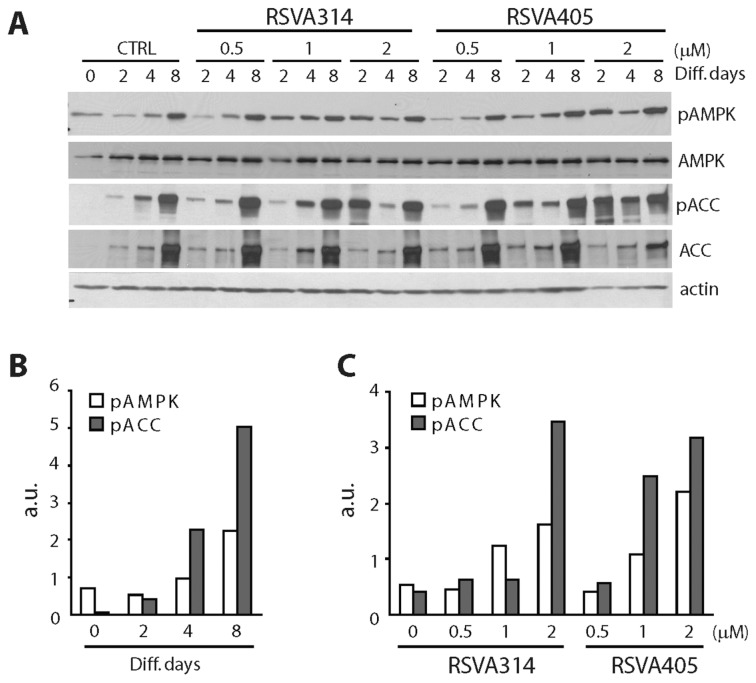
The phosphorylation status of AMPK and ACC were assessed at different time points during adipocyte differentiation. As reported before (10,12), AMPK and ACC phosphorylation gradually increased during 3T3-L1 adipocyte differentiation from day 4 to day 8 (Figure 4A, first four lanes, and Figure 4B), suggesting that AMPK activation occurs during and may be involved in the late steps of adipogenesis during the differentiation and terminal differentiation phases. Treatment with RSVA314 or RSVA405 resulted in a robust and dose-dependent increase in phosphorylated AMPK and ACC at an earlier time point (day 2; Figures 4A, C), showing that the RSVA compounds triggered early AMPK activation during the proliferation phase.
Figure 4.
Effect of RSVA314 and RSVA405 treatment on AMPK activation at the different phases of adipocyte differentiation. (A–C) Differentiating 3T3-L1 cells were treated with the indicated concentrations of RSVA314 and RSVA405 or vehicle (DMSO; CTRL). Phospho-AMPK, total AMPK, phospho-ACC, total ACC and actin protein levels were examined on day 0, 2, 4 and 8 of adipocyte differentiation (Diff. days; A). Densitometric analyses and quantification of the levels of phospho-AMPK and phospho-ACC at the different phases of adipocyte differentiation in control cells (B) and on day 2 of differentiation in cells treated as in (A) were performed (C). a.u., arbitrary units.
Effect of RSVA314 and RSVA405 Treatment on Adipogenesis-Induced Gene Expression Changes
Expression of several proteins involved in adipogenesis is changed in differentiated 3T3-L1 cells. These proteins include several transcription factors, enzymes, binding proteins and cytokines (7,8). Among them, two transcription factors, PPARγ and C/EBPα, are strongly elevated during adipocyte differentiation to orchestrate the expression of several other adipogenic effectors, including aP2 and FAS, or the cytokines RANTES and resistin. Using Western blot and real-time PCR analyses, we confirmed the robust elevation of expression of several adipogenic effectors and markers, including PPARγ, C/EBPα, FAS, aP2, RANTES or resistin during 3T3-L1 adipocyte differentiation (Figure 5A, Table 1). Strikingly, treatments with RSVA314 or RSVA405 at concentrations as low as 0.5 μmol/L resulted in a significant inhibition of the expression of PPARγ, FAS and aP2 (Figure 5B, Table 1). Inhibition of the expression changes of other adipocyte markers and effectors by the RSVAs, including glucose transporter (GLUT)-4 (Slc2a4) and phosphatidylinositol 3-kinase regulatory subunit α (Pik3r1; see Table 1), two genes involved in glucose transport (23), were also observed and further supported the antiadipogenic properties of these compounds. Taken together, these data show that RSVA314 and RSVA405 inhibited the expression changes of key adipogenesis-related transcription factors and effectors, thereby confirming their antiadipogenic properties.
Figure 5.
Effect of RSVA314 and RSVA405 treatment on the expression of PPARγ, FAS and aP2 during adipocyte differentiation. (A) PPARγ, FAS, aP2 and actin protein levels were determined at different days of differentiation (Diff. days) in 3T3-L1 cells. (B) Protein extracts from 3T3-L1 cells treated with the indicated concentrations of RSVA314 or RSVA405 were analyzed for PPARγ, FAS, aP2 and actin protein levels on day 2 of differentiation.
Table 1.
Effect of RSVA314 and RSVA405 treatment on adipogenesis-induced gene expression changes in 3T3-L1 cells.
| Gene | Differentiated | RSVA314 | RSVA405 |
|---|---|---|---|
| Ceacam1 | 3.04 ± 0.37a | 0.60 ± 0.12b | 0.81 ± 0.26b |
| Trib3 | 0.08 ± 0.03a | 0.72 ± 0.14b | 0.69 ± 0.08b |
| PPARg | 1.57 ± 0.05c | 0.72 ± 0.04b | 0.67 ± 0.06b |
| Agt | 5.46 ± 2.17c | 0.36 ± 0.17b | 0.36 ± 0.12b |
| Pik3r1 | 1.59 ± 0.12c | 0.87 ± 0.07b | 0.84 ± 0.06b |
| Cebpa | 2.79 ± 0.21c | 0.70 ± 0.07d | 0.74 ± 0.06d |
| Retn (resistin) | 4.95 ± 0.45c | 1.12 ± 0.05d | 1.24 ± 0.22d |
| Slc2a4 (GLUT4) | 11.20 ± 7.41c | 2.48 ± 1.55d | 1.47 ± 0.28d |
| Snap25 | 4.94 ± 1.23c | 1.11 ± 0.04d | 1.23 ± 0.21d |
| Srebf1 | 1.79 ± 0.04c | 0.96 ± 0.08d | 0.91 ± 0.05d |
| Foxc2 | 1.94 ± 0.06c | 1.13 ± 0.16d | 1.13 ± 0.10d |
| Ccl5 (RANTES) | 0.54 ± 0.07e | 1.12 ± 0.17b | 1.74 ± 0.20b |
| Gpd1 | 1.67 ± 0.31e | 0.78 ± 0.16b | 0.39 ± 0.18b |
| Vamp2 | 0.76 ± 0.09e | 1.42 ± 0.13b | 1.18 ± 0.19d |
| Pfkfb3 | 2.45 ± 0.91e | 1.04 ± 0.51d | 1.33 ± 0.45d |
| Il4ra | 1.53 ± 0.01e | 1.03 ± 0.07f | 0.75 ± 0.09d |
| Igfbp5 | 3.73 ± 0.87e | 1.12 ± 0.05f | 1.24 ± 0.22f |
| Nrf1 | 0.73 ± 0.05e | 0.97 ± 0.03f | 0.96 ± 0.03f |
| Ace | 2.49 ± 0.46e | 1.87 ± 0.51 | 1.24 ± 0.22f |
| Icam1 | 0.31 ± 0.13 | 2.03 ± 0.51 | 2.23 ± 0.35f |
Gene expression was analyzed by real-time PCR using RNA from 3T3-L1 cells differentiated for 2 d and treated or not with the RSVAs (2 μmol/L). Data are expressed as fold-change compared with nondifferentiated control condition ± standard deviation (SD) of three independent experiments.
P < 0.001,
P < 0.01,
P < 0.05, versus nondifferentiated condition.
P < 0.001,
P < 0.01,
P < 0.05, versus differentiated condition (Student t test).
Effect of RSVA405 Administration on HFD-Induced Obesity in Mice
In mice with HFD-induced obesity, resveratrol was found to reduce fat accumulation and to improve the metabolic deterioration observed in this model (21,24). Recently, studies in AMPK knockout mice revealed that AMPK activation is required for the metabolic effects of resveratrol against obesity (16). In this context and on the basis of our in vitro data in 3T3-L1 cells, we then tested the ability of RSVA405 to prevent obesity in mice fed an HFD. As expected, mice fed the HFD for 11 wks steadily and significantly gained weight (Table 2). Supplementation of the HFD with 20 or 100 mg/kg/d significantly prevented body weight gain during this period (Table 2). No difference in food intake was observed (2.45 ± 0.08 g [HFD]; 2.4 ± 0.1 g [HFD ± RSVA405 20 mg/kg/d]; 2.4 ± 0.08 [HFD ± RSVA405 100 mg/kg/d], n = 5), indicating that the weight loss cannot be explained by reduced consumption of the diet. No significant amelioration in blood glucose, triglycerides and cholesterol at the end of the treatment period was observed in the treated mice (Table 3). However, a dose-dependent trend of reduction in blood glucose was observed in RSVA405-treated obese mice (Table 3).
Table 2.
Effect of RSVA405 administration on body weight of mice fed an HFD.
| Week(s) of treatment | Standard diet | HFD | HFD + RSVA405 (20 mg/kg/d) | HFD + RSVA405 (100 mg/kg/d) |
|---|---|---|---|---|
| 1 | 20 ± 1.6 | 18.2 ± 0.9 | 18.4 ± 1.1 | 18.5 ± 1.1 |
| 2 | 22.2 ± 1.8 | 23 ± 0.7 | 22 ± 1 | 22.3 ± 1 |
| 3 | 23.4 ± 1.8 | 27.3 ± 0.5a | 24.3 ± 1.36b | 24.6 ± 1.4c |
| 4 | 24.5 ± 2.4 | 29 ± 0.4a | 27.1 ± 2.2c | 25.9 ± 1.9c |
| 5 | 25.8 ± 2.9 | 31.4 ± 0.8a | 30.2 ± 2.2 | 28.4 ± 1.9c |
| 6 | 27.6 ± 3.5 | 34.7 ± 1.1d | 33 ± 2.3c | 30.4 ± 2.1c |
| 7 | 28.4 ± 3.2 | 37.2 ± 1.1d | 34.8 ± 3.1c | 32.8 ± 2.2c |
| 8 | 28.7 ± 3.2 | 40.7 ± 1.4e | 36.4 ± 2.3c | 35.4 ± 2.3c |
| 9 | 29.7 ± 3.2 | 42.8 ± 1.6e | 38.4 ± 2.3c | 36.8 ± 2.6b |
| 10 | 31 ± 3.2 | 45.2 ± 1.4e | 39.9 ± 2.5c | 38.6 ± 2.6b |
| 11 | 32.4 ± 3.2 | 46.5 ± 1.2e | 42 ± 2.4c | 40.8 ± 3c |
Data are mean ± SD. n = 5.
P < 0.05,
P < 0.01,
P < 0.001, versus standard diet condition.
P < 0.01,
P < 0.05, versus HFD condition.
Table 3.
Effect of RSVA405 administration on fasted blood glucose, triglycerides and cholesterol at the end of the treatment period in mice fed an HFD.
| Standard diet | HFD | HFD + RSVA405 (20 mg/kg/d) | HFD + RSVA405 (100 mg/kg/d) | |
|---|---|---|---|---|
| Glucose (mg/dL) | 199 ± 31 | 238 ± 30 | 204 ± 56 | 187 ± 29 |
| Triglycerides (mg/dL) | 101 ± 5 | 90 ± 8 | 117 ± 10 | 130 ± 26 |
| Cholesterol (mg/dL) | 138 ± 77 | 291 ± 34 | 295 ± 22 | 276 ± 23 |
Data are mean ± SD. n = 5.
DISCUSSION
In this report, we show that the novel small-molecule agonists of AMPK (RSVA314 and RSVA405) inhibited 3T3-L1 adipocyte differentiation by interfering with preadipocyte MCE during the proliferation phase (IC50 ~ 0.5 μmol/L). The RSVAs acted without inducing toxicity and by triggering early activation of AMPK in proliferating preadipocytes. Importantly, the RSVA compounds blocked the adipogenesis-induced transcriptional changes of several genes critically involved in the adipogenic process, including PPARγ, C/EBPα, FAS, aP2, RANTES or resistin, at concentrations consistent with the effect of these compounds on adipocyte differentiation. Furthermore, oral administration of RSVA405 was found to significantly reduce body weight gain in HFD-induced obese mice.
AMPK is a critical cellular energy sensor and regulator and thus represents a promising drug target for metabolic disorders. Overwhelming evidence using pharmacological and genetic approaches now indicates that AMPK activation improves the deterioration of lipid and glucose metabolism observed in models of type 2 diabetes and obesity (5,25–27). In animal models, AMPK signaling is reduced in several organs and tissues by chronic exposure to fatty acid, a situation that occurs during obesity and could contribute to the pathogenesis of this condition. Furthermore, whole-body AMPKα2 knockdown in mice was found to exacerbate the increase in body weight and fat mass in mice fed an HFD (28). AMPK is activated during the late phases of adipocyte differentiation, further suggesting that this kinase is involved in the adipogenic process. However, the role played by AMPK in white adipose tissue maintenance remains uncertain. Studies in animal models and cultured adipocytes have showed that, in mature adipocytes, AMPK has antilipolytic effects via a mechanism implicating hormone-sensitive lipase inhibition (29,30). It is therefore unclear whether pharmacological activation of AMPK in adipocytes could improve lipid metabolism in a cell-autonomous manner and reduce white adipose tissue excess in obesity.
Several pharmacological activators of AMPK were identified and found to have beneficial effects against the metabolic deterioration observed in metabolic diseases. These include the highly prescribed antidiabetic drugs biguanides (for example, metformin) and thiazolidinediones, which might act, at least in part, via activation of AMPK (25). Some of these activators are natural molecules, such as resveratrol, a polyphenol found in abundance in grape skin and wine. In mammals, resveratrol was found to mitigate diet-induced obesity (21,24). The direct molecular target of resveratrol remains unknown, but this polyphenol indirectly activates AMPK by promoting the calcium/CaMKKβ-dependent phosphorylation of AMPK (13) and by facilitating the AMP-dependent activation of AMPK (14). In the current study, we report the antiadipogenic effect of two structurally related synthetic small-molecule analogs of resveratrol, RSVA314 and RSVA405. These molecules were selected on the basis of their improved potency to activate AMPK in different cell lines (20). Like resveratrol, the RSVA compounds were previously found to indirectly activate AMPK via a mechanism implicating CaMKKβ (20). These compounds were also found to significantly lower ATP levels in treated fibroblasts and neuroblastoma cells, suggesting that the RSVAs may also activate AMPK via a mechanism implicating an elevation of the AMP:ATP ratio (20). Although the RSVAs indirectly target AMPK and may involve different mechanisms of activation, a kinase array performed in cell-based assays showed that these compounds are relatively specific for AMPK (20). Altogether these studies support the notion that RSVA314 and RSVA405 inhibit adipogenesis by triggering early AMPK activation during the proliferation phase, a phenomenon already observed for other pharmacological activators of AMPK, such as A-769662 or AICAR (10,12). In this context, it is tempting to speculate that early activation of AMPK could be sensed by the adipocytes as a signal of energy depletion, thus leading to inhibition of ATP-consuming pathways, such as lipogenesis. In future studies, it will be important to identify the direct molecular target of the RSVAs and to confirm, by pharmacological and genetic approaches targeting AMPK, the requirement for AMPK activation in the antiadipogenic effect of these compounds.
The current study shows that RSVA314 and RSVA405 acted during the proliferation phase to potently block preadipocyte MCE. Compelling evidence supports the key role played by AMPK activation in cell growth inhibition. Cell growth and proliferation are highly energy demanding, and AMPK acts as a molecular checkpoint to control whether the cellular energy status permits the energy expenditure required for the anabolic process (25). AMPK controls cell growth by directly phosphorylating and activating tuberous sclerosis complex 2 (TSC2), a tumor suppressor that inhibits the mammalian target of rapamycin C1 (mTORC1). mTOR is a protein kinase critically involved in cellular homeostasis (31,32). mTOR promotes cell growth and anabolism by increasing protein and lipid synthesis via activation of p70-S6 kinase (p70S6K) and by decreasing autophagic catabolism through phosphorylation-mediated inhibition of the autophagy-initiating kinase Ulk1 (32). AMPK can also promote autophagy by directly phosphorylating and activating Ulk1 (33,34). Autophagy is an evolutionary conserved lysosomal pathway involved in protein and organelle degradation (35). Our previous data have shown that resveratrol and its analogs, RSVA314 and RSVA405, are potent inhibitors of mTOR signaling via a mechanism implicating AMPK activation (13,20). Specifically, we showed that resveratrol and the RSVAs promoted the inactivation of several downstream effectors of mTOR involved in mRNA translation, including p70S6K (13,20). Thus, both resveratrol and the RSVA compounds by activating AMPK are potent inhibitors of mTOR and thus have strong antiproliferative properties. Altogether, these data strongly suggest that the antiadipogenic effects of the RSVAs in 3T3-L1 cells were mediated, at least in part, by early AMPK activation and cell growth inhibition in preadipocytes during MCE. Additional studies are required to determine the precise mechanism by which RSVA314 and RSVA405 block preadipocyte MCE and whether mTOR or autophagy is involved in this mechanism.
Our results also show that RSVA405 administration at 20 or 100 mg/kg/day significantly lowered the weight gain observed in mice fed an HFD. We failed, however, to see any significant changes in glucose or lipid levels in RSVA405-treated HFD mice. It is important to note that although the effect of an HFD (60% of fat) on weight gain in mice is robust, its consequences on blood glucose and cholesterol levels are often modest, and triglyceride levels are usually not changed by this intervention (21). This lack of marked difference between the lean control mice and the HFD mice may explain the apparent absence of effect of RSVA405 on these blood parameters. Nevertheless, a trend toward an increase in fasted glucose levels in HFD mice and toward a decrease in this elevated glucose level in RSVA405-treated HFD mice was found in our study, suggesting that RSVA405 treatment may also influence glucose metabolism in obese mice. Further studies including larger sample sizes of treated mice will be required to assess in more detail the effect of the RSVA compounds on blood glucose and lipid deregulations during obesity development. Moreover, because AMPK and resveratrol were both proposed to have beneficial effects by controlling energy expenditure in vivo via mechanisms facilitating mitochondrial function, aerobic capacity and endurance (24,36), it will be interesting to determine whether the RSVA compounds might influence weight gain by also controlling energy expenditure.
In conclusion, the novel small-molecule agonists of AMPK (RSVA314 and RSVA405) are potent inhibitors of adipocyte differentiation. These compounds triggered early activation of AMPK in proliferating preadipocytes and acted by interfering with preadipocyte MCE during the proliferation phase. Furthermore, RSVA405 was found to have in vivo efficacy against body weight gain in HFD-induced obese mice. This work suggests that the RSVA compounds and activation of AMPK in preadipocytes have therapeutic potential against obesity.
ACKNOWLEDGMENTS
This work was supported in part by the National Institutes of Health (grant PO1 AT004511; National Center for Complementary and Alternative Medicine [NCCAM] Project 2 to P Marambaud).
Footnotes
Online address: http://www.molmed.org
DISCLOSURE
The authors declare that they have no competing interests as defined by Molecular Medicine, or other interests that might be perceived to influence the results and discussion reported in this paper.
REFERENCES
- 1.Hardie DG. AMP-activated/SNF1 protein kinases: conserved guardians of cellular energy. Nat Rev Mol Cell Biol. 2007;8:774–85. doi: 10.1038/nrm2249. [DOI] [PubMed] [Google Scholar]
- 2.Fogarty S, et al. Calmodulin-dependent protein kinase kinase-beta activates AMPK without forming a stable complex: synergistic effects of Ca2+ and AMP. Biochem J. 2010;426:109–18. doi: 10.1042/BJ20091372. [DOI] [PMC free article] [PubMed] [Google Scholar]
- 3.Carling D, Sanders MJ, Woods A. The regulation of AMP-activated protein kinase by upstream kinases. Int. J. Obes. (Lond) 2008;32(Suppl 4):S55–9. doi: 10.1038/ijo.2008.124. [DOI] [PubMed] [Google Scholar]
- 4.Viollet B, et al. Targeting the AMPK pathway for the treatment of type 2 diabetes. Front Biosci. 2009;14:3380–400. doi: 10.2741/3460. [DOI] [PMC free article] [PubMed] [Google Scholar]
- 5.Zhang BB, Zhou G, Li C. AMPK: an emerging drug target for diabetes and the metabolic syndrome. Cell Metab. 2009;9:407–16. doi: 10.1016/j.cmet.2009.03.012. [DOI] [PubMed] [Google Scholar]
- 6.Spiegelman BM, Flier JS. Obesity and the regulation of energy balance. Cell. 2001;104:531–43. doi: 10.1016/s0092-8674(01)00240-9. [DOI] [PubMed] [Google Scholar]
- 7.Farmer SR. Transcriptional control of adipocyte formation. Cell Metab. 2006;4:263–73. doi: 10.1016/j.cmet.2006.07.001. [DOI] [PMC free article] [PubMed] [Google Scholar]
- 8.Rosen ED, Spiegelman BM. Molecular regulation of adipogenesis. Annu Rev Cell Dev Biol. 2000;16:145–71. doi: 10.1146/annurev.cellbio.16.1.145. [DOI] [PubMed] [Google Scholar]
- 9.Ahn J, Lee H, Kim S, Park J, Ha T. The anti-obesity effect of quercetin is mediated by the AMPK and MAPK signaling pathways. Biochem Biophys Res Commun. 2008;373:545–9. doi: 10.1016/j.bbrc.2008.06.077. [DOI] [PubMed] [Google Scholar]
- 10.Giri S, et al. AICAR inhibits adipocyte differentiation in 3T3L1 and restores metabolic alterations in diet-induced obesity mice model. Nutr. Metab. (Lond) 2006;3:31. doi: 10.1186/1743-7075-3-31. [DOI] [PMC free article] [PubMed] [Google Scholar]
- 11.Hwang JT, et al. Anti-obesity effects of ginsenoside Rh2 are associated with the activation of AMPK signaling pathway in 3T3-L1 adipocyte. Biochem Biophys Res Commun. 2007;364:1002–8. doi: 10.1016/j.bbrc.2007.10.125. [DOI] [PubMed] [Google Scholar]
- 12.Zhou Y, et al. Inhibitory effects of A-769662, a novel activator of AMP-activated protein kinase, on 3T3-L1 adipogenesis. Biol Pharm Bull. 2009;32:993–8. doi: 10.1248/bpb.32.993. [DOI] [PubMed] [Google Scholar]
- 13.Vingtdeux V, et al. AMP-activated protein kinase signaling activation by resveratrol modulates amyloid-beta peptide metabolism. J Biol Chem. 2010;285:9100–13. doi: 10.1074/jbc.M109.060061. [DOI] [PMC free article] [PubMed] [Google Scholar]
- 14.Hawley SA, et al. Use of cells expressing gamma subunit variants to identify diverse mechanisms of AMPK activation. Cell Metab. 2010;11:554–65. doi: 10.1016/j.cmet.2010.04.001. [DOI] [PMC free article] [PubMed] [Google Scholar]
- 15.Canto C, Auwerx J. PGC-1alpha, SIRT1 and AMPK, an energy sensing network that controls energy expenditure. Curr Opin Lipidol. 2009;20:98–105. doi: 10.1097/MOL.0b013e328328d0a4. [DOI] [PMC free article] [PubMed] [Google Scholar]
- 16.Um JH, et al. AMPK-deficient mice are resistant to the metabolic effects of resveratrol. Diabetes. 2010;59:554–63. doi: 10.2337/db09-0482. [DOI] [PMC free article] [PubMed] [Google Scholar]
- 17.Baile CA, et al. Effect of resveratrol on fat mobilization. Ann N Y Acad Sci. 2011;1215:40–7. doi: 10.1111/j.1749-6632.2010.05845.x. [DOI] [PubMed] [Google Scholar]
- 18.Marambaud P, Zhao H, Davies P. Resveratrol promotes clearance of Alzheimer’s disease amyloid-beta peptides. J Biol Chem. 2005;280:37377–82. doi: 10.1074/jbc.M508246200. [DOI] [PubMed] [Google Scholar]
- 19.Vingtdeux V, Dreses-Werringloer U, Zhao H, Davies P, Marambaud P. Therapeutic potential of resveratrol in Alzheimer’s disease. BMC Neurosci. 2008;9(Suppl 2):S6. doi: 10.1186/1471-2202-9-S2-S6. [DOI] [PMC free article] [PubMed] [Google Scholar]
- 20.Vingtdeux V, et al. Novel synthetic small-molecule activators of AMPK as enhancers of autophagy and amyloid-beta peptide degradation. FASEB J. 2011;25:219–31. doi: 10.1096/fj.10-167361. [DOI] [PMC free article] [PubMed] [Google Scholar]
- 21.Baur JA, et al. Resveratrol improves health and survival of mice on a high-calorie diet. Nature. 2006;444:337–42. doi: 10.1038/nature05354. [DOI] [PMC free article] [PubMed] [Google Scholar]
- 22.Rayalam S, Yang JY, Ambati S, Della-Fera MA, Baile CA. Resveratrol induces apoptosis and inhibits adipogenesis in 3T3-L1 adipocytes. Phytother Res. 2008;22:1367–71. doi: 10.1002/ptr.2503. [DOI] [PubMed] [Google Scholar]
- 23.Fujii N, Jessen N, Goodyear LJ. AMP-activated protein kinase and the regulation of glucose transport. Am J Physiol Endocrinol Metab. 2006;291:E867–77. doi: 10.1152/ajpendo.00207.2006. [DOI] [PubMed] [Google Scholar]
- 24.Lagouge M, et al. Resveratrol improves mitochondrial function and protects against metabolic disease by activating SIRT1 and PGC-1alpha. Cell. 2006;127:1109–22. doi: 10.1016/j.cell.2006.11.013. [DOI] [PubMed] [Google Scholar]
- 25.Fogarty S, Hardie DG. Development of protein kinase activators: AMPK as a target in metabolic disorders and cancer. Biochim Biophys Acta. 20101804:581–91. doi: 10.1016/j.bbapap.2009.09.012. [DOI] [PubMed] [Google Scholar]
- 26.Viollet B, et al. AMPK inhibition in health and disease. Crit Rev Biochem Mol Biol. 2010;45:276–95. doi: 10.3109/10409238.2010.488215. [DOI] [PMC free article] [PubMed] [Google Scholar]
- 27.Musi N, Goodyear LJ. Targeting the AMP-activated protein kinase for the treatment of type 2 diabetes. Curr Drug Targets Immune Endocr Metabol Disord. 2002;2:119–27. [PubMed] [Google Scholar]
- 28.Villena JA, et al. Induced adiposity and adipocyte hypertrophy in mice lacking the AMP-activated protein kinase-alpha2 subunit. Diabetes. 2004;53:2242–9. doi: 10.2337/diabetes.53.9.2242. [DOI] [PubMed] [Google Scholar]
- 29.Daval M, et al. Anti-lipolytic action of AMP-activated protein kinase in rodent adipocytes. J Biol Chem. 2005;280:25250–7. doi: 10.1074/jbc.M414222200. [DOI] [PubMed] [Google Scholar]
- 30.Corton JM, Gillespie JG, Hawley SA, Hardie DG. 5-aminoimidazole-4-carboxamide ribonucleoside: a specific method for activating AMP-activated protein kinase in intact cells. Eur J Biochem. 1995;229:558–65. doi: 10.1111/j.1432-1033.1995.tb20498.x. [DOI] [PubMed] [Google Scholar]
- 31.Wullschleger S, Loewith R, Hall MN. TOR signaling in growth and metabolism. Cell. 2006;124:471–84. doi: 10.1016/j.cell.2006.01.016. [DOI] [PubMed] [Google Scholar]
- 32.Chan EY. mTORC1 phosphorylates the ULK1-mAtg13-FIP200 autophagy regulatory complex. Sci. Signal. 2009;2:pe51. doi: 10.1126/scisignal.284pe51. [DOI] [PubMed] [Google Scholar]
- 33.Kim J, Kundu M, Viollet B, Guan KL. AMPK and mTOR regulate autophagy through direct phosphorylation of Ulk1. Nat Cell Biol. 2011;13:132–41. doi: 10.1038/ncb2152. [DOI] [PMC free article] [PubMed] [Google Scholar]
- 34.Egan DF, et al. Phosphorylation of ULK1 (hATG1) by AMP-activated protein kinase connects energy sensing to mitophagy. Science. 2011;331:456–61. doi: 10.1126/science.1196371. [DOI] [PMC free article] [PubMed] [Google Scholar]
- 35.Maiuri MC, Zalckvar E, Kimchi A, Kroemer G. Self-eating and self-killing: crosstalk between autophagy and apoptosis. Nat Rev Mol Cell Biol. 2007;8:741–52. doi: 10.1038/nrm2239. [DOI] [PubMed] [Google Scholar]
- 36.Narkar VA, et al. AMPK and PPARdelta agonists are exercise mimetics. Cell. 2008;134:405–15. doi: 10.1016/j.cell.2008.06.051. [DOI] [PMC free article] [PubMed] [Google Scholar]


