Abstract
Duchenne muscular dystrophy (DMD) is an X-linked, lethal, degenerative disease that results from mutations in the dystrophin gene, causing necrosis and inflammation in skeletal muscle tissue. Treatments that reduce muscle fiber destruction and immune cell infiltration can ameliorate DMD pathology. We treated the mdx mouse, a model for DMD, with the immunosuppressant drug rapamycin (RAPA) both locally and systemically to examine its effects on dystrophic mdx muscles. We observed a significant reduction of muscle fiber necrosis in treated mdx mouse tibialis anterior (TA) and diaphragm (Dia) muscles 6 wks post-treatment. This effect was associated with a significant reduction in infiltration of effector CD4+ and CD8+ T cells in skeletal muscle tissue, while Foxp3+ regulatory T cells were preserved. Because RAPA exerts its effects through the mammalian target of RAPA (mTOR), we studied the activation of mTOR in mdx TA and Dia with and without RAPA treatment. Surprisingly, mTOR activation levels in mdx TA were not different from control C57BL/10 (B10). However, mTOR activation was different in Dia between mdx and B10; mTOR activation levels did not rise between 6 and 12 wks of age in mdx Dia muscle, whereas a rise in mTOR activation level was observed in B10 Dia muscle. Furthermore, mdx Dia, but not TA, muscle mTOR activation was responsive to RAPA treatment.
INTRODUCTION
Duchenne muscular dystrophy (DMD) is a fatal, X-linked disorder affecting 1:3,600 to 1:6,000 male births worldwide that causes degeneration of striated muscle (1–3). This progressive disease is due to mutations in the dystrophin gene that result in absence of expression of a functional dystrophin protein. Dystrophin is an essential structural muscle protein that links the intracellular filamentous actin cytoskeleton and the extracellular basal lamina of muscle fibers (4–6). Absence of dystrophin protein in muscle fibers alters muscle fiber membrane structure, leading to membrane damage during muscle contraction and disrupting signaling across the membrane. The mdx mouse is a disease model of DMD (7) in which a mutation in the dystrophin gene causes absence of dystrophin protein in muscles and leads to necrosis and inflammation in muscle tissue.
The ongoing necrosis of dystrophic skeletal muscle leads to infiltration in the diseased muscle of immune cells that may be, in part, autoreactive. In previous studies, depletion of CD4+ and CD8+ T cells in the mdx mouse resulted in a significantly lower level of muscle fiber necrosis and fibrosis (8,9). Furthermore, the only proven treatment for human DMD, prednisone, is a known immunosuppressant (10), suggesting that down-regulation of the immune system in a dystrophic setting may have therapeutic effects.
Rapamycin (RAPA), also called sirolimus, has been used widely for immune suppression in the setting of allo-graft organ or tissue transplantation (11–13). Studies have shown that RAPA has lower toxicity and leads to a greater decrease in T-cell immunity when compared to other immunosuppressant drugs, such as cyclosporine A (14). One of the mechanisms of the selective effect of RAPA is an increase in Foxp3+ regulatory T (Treg) cell survival and function, which has been shown in vitro and in vivo (15,16). Therefore, we hypothesized that RAPA treatment of the mdx mouse would diminish dystrophic pathology of the diseased muscle tissue by providing a more favorable balance between effector T cells and Treg cells.
In mammals, RAPA binds to the mammalian target of rapamycin (mTOR), which is a highly conserved serine/threonine protein kinase that regulates cell growth and protein synthesis (17–19). There are two distinct mTOR complexes, complex 1 and complex 2 (mTORC1 and mTORC2, respectively), that vary in both structure and function and respond differentially to nutrients, cellular energy and growth factors, as reviewed by Zhou and Huang (20). The relative importance of mTORC1 and mTORC2 differs among various tissues. In skeletal muscle, mTORC1, which is RAPA-sensitive, has been shown to play an important role in muscle growth (21,22). Skeletal muscle mTOR activation levels have been followed longitudinally in the mdx mouse (23). However, comparison of mTOR activation between age-matched mdx and wild-type C57BL/10 (B10) mice has not been reported. Findings of these studies are important for a better molecular understanding of DMD pathology, and can lead to potential therapeutic approaches for this disease.
Because RAPA is a potent immunosuppressant drug and has the potential to modulate muscle growth and regeneration, we studied its effect on the pathology of dystrophic muscle. We examined the effects of RAPA on mdx muscle pathology and mTOR activation when administered either locally or systemically during the peak of mdx disease- related muscle degeneration and regeneration. Given that systemic immune suppression may be associated with adverse outcomes, we used two strategies to treat mdx mice with RAPA: 1) local treatment of mdx tibialis anterior (TA) muscles by injection of RAPA-containing microparticles and 2) systemic treatment of mdx mice through RAPA-containing water. Systemic RAPA administration facilitated the analysis of treatment effects on the diaphragm (Dia) muscle, which is not easily accessible by direct intramuscular injection.
MATERIALS AND METHODS
Mice
Wild-type C57BL/10J (B10) and C57BL/10ScSnDmdmdx/J (mdx) mice were obtained from The Jackson Laboratory (Bar Harbor, ME, USA). All animal studies were performed according to the Institutional Animal Care and Use Committee (IACUC) Guidelines for the Care and Use of Laboratory Animals.
Total Muscle Protein Extract (TMPE) Preparation
Freshly isolated mouse muscle was cut into small pieces in lysis buffer (50 mmol/L Tris-HCl pH 8.0, 5 mmol/L EGTA pH 7.4, 5 mmol/L EDTA pH 8.0, 5% SDS) and incubated on ice for 45 min. Samples were then sonicated briefly and centrifuged at 16,100g for 20 min at 4°C. Supernatant was collected and stored at −80°C.
Rapamycin-Containing PLGA Microparticle Preparation
RAPA-releasing microparticles were produced in our laboratory as described previously (24). In brief, RAPA microparticles were produced by using the single emulsion-evaporation technique. An emulsion of an organic solution containing RAPA and PLGA was formed in a bulk aqueous solution through high-speed homogenization at 3,000 rpm by using a homogenizer (Silverson L4RT-A, Silverson, Chesham Bucks, UK). Following evaporation of the organic solvent (dichloromethane), microparticles formed in the aqueous solution were freeze-dried for subsequent use in experiments. Microparticles were fabricated to be ~15 μm in size so that they do not move from the site of injection. The amount of RAPA encapsulated in 1 mg of microparticles was measured to be approximately 4 μg.
Intramuscular Microparticle Injection
Six-wk-old mdx mice were injected intramuscularly (IM) with rapamycin- containing microparticles (RAPA beads) in sterile PBS in the TA muscle (30 μL per TA muscle). Control mice either received empty microparticles (blank beads) or were left untreated. Microparticles were injected into the TA muscles with a high-dose injection (1 mg RAPA beads per injection) initially, followed by a half-dose injection (0.5 mg RAPA beads per injection) 2 wks later. Mice were euthanized 6 wks after initiation of treatment, thus allowing for complete degradation of PLGA micro particles by the time of analysis.
RAPA/H2O Preparation and Administration
To prepare the mdx mouse drinking water, containing RAPA (LC Laboratories, Woburn, MA, USA), RAPA was dissolved in autoclaved water to achieve administration of 1.5 mg/kg mouse (about 0.04 mg/mouse/day). RAPA-containing water was prepared fresh every 7 to 10 d. To generate the aqueous solution of RAPA, RAPA powder was first dissolved in dimethyl sulfoxide (DMSO) to a stock concentration of 10 mg/mL. The final concentration of DMSO in RAPA-containing water was 0.1% vol/vol. The amount of RAPA added to the mdx mouse drinking water was calculated such that the amount of RAPA administered by RAPA drinking water approximately matched the amount of RAPA administered locally by the RAPA-containing microparticles on a daily basis.
Gel Electrophoresis and Western Blotting
Gel electrophoresis and Western blotting were done according to standard protocols. Briefly, TMPE from muscle samples from all mouse groups was electrophoresed on 5% Acrylamide gel (Bio-Rad; Hercules, CA, USA) for 3 h at 110V. Protein samples were transferred from the gel to nitrocellulose membrane (GE Healthcare; Piscataway, NJ, USA) for 1.5 h at 110V at 4°C. The membrane was blocked in 5% milk/1% sheep serum/TBST (10 mmol/L Tris pH 8.0, 150 mmol/L NaCl, 0.5 mmol/L Tween-20) overnight at 4°C. The membrane containing TMPE was incubated with primary rabbit anti–mouse total mTOR and phosphorylated mTOR antibodies (Cell Signaling Technology; Danvers, MA, USA) in 5% BSA/TBST for 1.5 h, then with HRP-conjugated donkey anti–rabbit IgG (GE Healthcare) diluted in TBST for 45 min at room temperature. ECL detection reagent (GE Healthcare) was used to detect the chemiluminescent signal and Kodak film was used for visualizing the signal. The initial exposure time was 5–15 min. To confirm the absence of a band, an additional film was exposed to each membrane overnight. The bands then were analyzed using MCID image analysis software (Interfocus Imaging, Cambridge, England) to find the density of each band. Values for phospho-mTOR were then divided by total mTOR values for evaluation of the level of mTOR activation for each mouse tissue sample. GAPDH labeling was used as a protein loading control.
Muscle Tissue Processing
Freshly dissected muscle was incubated in 2% paraformaldehyde/PBS on ice for 2 h, then transferred into 30% sucrose overnight at 4°C. The next day the muscle tissue was snap-frozen in 2-methylbutane cooled with dry ice and stored at −80°C.
Immunohistochemistry of Inflammatory Cells
Ten-μm cryosections of muscle samples were prepared. Sections were rehydrated in PBS, blocked in peroxidase-blocking reagent (DAKO Cytomation; Carpinteria, CA, USA) for 5 min and then blocked in 10% goat serum/PBS for 1 h at room temperature. The primary antibody incubation using rat anti-CD4 and Foxp3 (ebiosciences; San Diego, CA, USA; 16-0041-81 and 14-4771-80) and rat anti-CD8 (Pharmingen; San Jose, CA, USA) purified antibodies diluted in 10% goat serum/PBS was performed for 1.5 h at room temperature. Appropriate isotype controls (Pharmingen) also were used to approximate background antibody binding. Sections were incubated for 1 h with secondary biotinylated goat anti–rat IgG (Pharmingen) diluted in DAKO antibody diluent (DAKO Cytomation). Sections were incubated in ABC Vectastain avidin-HRP detection solution (Vector Laboratories; Burlingame, CA, USA) for 30 min at room temperature and DAB peroxidase substrate solution (Vector Laboratories) for 4 min. Eosin counter-staining was performed to visualize muscle fibers. To analyze infiltrating cells in each group, the total number of cells per cross-section of vector-injected TA muscles was counted and the average number of cells from sections from different mice was calculated.
Immunofluoresence Staining for Necrotic Fibers
Muscle fiber necrosis was evaluated by incubating muscle cryosections with fluorescently labeled IgG. In brief, muscle cryosections (10 μm thick) were rehydrated with PBS, then blocked with 1% gelatin/1% rabbit serum/PBS for 15 min. Samples next were washed with PBS-G wash buffer (0.2% gelatin/PBS) and incubated with Alexa 488–conjugated rabbit anti–mouse IgG (Invitrogen; Carslbad, CA, USA) for 1 h, following with PBS-G washes. IgG-labeled fibers were counted on representative muscle sections from all experimental mice.
Immunofluorescence Staining for Regenerating Fibers
Muscle fiber regeneration was evaluated by immunoreactivity for embryonic myosin heavy chain (eMyoHC). Ten-μm cryosections were rehydrated with PBS, blocked first with avidin and biotin block (Vector Laboratories) and then with mouse IgG block (Vector Laboratories), according to the manufacturer’s instructions. Samples also were blocked briefly with 10% goat serum/PBS for 15 min at room temperature. Incubation with primary anti-eMyoHC antibody F1.652 (Developmental Studies Hybridoma Bank (Iowa City, IA, USA) was done for 3 h at room temperature. Appropriate isotype control (Pharmingen) also was used to approximate background antibody binding. Sections then were incubated with biotinylated goat anti–mouse IgG (Pharmingen) and tertiary FITC- conjugated donkey anti–goat IgG. Muscle fibers expressing eMyoHC were counted on representative muscle sections from all experimental mice.
Statistical Analysis
In all performed studies, the statistical analysis was performed by Student t test, in which a treatment group and a control group or two treatment groups were compared as unpaired sets. In all experiments, P < 0.05 were considered significant.
RESULTS
RAPA Lowers Infiltrating Effector T cells, but not Foxp3+ Regulatory T Cells in mdx Muscles
Given that infiltration of effector T cells in dystrophic muscles of mdx mice plays an important role in the pathology associated with the disease, we hypothesized that RAPA would improve the pathology of mdx muscle by reducing the level of infiltrating T cells. Both local and systemic treatments began at 6 wks of age and were continued as described during the 6-wk treatment period prior to euthanization and analysis. TA and Dia muscles were analyzed in the systemic RAPA-treated group. For the microparticle-administered RAPA studies only TA muscles were analyzed because transfer of microparticles does not provide systemic RAPA delivery.
To assess the immunosuppressant effects of RAPA on dystrophic muscle tissue, infiltrating CD4+ and CD8+ T cells in TA muscles of mdx mice were counted. There was a significant decrease in total infiltrating CD4+ T cells in mdx muscles treated with local or systemic RAPA compared with untreated or blank bead-injected mdx muscles (Figure 1). A significant decrease in CD8+ T cells also was observed in mdx TA muscles with systemic RAPA-treatment, but not with local treatment (Figure 1A). Infiltrating CD4+ and CD8+ T cells were observed at highest levels in areas of necrosis in untreated mdx muscle tissue (Figure 1B). In mdx muscles treated with RAPA, however, infiltration of both CD4+ and CD8+ T cells was scattered in the tissue. In contrast to the effects on CD8+ and total CD4+ T cell levels, Foxp3+ Treg cells were not reduced significantly in mdx TA muscles treated with local or systemic RAPA compared with the untreated or blank-bead-treated age-matched muscles.
Figure 1.
T-cell infiltration in RAPA-treated mdx muscles. Six wks following either local rapamycin (RAPA) microparticle (Rapa Bead) administration to the tibialis anterior (TA) muscle or systemic RAPA administration in drinking water (Rapa H2O) of mdx mice beginning at 6 wk of age, TA muscles were collected for analysis. Controls included untreated wild-type C57BL/10 muscle (B10) and mdx muscle from mice that were treated with blank beads (Blank Bead) or untreated. T-cell infiltration was evaluated by immunohistochemistry of TA muscle cross-sections to detect CD4+ and CD8+ effector T cells and Foxp3+ T regulatory cells. Muscle sections incubated with isotype control antibody (Iso) in place of the primary antibody serve as negative controls for immunohistochemistry. Data is presented as the average number of cells (± SE) per cross-section (A). Arrows in panel (B) indicate T-cell infiltrates. n = 5 for RAPA-treated and B10 groups and n = 4 for the blank bead and untreated group. *P < 0.05 and **P < 0.01.
RAPA Lowers Muscle Fiber Necrosis and Regeneration in mdx Muscles
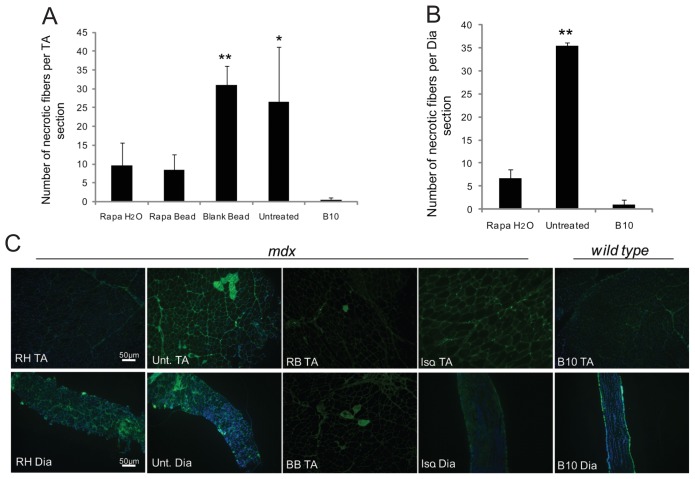
Infiltrating T cells contribute to the pathologic process of muscle fiber necrosis in dystrophic muscles of the mdx mouse and DMD patients (9,25). Therefore, we quantitatively examined the level of muscle fiber necrosis in RAPA-treated and age-matched untreated mdx muscles (Figure 2). The total number of necrotic fibers per TA muscle cross- section, as determined by incubation with fluorescently labeled IgG, in mice with systemic or local RAPA treatment was significantly less compared with age-matched untreated mdx muscle (Figures 2A, C). Furthermore, with systemic RAPA treatment, we also observed significantly decreased necrosis in the diaphragm (Figures 2B, C). The necrotic muscle fibers in untreated mdx mice were observed in large groups of fibers in muscle tissue, whereas, in the RAPA-treated mice, the fewer necrotic fibers were observed as scattered small groups or individual fibers within the muscle tissue (see Figure 2C).
Figure 2.
RAPA treatment lowers muscle fiber necrosis in mdx muscles. Six wk after treatment of 6-wk-old mdx mice by either rapamycin (RAPA)-containing microparticles (Rapa Bead or RB) or RAPA-containing drinking water (Rapa H2O or RH), tibialis anterior (TA) muscles of all of the mice and diaphragm (Dia) muscles of Rapa H2O-treated mice and control mice were collected for analysis. Control groups included age-matched wild-type C57BL/10 (B10) mice, untreated mdx (Unt) mice and blank bead (BB)-treated mice. Muscle sections incubated with isotype control antibody (Iso) in place of the primary antibody serve as negative controls for immunohistochemistry. Graphs show data of TA (A) and Dia (B) muscle tissues. Graphs show average number of necrotic muscle fibers per TA or Dia muscle cross-sections ± SE. n = 5 for RAPA-treated and B10 groups and n = 4 for untreated group. *P < 0.05; **P < 0.01. Representative sections of TA and Dia muscle incubated with an Alexa 488–conjugated anti–mouse IgG is shown (C).
We also observed a significantly lower level of muscle fiber regeneration, as indicated by fewer TA and Dia muscle fibers that expressed eMyoHC, in systemic RAPA-treated mdx mice compared with age-matched untreated mdx mice (Figures 3A, B). There also were fewer fibers that expressed eMyoHC in TA muscle of local RAPA-treated mdx mice compared with blank bead-treated mdx mice (data not shown).
Figure 3.
Muscle fiber regeneration in RAPA-treated mdx muscles. Tibialis anterior (TA) and diaphragm (Dia) muscles of mdx mice systemically treated with rapamycin (RAPA) were collected for analysis 6 wks following treatment. Panel A shows the average number of muscle fibers expressing embryonic myosin heavy chain (± SE) per TA or Dia muscle cross-section. n = 5 for RAPA-treated and B10 groups and n = 4 for untreated group.*P < 0.05; **P < 0.01. Panel B shows representative sections as labeled.
mTOR Activation Differs between TA and Dia Muscles of mdx Mouse
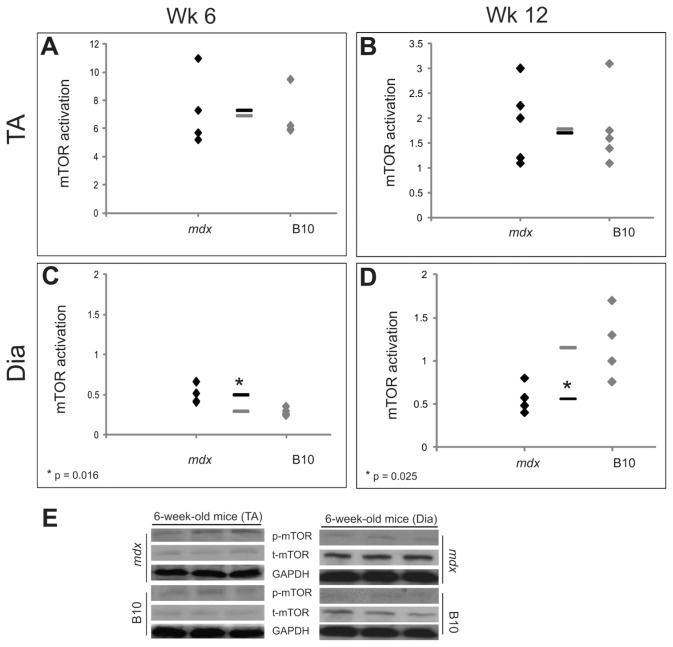
The activation of mTOR by phospho-rylation in the presence of energy, nutrients or growth factors is known to be important for muscle fiber growth (26), and RAPA can block this activation. Therefore, we examined the level of mTOR activation in TA and Dia muscles of 6-wk-old (during active necrosis) and 12-wk-old (after the peak of active necrosis) age-matched untreated mdx and wild-type B10 mice. Surprisingly, mTOR activation was not significantly different between TA muscle of untreated mdx and age-matched B10 mice at 6 and 12 wks of age (Figures 4A, B). In both mdx and B10 TA muscles the level of mTOR activation decreased between 6 and 12 wks of age.
Figure 4.
mTOR phosphorylation in muscle of 6- and 12-wk-old mdx and B10 mice. Phosphorylation of the mammalian target of rapamycin (mTOR), which is an indicator of its activation, was analyzed by Western blot of total muscle protein extracts of tibialis anterior (TA) and diaphragm (Dia) of 6- and 12-wk-old mdx mice and was compared to age-matched wild-type C57BL/10 (B10) mice. Ratios of phosphorylated mTOR (p-mTOR) to total mTOR (t-mTOR) are shown for 6-wk-old (A) and 12-wk-old (B) TA muscle and for 6-wk-old (C) and 12-wk-old (D) Dia muscle. Values presented in graphs (A–D) were calculated by measuring average blot intensity of the labeling for each tissue type. A representative image of Western blot labeling of p-mTOR and t-mTOR in TA and Dia muscles of 6-wk-old mdx and B10 mice is shown (E). GAPDH is shown as a loading control. n = 5 for B10 groups and n = 4 for mdx groups. *P < 0.05.
However, mTOR activation was significantly different between mdx and B10 mice in Dia muscle at both 6 and 12 wks of age (Figures 4C–E). At 6 wks of age, mTOR activation was lower in B10 Dia muscle than in mdx Dia muscle. mTOR activation in Dia muscles of B10 mice increased from 6 to 12 wks of age. However, mTOR activation remained at similar levels in mdx Dia muscles over the same time frame.
mTOR Activation Is Affected in Dia but Not TA Muscles of RAPA-Treated Mice
RAPA directly interacts with mTOR in skeletal muscles. Therefore, we compared activation of mTOR in skeletal muscles of mdx mice that received systemic and local RAPA treatment with age-matched untreated or blank bead–treated mdx mice. RAPA treatment led to a significantly lower level of mTOR activation in mdx Dia muscle compared with untreated age-matched mdx Dia muscle (Figure 5A). However, surprisingly, neither local nor systemic RAPA treatment resulted in a significant change in mTOR activation in TA muscles of treated mdx mice compared with untreated age-matched mdx mice (Figures 5B, C).
Figure 5.
mTOR phosphorylation following RAPA treatment. The activation of the mammalian target of rapamycin (mTOR), as determined by ratios of phosphorylated mTOR to total mTOR measured by Western blot, are shown for diaphragm (Dia) (A) and tibialis anterior (TA) (B) muscles of systemic rapamycin (RAPA)-treated (Rapa H2O) mdx mice and TA muscles of local RAPA-treated mdx mice (Rapa Bead) (C). n = 5 for RAPA-treated groups and n = 4 for untreated groups. **P < 0.01.
DISCUSSION
In the study presented here, we explored the effect of RAPA on inflammation, necrosis, regeneration and mTOR activation in dystrophic mdx mouse muscle. The effects of different immunosuppressant drugs, including cyclosporine A, prednisone and deflazacort, have been investigated in dystrophin-deficient muscles, as reviewed by Iannitti et al. (27) The study presented here, however, is the first report of RAPA treatment for dystrophin-deficient skeletal muscle of the mdx murine model for DMD, a model in which a genetic defect in dystrophin expression promotes inflammatory cell infiltration in muscle tissue.
RAPA has proven benefit for promoting a higher rate of graft survival in organ transplantation (28–30). Furthermore, RAPA has been used widely in various studies to expand or select for Foxp3+ Tregs (15,16,31–33). In contrast to the inhibitory effect of RAPA on proliferation of effector T cells, Tregs proliferate and function in the presence of RAPA (15,16,26,33). We show here that Foxp3+ cells survived in dystrophic skeletal muscle when RAPA was administered for 6 weeks to mdx mice, whereas total CD4+ and CD8+ T cells decreased significantly. The most prominent effect was observed with systemic RAPA treatment.
Because the selective proliferation of Foxp3+ Treg cells coupled with a decrease in effector T cells could lead to improvements in morphology and function of dystrophic skeletal muscle, we examined levels of necrosis and regeneration in RAPA-treated mdx Dia and TA muscle. Furthermore, based on prior data showing a beneficial effect of RAPA for organ transplantation and the known improvement in dystrophic phenotype of muscle in muscular dystrophy with other immunosuppressant drugs, we hypothesized that RAPA would decrease necrosis of dystrophic skeletal muscle. We demonstrated that RAPA administration ameliorated the dystrophic phenotype of mdx muscle. There was a significant reduction of muscle fiber necrosis in both TA and Dia muscles of the mdx mouse. The finding in the Dia is of particular importance because the level of necrosis in untreated Dia muscle is higher compared with that in untreated TA muscle (34).
The lower muscle fiber regeneration observed in RAPA-treated muscles may be a direct result of the reduction in muscle fiber necrosis, because a lower level of necrosis could diminish the need for muscle tissue regeneration. In addition, RAPA may inhibit protein synthesis in the treated muscle tissue (35). Previous work suggests, however, that when mTOR was selectively knocked out in skeletal muscles, muscle fiber regeneration persisted in affected muscles (36).
A previous study of mTOR activation in the adult mdx mouse correlated advancing age with a reduction in mTOR signaling in mdx muscle between 18 and 24 months of age (23). However, this previously reported study did not compare mdx with age-matched wild-type B10 mice. We hypothesized that mTOR activation would be altered in the active phase of degeneration in dystrophic muscle because of ongoing muscle fiber degeneration and regeneration that characterizes the pathologic process of muscular dystrophy. Furthermore, mTOR has been shown to be a crucial regulator of protein synthesis, which is required for effective muscle fiber regeneration. In this study, mTOR activation in Dia muscles of 6-week-old and 12-week-old mdx versus age-matched wild-type B10 mice was significantly different at both time points. mTOR activation was significantly higher in mdx mice at 6 weeks of age, when the dystrophic muscle fibers were undergoing a high level of regeneration. At 12 weeks of age, when the pathological processes of degeneration and regeneration had decreased in mdx dystrophic muscle tissue, however, there was a significantly lower level of mTOR activation in Dia muscles of mdx mice compared with age-matched wild-type mice. In fact, mTOR activation increased by age in Dia muscle of B10 mice, but not in mdx mice, raising the possibility that the early reduction in muscle fiber regeneration that was observed in Dia muscles of mdx mice (37) could be related to a failure of mTOR activity to increase. In contrast to Dia muscle, mTOR activation was not significantly different in the TA muscle of dystrophic and wild-type mice at either 6- or 12-weeks of age. This suggests a difference between the patterns of mTOR activation in TA versus Dia muscles of mdx mice. This is a novel finding that contributes to our understanding of the differences in function and progression of pathology between Dia and limb muscle of mdx mice (38–40).
In addition to the differences observed in untreated mdx TA and Dia muscles, the findings of this study also showed a significant difference between mdx TA and Dia muscles in response to treatment with the immunosuppressant drug RAPA. The data suggest that Dia muscle was more sensitive to the effects of RAPA treatment, supported by a more significant decrease in muscle fiber necrosis and a significant reduction in mTOR activation following RAPA treatment compared with TA muscles. Interestingly, in previous murine studies in which mTOR was knocked out in skeletal muscles, Dia muscle showed a more severe pathological response with a higher level of muscle fiber damage to the absence of mTOR compared with other muscles (36). It also has been shown in other studies that protein synthesis in rat hind-limb muscles was independent of RAPA-sensitive pathways (41), indicating that RAPA may not have the same effect in Dia and TA muscles. Our studies suggest that the decrease in necrosis and regeneration in mdx TA muscle induced by RAPA is independent of mTOR. Additional studies of molecules in the mTOR pathway may further elucidate the differences between Dia and limb muscle in the mdx mouse.
In addition to the general effect of RAPA on muscle pathology in mdx mice, we compared local versus systemic administration of RAPA on both disease pathology and mTOR activation. RAPA-containing microparticles have previously been used in altering dendritic cell behavior (24). The systemic route of RAPA administration was tested in studies of cancer (42) and aging (43). The findings of this 6-week study suggested that although there were more significant differences in lowering effector T-cell infiltration and muscle fiber necrosis when RAPA was given systemically compared with when it was given locally, the positive effects were comparable between the two administration strategies. Nonetheless, it is important to realize the limitations of each strategy as a systemic treatment affects tissues other than muscle and local treatment is more difficult to perform because it requires multiple and repeated injections. Provided the beneficial effects of the systemic and local RAPA treatments are comparable, in a disease such as DMD, in which every muscle tissue is affected, a systemic treatment may provide a more beneficial clinical outcome. However, local RAPA treatment may find greater clinical applicability in situations where preservation of individual muscles could improve quality of life of DMD patients.
In conclusion, the results of the present study demonstrating the effect of RAPA on decreasing inflammation, preserving Foxp3+ T cells and decreasing necrosis in dystrophic mdx muscle tissue could lead to the further development of treatments for DMD. In addition, the findings of novel differences of mTOR activation between TA and Dia muscles in mdx mice, both untreated and with RAPA treatment, add to the molecular understanding of the dystrophic phenotype.
ACKNOWLEDGMENTS
We thank Daniel P Reay for providing valuable technical support and for breeding mice used in these studies. This work was supported by F31-NS056780-01A2 (S Eghtesad) from the National Institutes of Health (NIH), KL2 RR024154 (SR Little) from the National Center for Research Resources (NCRR), a component of the NIH and grant W81XWH-05-1-0334 (PR Clemens) from the United States Army Medical Research and Materiel Command. The myosin (embryonic) monoclonal antibody developed by HM Blau was obtained from the Developmental Studies Hybridoma Bank developed under the auspices of the NICHD and maintained by The University of Iowa, Department of Biology, Iowa City, IA, USA.
Footnotes
Online address: http://www.molmed.org
DISCLOSURE
The authors take full responsibility for the contents of this paper, which do not represent the views of NCCR, the NIH, the Department of Veterans Affairs or the United States Government.
REFERENCES
- 1.Drousiotou A, et al. Neonatal screening for Duchenne muscular dystrophy: a novel semi-quantitative application of the bioluminescence test for creatine kinase in a pilot national program in Cyprus. Genet Test. 1998;2:55–60. doi: 10.1089/gte.1998.2.55. [DOI] [PubMed] [Google Scholar]
- 2.Emery AE. Population frequencies of inherited neuromuscular diseases—a world survey. Neuromuscul Disord. 1991;1:19–29. doi: 10.1016/0960-8966(91)90039-u. [DOI] [PubMed] [Google Scholar]
- 3.Parsons EP, Bradley DM, Clarke AJ. Newborn screening for Duchenne muscular dystrophy. Arch Dis Child. 2003;88:91–2. doi: 10.1136/adc.88.1.91-b. [DOI] [PMC free article] [PubMed] [Google Scholar]
- 4.Emery AE. Dystrophin function. Lancet. 1990;335:1289. doi: 10.1016/0140-6736(90)91364-g. [DOI] [PubMed] [Google Scholar]
- 5.Hoffman EP, Brown RH, Jr, Kunkel LM. Dystrophin: the protein product of the Duchenne muscular dystrophy locus. Cell. 1987;51:919–28. doi: 10.1016/0092-8674(87)90579-4. [DOI] [PubMed] [Google Scholar]
- 6.Hoffman EP, Schwartz L. Dystrophin and disease. Mol Aspects Med. 1991;12:175–94. doi: 10.1016/0098-2997(91)90001-3. [DOI] [PubMed] [Google Scholar]
- 7.Bulfield G, Siller W, Wight P, Moore K. X Chromosome-linked muscular dystrophy (mdx) in the mouse. Proc Natl Acad Sci. 1984;81:1189–92. doi: 10.1073/pnas.81.4.1189. [DOI] [PMC free article] [PubMed] [Google Scholar]
- 8.Farini A, et al. T and B lymphocyte depletion has a marked effect on the fibrosis of dystrophic skeletal muscles in the scid/mdx mouse. J Pathol. 2007;213:229–38. doi: 10.1002/path.2213. [DOI] [PubMed] [Google Scholar]
- 9.Spencer MJ, Montecino-Rodriguez E, Dorshkind K, Tidball JG. Helper (CD4(+)) and cytotoxic (CD8(+)) T cells promote the pathology of dystrophin-deficient muscle. Clin Immunol. 2001;98:235–43. doi: 10.1006/clim.2000.4966. [DOI] [PubMed] [Google Scholar]
- 10.Burleson RL, Marbarger PD, Jermanovich N, Brennan AM, Scruggs BF. A prospective study of methylprednisolone and prednisone as immunosuppressive agents in clinical renal transplantation. Transplant Proc. 1981;13:339–43. [PubMed] [Google Scholar]
- 11.Liu Y, Chen Y, Liu FQ, Lamb JR, Tam PK. Combined treatment with triptolide and rapamycin prolongs graft survival in a mouse model of cardiac transplantation. Transpl Int. 2008;21:483–94. doi: 10.1111/j.1432-2277.2007.00630.x. [DOI] [PubMed] [Google Scholar]
- 12.Vilquin JT, et al. Successful myoblast allo-transplantation in mdx mice using rapamycin. Transplantation. 1995;59:422–6. [PubMed] [Google Scholar]
- 13.Thomson AW, Turnquist HR, Raimondi G. Immunoregulatory functions of mTOR inhibition. Nat Rev Immunol. 2009;9:324–37. doi: 10.1038/nri2546. [DOI] [PMC free article] [PubMed] [Google Scholar]
- 14.Libetta C, et al. The effect of sirolimus- or cyclosporine-based immunosuppression effects on T-cell subsets in vivo. Kidney Int. 2007;72:114–20. doi: 10.1038/sj.ki.5002255. [DOI] [PubMed] [Google Scholar]
- 15.Battaglia M, et al. Rapamycin promotes expansion of functional CD4+CD25+FOXP3+ regulatory T cells of both healthy subjects and type 1 diabetic patients. J Immunol. 2006;177:8338–47. doi: 10.4049/jimmunol.177.12.8338. [DOI] [PubMed] [Google Scholar]
- 16.Battaglia M, Stabilini A, Roncarolo MG. Rapamycin selectively expands CD4+CD25+ FoxP3+ regulatory T cells. Blood. 2005;105:4743–8. doi: 10.1182/blood-2004-10-3932. [DOI] [PubMed] [Google Scholar]
- 17.Hedhli N, Pelat M, Depre C. Protein turnover in cardiac cell growth and survival. Cardiovasc Res. 2005;68:186–96. doi: 10.1016/j.cardiores.2005.06.025. [DOI] [PubMed] [Google Scholar]
- 18.Ikenoue T, Hong S, Inoki K. Monitoring mammalian target of rapamycin (mTOR) activity. Methods Enzymol. 2009;452:165–80. doi: 10.1016/S0076-6879(08)03611-2. [DOI] [PubMed] [Google Scholar]
- 19.Yang X, et al. The mammalian target of rapamycin-signaling pathway in regulating metabolism and growth. J Anim Sci. 2008;86:E36–50. doi: 10.2527/jas.2007-0567. [DOI] [PubMed] [Google Scholar]
- 20.Zhou H, Huang S. The complexes of mammalian target of rapamycin. Curr Protein Pept Sci. 2010;11:409–24. doi: 10.2174/138920310791824093. [DOI] [PMC free article] [PubMed] [Google Scholar]
- 21.Bodine SC, et al. Akt/mTOR pathway is a crucial regulator of skeletal muscle hypertrophy and can prevent muscle atrophy in vivo. Nat Cell Biol. 2001;3:1014–9. doi: 10.1038/ncb1101-1014. [DOI] [PubMed] [Google Scholar]
- 22.Rommel C, et al. Mediation of IGF-1-induced skeletal myotube hypertrophy by PI(3)K/Akt/mTOR and PI(3)K/Akt/GSK3 pathways. Nat Cell Biol. 2001;3:1009–13. doi: 10.1038/ncb1101-1009. [DOI] [PubMed] [Google Scholar]
- 23.Mouisel E, Vignaud A, Hourde C, Butler-Browne G, Ferry A. Muscle weakness and atrophy are associated with decreased regenerative capacity and changes in mTOR signaling in skeletal muscles of venerable (18–24-month-old) dystrophic mdx mice. Muscle Nerve. 2010;41:809–18. doi: 10.1002/mus.21624. [DOI] [PubMed] [Google Scholar]
- 24.Jhunjhunwala S, Raimondi G, Thomson AW, Little SR. Delivery of rapamycin to dendritic cells using degradable microparticles. J Control Release. 2009;133:191–7. doi: 10.1016/j.jconrel.2008.10.011. [DOI] [PMC free article] [PubMed] [Google Scholar]
- 25.Spencer MJ, Tidball JG. Do immune cells promote the pathology of dystrophin-deficient myopathies. Neuromuscul Disord. 2001;11:556–64. doi: 10.1016/s0960-8966(01)00198-5. [DOI] [PubMed] [Google Scholar]
- 26.Lange CM, et al. Increased frequency of regulatory T cells and selection of highly potent CD62L+ cells during treatment of human lung transplant recipients with rapamycin. Transpl Int. 2010;23:266–76. doi: 10.1111/j.1432-2277.2009.00973.x. [DOI] [PubMed] [Google Scholar]
- 27.Iannitti T, Capone S, Feder D, Palmieri B. Clinical use of immunosuppressants in Duchenne muscular dystrophy. J Clin Neuromuscul Dis. 2010;12:1–21. doi: 10.1097/CND.0b013e3181d4a4f9. [DOI] [PubMed] [Google Scholar]
- 28.Charfi S, et al. Successful treatment of post-renal transplant gastric and pulmonary Kaposi’s sarcoma with conversion to rapamycin treatment. Saudi J Kidney Dis Transpl. 2007;18:617–20. [PubMed] [Google Scholar]
- 29.Iglesias J, et al. Rapamycin-based rescue therapy after chronic rejection in a pediatric liver transplant patient. Transpl Int. 2003;16:765–7. doi: 10.1007/s00147-003-0608-0. [DOI] [PubMed] [Google Scholar]
- 30.Kerkar N, et al. Rapamycin successfully treats post-transplant autoimmune hepatitis. Am J Transplant. 2005;5:1085–9. doi: 10.1111/j.1600-6143.2005.00801.x. [DOI] [PubMed] [Google Scholar]
- 31.Haxhinasto S, Mathis D, Benoist C. The AKT-mTOR axis regulates de novo differentiation of CD4+Foxp3+ cells. J Exp Med. 2008;205:565–74. doi: 10.1084/jem.20071477. [DOI] [PMC free article] [PubMed] [Google Scholar]
- 32.Sauer S, et al. T cell receptor signaling controls Foxp3 expression via PI3K, Akt, and mTOR. Proc Natl Acad Sci U S A. 2008;105:7797–802. doi: 10.1073/pnas.0800928105. [DOI] [PMC free article] [PubMed] [Google Scholar]
- 33.Strauss L, et al. Selective Survival of Naturally Occurring Human CD4+CD25+Foxp3+ Regulatory T Cells Cultured with Rapamycin. J Immunol. 2007;178:320–9. doi: 10.4049/jimmunol.178.1.320. [DOI] [PubMed] [Google Scholar]
- 34.Stedman HH, et al. The mdx mouse diaphragm reproduces the degenerative changes of Duchenne muscular dystrophy. Nature. 1991;352:536–9. doi: 10.1038/352536a0. [DOI] [PubMed] [Google Scholar]
- 35.Drummond MJ, et al. Rapamycin administration in humans blocks the contraction-induced increase in skeletal muscle protein synthesis. J Physiol. 2009;587:1535–46. doi: 10.1113/jphysiol.2008.163816. [DOI] [PMC free article] [PubMed] [Google Scholar]
- 36.Risson V, et al. Muscle inactivation of mTOR causes metabolic and dystrophin defects leading to severe myopathy. J Cell Biol. 2009;187:859–74. doi: 10.1083/jcb.200903131. [DOI] [PMC free article] [PubMed] [Google Scholar]
- 37.Wu B, et al. Dose-dependent restoration of dystrophin expression in cardiac muscle of dystrophic mice by systemically delivered morpholino. Gene Ther. 2010;17:132–40. doi: 10.1038/gt.2009.120. [DOI] [PubMed] [Google Scholar]
- 38.Esser K. Regulation of mTOR signaling in skeletal muscle hypertrophy. J Musculoskelet Neuronal Interact. 2008;8:338–9. [PubMed] [Google Scholar]
- 39.Ge Y, et al. mTOR regulates skeletal muscle regeneration in vivo through kinase- dependent and kinase-independent mechanisms. Am J Physiol Cell Physiol. 2009;297:C1434–44. doi: 10.1152/ajpcell.00248.2009. [DOI] [PMC free article] [PubMed] [Google Scholar]
- 40.Pende M. mTOR, Akt, S6 kinases and the control of skeletal muscle growth. Bull Cancer. 2006;93:E39–43. [PubMed] [Google Scholar]
- 41.Fluckey JD, et al. Insulin facilitation of muscle protein synthesis following resistance exercise in hindlimb-suspended rats is independent of a rapamycin-sensitive pathway. Am J Physiol Endocrinol Metab. 2004;287:E1070–5. doi: 10.1152/ajpendo.00329.2004. [DOI] [PubMed] [Google Scholar]
- 42.Robinson J, et al. Oral rapamycin reduces tumour burden and vascularization in Lkb1(+/−) mice. J Pathol. 2009;219:35–40. doi: 10.1002/path.2562. [DOI] [PubMed] [Google Scholar]
- 43.Harrison DE, et al. Rapamycin fed late in life extends lifespan in genetically heterogeneous mice. Nature. 2009;460:392–5. doi: 10.1038/nature08221. [DOI] [PMC free article] [PubMed] [Google Scholar]


