Abstract
Background:
Breast cancer risk for postmenopausal women is positively associated with circulating concentrations of oestrogens and androgens, but the determinants of these hormones are not well understood.
Methods:
Cross-sectional analyses of breast cancer risk factors and circulating hormone concentrations in more than 6000 postmenopausal women controls in 13 prospective studies.
Results:
Concentrations of all hormones were lower in older than younger women, with the largest difference for dehydroepiandrosterone sulphate (DHEAS), whereas sex hormone-binding globulin (SHBG) was higher in the older women. Androgens were lower in women with bilateral ovariectomy than in naturally postmenopausal women, with the largest difference for free testosterone. All hormones were higher in obese than lean women, with the largest difference for free oestradiol, whereas SHBG was lower in obese women. Smokers of 15+ cigarettes per day had higher levels of all hormones than non-smokers, with the largest difference for testosterone. Drinkers of 20+ g alcohol per day had higher levels of all hormones, but lower SHBG, than non-drinkers, with the largest difference for DHEAS. Hormone concentrations were not strongly related to age at menarche, parity, age at first full-term pregnancy or family history of breast cancer.
Conclusion:
Sex hormone concentrations were strongly associated with several established or suspected risk factors for breast cancer, and may mediate the effects of these factors on breast cancer risk.
Keywords: breast cancer, hormones, oestrogens, androgens, sex hormone-binding globulin
Breast cancer risk for postmenopausal women is positively associated with the circulating concentrations of oestrogens and androgens (Endogenous Hormones and Breast Cancer Collaborative Group, 2002; Missmer et al, 2004; Kaaks et al, 2005). The increase in risk of postmenopausal breast cancer associated with obesity may be largely explained by the relatively high circulating concentrations of free oestradiol in obese postmenopausal women (Endogenous Hormones and Breast Cancer Collaborative Group, 2003a; Rinaldi et al, 2006), but it is unclear to what extent the effects of other risk factors may be mediated by their associations with circulating sex hormones. A number of previous studies have examined the associations of circulating sex hormones with known and possible breast cancer risk factors, such as age at menarche, parity, age at first full-term pregnancy, type of menopause, smoking and alcohol, but they have not been large enough to provide definitive results (see Discussion). The Endogenous Hormones and Breast Cancer Collaborative Group was established to conduct collaborative analyses of individual data from prospective studies of endogenous hormones and breast cancer. We report here the cross-sectional associations of circulating concentrations of sex hormones and sex hormone-binding globulin (SHBG) with 12 demographic, reproductive and lifestyle factors.
Materials and methods
Data collection
Published studies were eligible for the collaborative reanalysis if they included data on endogenous hormones and breast cancer risk using prospectively collected blood samples from postmenopausal women, as described previously (Endogenous Hormones and Breast Cancer Collaborative Group, 2002). Studies were identified by computer-aided literature searches, within relevant review articles, and through our discussions with colleagues. Studies were eligible for inclusion if they had published peer-reviewed data on endogenous hormone concentrations and breast cancer risk using prospectively collected blood samples from postmenopausal women. The data reported here are for the standardised database as available for analysis by the collaborative group in late 2010. The studies included were: CLUE I, Washington County, MD, USA (Wysowski et al, 1987; Gordon et al, 1990; Helzlsouer et al, 1994); Columbia, MO, USA (Dorgan et al, 1996, 1997); European Prospective Investigation into Cancer and Nutrition (EPIC), Europe (Kaaks et al, 2005); Guernsey, UK (Thomas et al, 1997); Malmö/Umeå, Sweden (Manjer et al, 2003); the Melbourne Collaborative Cohort Study (MCCS), Australia (Baglietto et al, 2009); Nurses’ Health Study, USA (Hankinson et al, 1998); New York University Women's Health Study (NYU WHS), USA (Toniolo et al, 1995; Zeleniuch-Jacquotte et al, 1997, 2004); Study of Hormones and Diet in the Etiology of Breast Tumors (ORDET), Italy (Berrino et al, 1996); Rancho Bernardo, USA (Barrett-Connor et al, 1990; Garland et al, 1992); Radiation Effects Research Foundation (RERF), Japan (Kabuto et al, 2000); Study of Osteoporotic Fractures (SOF), USA (Cauley et al, 1999); and the Women's Health Initiative, Observational Study (WHI-OS), USA (Gunter et al, 2009). Details of the recruitment of participants, informed consent and definitions of reproductive variables are in the original publications. Height and weight were measured in eight studies (EPIC (most centres), Guernsey, Malmö/Umeå, MCCS, ORDET, Rancho Bernardo, SOF and WHI-OS) and were self-reported in four studies (Columbia, Nurses’ Health Study, NYU WHS and RERF). With the exception of the RERF Study in Japan, the majority of the women in these studies were of white European ethnic origin.
Collaborators were asked to provide data on concentrations of the hormones oestradiol (total), free oestradiol, oestrone, androstenedione, dehydroepiandrosterone sulphate (DHEAS), testosterone, free testosterone and SHBG, where available. Details of the assays and median hormone concentrations for the data included in our analyses are in Table 1. Data from CLUE I were analysed as two substudies because of changes in the assay method for oestradiol, oestrone and androstenedione between the first phase and the second phase. Data for oestradiol from the Malmö/Umeå Study were not included because over 50% of recorded values were below the detection limit of the assay. The majority of studies measured hormone concentrations in serum, whereas some (Malmö/Umeå, MCCS, Nurses’ Health Study and Rancho Bernardo) used plasma. Circulating concentrations of free oestradiol and free testosterone were calculated from the concentrations of oestradiol and testosterone, respectively, and SHBG, with albumin assumed to be constant (40 g l−1), according to the law of mass action (Sodergard et al, 1982; Endogenous Hormones and Breast Cancer Collaborative Group, 2003b); in the subset of studies that had measured free oestradiol and free testosterone, the correlations of these values with the calculated values were 0.98 and 0.77, respectively, and using calculated values for all participants increased the sample size for these analyses, while standardizing the estimation approach.
Table 1. Assay methods and median hormone and SHBG concentrations.
|
Oestradiol
|
Oestrone
|
Androstenedione
|
DHEAS
|
Testosterone
|
SHBG
|
|||||||||||||
|---|---|---|---|---|---|---|---|---|---|---|---|---|---|---|---|---|---|---|
| Study, country | Method | CV, % | Median (dl), pmol l−1 | Method | CV, % | Median (dl), pmol l−1 | Method | CV, % | Median (dl), nmol l−1 | Method | CV, % | Median (dl), nmol l−1 | Method | CV, % | Median (dl), nmol l−1 | Method | CV, % | Median (dl), nmol l−1 |
| CLUE I, USA phase 1 | CP RIA | — | 161.5 | CP RIA | — | 183 | CP RIA | 5.48 | CP RIA | 0.81 | ||||||||
| CLUE I, USA phase 2 | E RIA | 7.1 | 58.7 | E RIA | 9.9 | 141 | E RIA | 10.0 | 2.74 | NE RIA | 2.4 | 1815 | IA | 10.9 | 55.8 | |||
| Columbia, MO, USA | NE RIA | 23.7 | 51.4 | NE RIA | 5.0 | 129 | NE RIA | 1.3 | 3.11 | NE RIA | 0.7 | 2204 | NE RIA | 5.8 | 0.59 | IRMA | 1.6 | 53.4 |
| EPIC, Europe | NE RIA | 5.8 | 89.1 | NE RIA | 10.2 | 145 | NE RIA | 4.8 | 3.04 | NE RIA | 7.0 | 2034 | NE RIA | 10.8 | 1.21 | IRMA | 8.0 | 34.5 |
| Guernsey, UK | E RIA | 15.1 | 35.0 (3.0) | NE RIA | 9 | 2041 | NE RIA | 4.5 | 0.91 (0.35) | IRMA | 4.1 | 63.0 | ||||||
| Malmö/Umeå, Sweden | NE RIA | 20.6 | 74 (4.44) | E RIA | ⩽15 | 2.33 (0.14) | NE RIA | ⩽15 | 2730 (200) | E RIA | ⩽15 | 1.22 (0.1) | IMF | ⩽15 | 55.4 | |||
| MCCS, Australia | NE ECIA | 10 | 57.0 (18) | NE RIA | 15 | 2.15 (0.02) | NE CIA | 10 | 1500 (200) | NE ECIA | 7 | 0.70 (0.1) | IRMA | 7 | 50.7(2) | |||
| Nurses’ Health Study, USA | E RIA | 11.0 | 22.0 (7.3) | E RIA | 12.1 | 89 (37.0) | E RIA | 12.1 | 1.99 (0.10) | NE RIA | 9.0 | 2313 (136) | E RIA | 12.3 | 0.76 (0.035) | IA | 15.5 | 47.7 |
| NYU WHS, USA | NE RIA | 13.2 | 81.8 | NE RIA | 14.4 | 99 | NE RIA | 13.5 | 2.46 | NE RIA | 14.7 | 2144 | NE RIA | 15.8 | 0.79 | IRMA | 13.5 | 48.1 |
| ORDET, Italy | E RIA | 9.6 | 21.7 | NE RIA | 3.7 | 2004 | NE RIA | 7.3 | 1.17 | IRMA | 4.4 | 41.5 | ||||||
| Rancho Bernardo, USA | E RIA | 7.1 | 40.4 (11) | E RIA | 7.7 | 113 (11) | E RIA | 4.3 | 1.92 (0.06) | NE RIA | 10 | 1755 (52) | E RIA | 4.9 | 0.75 (0.07) | Binding assay | 11.4 | 29.0 |
| RERF, Japan | NE RIA | 9.8 | 65.2 | NE RIA | — | 952 | RIA | 5.3 | 67.3 | |||||||||
| SOF, USA | E RIA | 11 | 22.0 (7.3) | E RIA | 17 | 74 (37.0) | E RIA | 16 | 1.29 (0.1) | NE RIA | 12 | 1742 (170) | CP RIA | 13 | 0.62 (0.03) | RIA | 7.8 | 42.3 (5) |
| WHI-OS, USA | NE IA | 5.9 | 40.4 (18.4) | |||||||||||||||
Abbreviations: CIA=competitive immunoassay; CLUE=Washington County, MD Study ‘Give us a clue to cancer and heart disease’ CP=chromatographic purification; CV=coefficient of variation: total if reported, otherwise between assay, otherwise within assay; dl=detection limit; E=extraction step; ECIA=electrochemiluminescence immunoassay; EPIC=European Prospective Investigation into Cancer and Nutrition; IA=immunoassay; IMF=immunofluorometry; IRMA=immunoradiometric assay; MCCS=Melbourne Collaborative Cohort Study; NE=no extraction step; NYU WHS=New York University Women's Health Study; ORDET=Study of Hormones and Diet in the Etiology of Breast Tumors; RIA=radioimmunoassay; RERF=Radiation Effects Research Foundation; SHBG=sex hormone-binding globulin; SOF=Study of Osteoporotic Fractures; WHI-OS=Women's Health Initiative, Observational Study.
Collaborators also provided data on reproductive and anthropometric factors for each woman in their study. Women who were using hormone replacement therapy or other exogenous sex hormones at the time of blood collection were excluded. All studies that contributed data to the analysis were cohorts in which blood samples were collected from women who were then followed to identify those subjects who developed breast cancer. The analyses in this paper use data only from the ‘control’ women who had not developed breast cancer during the follow-up period in each contributing study.
Statistical analysis
Hormone and SHBG concentrations were logarithmically transformed to normalise the distributions. A few hormone and SHBG values recorded as zero were reset to a suitably small non-zero value to allow logarithmic transformation: 1 pmol l−1, 0.01, 0.01 and 1 nmol l−1 for oestradiol (1 value), androstenedione (3 values), testosterone (16 values) and SHBG (2 values), respectively.
Partial correlations between the hormones and SHBG were computed using study-specific standardised values: (xjk-mj)/sj, where mj and sj denote the mean and standard deviation of the log-transformed hormone concentrations in study j and xjk is an observation from that study. These standardised values were adjusted for age at blood collection (continuous), type of menopause (natural, hysterectomy, bilateral ovariectomy) and body mass index (BMI; continuous) for women with known values for these variables. The use of standardisation effectively adjusts for the differences in mean hormone concentration between studies.
Geometric mean hormone and SHBG concentrations by categories of various factors, together with their 95% confidence intervals, were calculated using the predicted values from analysis of variance models scaled to the overall geometric mean concentration. The factors examined were age (<55, 55–59, 60–64, 65–69 and 70+ years), type of menopause (natural, hysterectomy, bilateral ovariectomy, other or unknown), BMI (calculated as weight in kilograms divided by the square of height in metres and categorised as <22.5, 22.5–24.9, 25.0–27.4, 27.5–29.9 and 30.0+ kg m−2), smoking (never, previous, current <15 cigarettes day and current 15+ cigarettes per day), alcohol (none, <10 g per day, 10–19 g per day and 20+ g per day (a 4 oz glass of wine typically contains 12 g of alcohol)), age at menarche (<12, 12–13 and 14+ years), number of full-term pregnancies (none, 1, 2, 3 and 4+), age at first full-term pregnancy (<25 and 25+ years), years since menopause (0–4, 5–14 and 15+ years), family history of breast cancer (no and yes), previous use of hormonal contraceptives (no and yes) and previous use of hormone therapy (no and yes).
The geometric mean hormone concentrations were adjusted for study, age at blood collection, type of menopause and BMI, as appropriate (categories as above, with an extra ‘unknown’ category for BMI, which was unknown for 4% of women with values for oestradiol). Heterogeneity between categories was assessed by F tests arising from the analysis of variance. Trends in hormone concentrations across categories of all quantitative variables except alcohol were tested by scoring the categories from 1 to the maximum number of categories and treating them as continuous variables in the analysis of variance. Alcohol was treated differently because of the highly skewed distribution of intakes, and the alcohol consumption categories were scored 0, 3, 12 and 30, respectively, in the tests for linear trend in accordance with the median daily alcohol intake in grams in those categories. Values for BMI, smoking, alcohol consumption and most of the other factors investigated were unknown for some women, and these individuals were excluded from the analyses of hormones in relation to these factors. Tests of heterogeneity between studies in the associations of hormones with other factors were obtained by adding a study-by-factor interaction term to the analysis of variance and using the F-test to assess its significance. For oestradiol (and free oestradiol), oestrone, androstenedione and testosterone (and free testosterone), the results were examined according to whether or not the hormone assay had included a purification step (usually by organic solvent extraction and column chromatography). CLUE I; Nurses’ Health Study; Rancho Bernardo; and SOF used purification steps in the assays for all of these analytes, and Columbia, MO; EPIC; NYU WHS; RERF; and WHI used all direct assays, whereas Guernsey and ORDET used extraction for oestradiol and direct assays for testosterone, and Malmö/Umeå used direct assays for oestrone and extraction for androstenedione and testosterone. All statistical tests were two-sided, and statistical significance was taken as P<0.05. All analyses were performed using Stata Statistical Software release 10 (Stata Corp., College Station, TX, USA).
Results
Collaborating studies and participant characteristics
A total of 13 studies contributed data, 7 in the United States of America, 1 each in Australia, Italy, Japan, Sweden and the United Kingdom, and the multi-centre European study EPIC (Table 2). In all, 6291 women contributed data to the analyses. Mean age at blood sampling ranged from 58.5 years in the Guernsey Study up to 71.7 years in SOF, with an overall mean of 61.7 years. Body mass index was available for all except one study (CLUE I), with an overall mean of 26.4 kg m−2. Type of menopause was available for all except one study (RERF), with most women in the other studies having had a natural (non-surgical) menopause. Smoking data were available for all studies and the proportion of current smokers ranged from 7.2% in SOF to 26.0% in Malmö/Umeå, with an overall proportion of 13.2%. Alcohol consumption data were available for eight studies and the proportion of current alcohol consumers ranged from 37.8% in NYU WHS to 70.0% in WHI-OS, with an overall proportion of 54.5%.
Table 2. Participant characteristics.
| Study, country | Number of womena | Mean age, years | Age range, years | Mean BMI, kg m−2 | Natural menopause, % | Current smokers, % | Alcohol consumers, % |
|---|---|---|---|---|---|---|---|
| CLUE I, USA | 194 | 61.4 | 38–96 | NA | 100.0 | 22.4 | NA |
| Columbia, MO, USA | 133 | 61.8 | 50–76 | 26.6 | 72.2 | 12.8 | NA |
| EPIC, Europeb | 1304 | 60.1 | 44–76 | 26.8 | 82.5 | 14.7 | 66.3 |
| Guernsey, UK | 177 | 58.5 | 45–79 | 25.6 | 94.9 | 18.8 | NA |
| Malmö/Umeå, Sweden | 436 | 60.0 | 46–73 | 26.0 | 96.1 | 26.0 | 69.0 |
| MCCS, Australia | 1146 | 60.4 | 40–70 | 27.4 | 79.4 | 8.5 | 40.8 |
| Nurses’ Health Study, USA | 641 | 61.4 | 46–69 | 26.2 | 74.8 | 12.5 | 58.1 |
| NYU WHS, USA | 563 | 58.9 | 44–65 | 25.4 | 76.7 | 8.1 | 37.8 |
| ORDET, Italy | 245 | 58.1 | 47–69 | 26.6 | 78.4 | 10.2 | 56.3 |
| Rancho Bernardo, USA | 582 | 66.7 | 50–79 | 24.6 | 72.5 | 22.6 | NA |
| RERF, Japan | 56 | 59.3 | 41–77 | 22.1 | NA | 16.7 | NA |
| SOF, USA | 378 | 71.7 | 65–87 | 26.6 | 86.2 | 7.2 | 41.0 |
| WHI-OS, USA | 436 | 64.5 | 50–80 | 27.7 | 65.1 | 7.4 | 70.0 |
| All studies | 6291 | 61.7 | 38–96 | 26.4 | 80.1 | 13.2 | 54.5 |
Abbreviations: BMI, body mass index; CLUE=Washington County, MD Study ‘Give us a clue to cancer and heart disease’ DHEAS=dehydroepiandrosterone sulphate; EPIC=European Prospective Investigation into Cancer and Nutrition; MCCS=Melbourne Collaborative Cohort Study; NA=not available; NYU WHS=New York University Women's Health Study; ORDET=Study of Hormones and Diet in the Etiology of Breast Tumors; RERF=Radiation Effects Research Foundation; SHBG=sex hormone-binding globulin; SOF=Study of Osteoporotic Fractures; WHI-OS=Women's Health Initiative, Observational Study.
Number of women with one or more sex hormone measurements. The numbers of women with measurements of oestradiol, free oestradiol, oestrone, androstenedione, DHEAS, testosterone, free testosterone and SHBG were 5614, 5003, 3848, 5181, 5389, 5666, 5494 and 5678, respectively.
France, Germany, Greece, Italy, The Netherlands, Spain and the United Kingdom.
Correlations between hormones
Concentrations of all the sex hormones were positively correlated with each other (Table 3); partial correlation coefficients ranged between 0.29 and 0.93. Free oestradiol was strongly correlated with total oestradiol (r=0.93) and inversely correlated with SHBG (r=−0.42). Free testosterone was strongly correlated with total testosterone (r=0.89) and inversely correlated with SHBG (r=−0.41). Sex hormone-binding globulin was weakly inversely correlated with each of the remaining sex hormones, except for testosterone. Similar correlations between hormones and SHBG were observed for studies that had used assays with a purification step for oestradiol, oestrone, androstenedione and testosterone and for studies that had used direct assays for these hormones (results not shown).
Table 3. Correlations between hormones and SHBG.
| Free oestradiol | Oestrone | Androstenedione | DHEAS | Testosterone | Free testosterone | SHBG | |
|---|---|---|---|---|---|---|---|
| Oestradiol | 0.93 | 0.50 | 0.29 | 0.29 | 0.35 | 0.35 | −0.08 |
| Free oestradiol | 0.50 | 0.29 | 0.31 | 0.32 | 0.47 | −0.42 | |
| Oestrone | 0.44 | 0.45 | 0.41 | 0.42 | −0.14 | ||
| Androstenedione | 0.60 | 0.59 | 0.56 | −0.08 | |||
| DHEAS | 0.56 | 0.56 | −0.11 | ||||
| Testosterone | 0.89 | 0.02 | |||||
| Free testosterone | −0.41 |
Abbreviations: DHEAS=dehydroepiandrosterone sulphate; SHBG=sex hormone-binding globulin.
Partial correlation coefficients between standardized log-transformed hormone and SHBG concentrations, adjusted for age at blood collection, type of menopause and body mass index.
The number of pairs of observations for each partial correlation is between 2945 and 5081.
All values are statistically significant at P<0.0001, except for the partial correlation between testosterone and SHBG.
Age
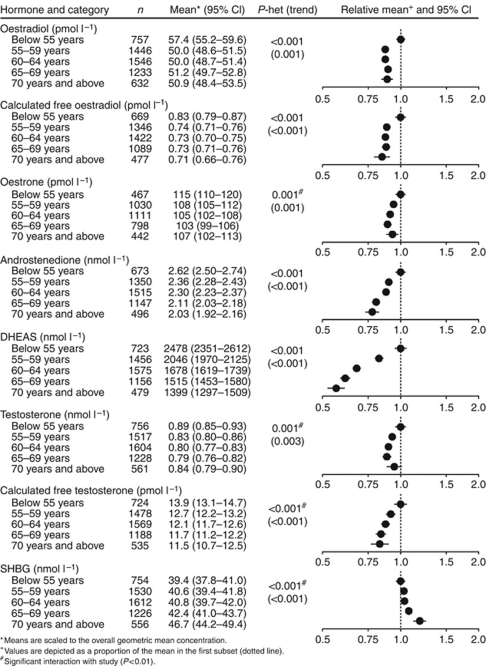
All the hormones and SHBG were significantly associated with age but not always in a linear pattern (Figure 1). Concentrations of oestradiol, oestrone and testosterone were highest in the youngest age group (below 55 years), after which they declined and did not vary much thereafter. Androstenedione and DHEAS were 23% and 44% lower, respectively, in women aged 70 years and above than in women aged below 55 years. Sex hormone-binding globulin was 21% higher in women aged 70 years and above than in women aged below 55 years. Calculated free oestradiol and calculated free testosterone were 16% and 18% lower, respectively, in women aged 70 years and above than in women aged below 55 years. There was significant heterogeneity (P<0.01) between studies in the associations of oestradiol, oestrone, testosterone, free testosterone and SHBG with age; for these hormones (but not for SHBG), there was no significant association with age from studies that had used assays with a purification step, but there were significant inverse associations with age from studies that had used direct assays (results not shown).
Figure 1.
Geometric mean concentration of selected hormones in postmenopausal women by categories of age at blood collection, adjusted for study, type of menopause and BMI.
Women who had a natural menopause within 5 years of blood collection had significantly higher circulating concentrations of oestradiol and free oestradiol than women who had a natural menopause at least 5 years previously (Table 4). Excluding women who had undergone menopause <5 years before blood collection, and women aged below 60 years at blood collection who had an unknown age at menopause, reduced but did not eliminate the difference in oestradiol, free oestradiol and oestrone concentrations between women aged <55 and older women, but had little impact on the associations of age with the other hormones and SHBG (results not shown).
Table 4. Relationships of circulating concentrations of sex hormones and SHBG in postmenopausal women with reproductive and other factors. Values are geometric means and 95% confidence intervals adjusted for study, age at blood collection, type of menopause and body mass index and scaled to the overall geometric mean concentration.
| Factor and subset | Oestradiol (pmol l−1) | Calculated free oestradiol (pmol l−1) | Oestrone (pmol l−1) | Androstenedione (nmol l−1) | DHEAS (nmol l−1) | Testosterone (nmol l−1) | Calculated free T (pmol l−1) | SHBG (nmol l−1) |
|---|---|---|---|---|---|---|---|---|
| Age at menarche (years) | ||||||||
| Below 12 | 53.6 (51.6–55.6) | 0.77 (0.74–0.81) | 109 (105–114) | 2.26 (2.16–2.37) | 1816 (1722–1914) | 0.82 (0.78–0.86) | 12.3 (11.7–13.0) | 41.5 (40.1–43.0) |
| 12–13 | 51.2 (50.2–52.3) | 0.74 (0.72–0.75) | 105 (102–107) | 2.30 (2.24–2.36) | 1795 (1743–1848) | 0.82 (0.80–0.84) | 12.3 (11.9–12.7) | 41.9 (41.1–42.7) |
| 14 and above | 50.5 (49.3–51.7) | 0.74 (0.72–0.76) | 109 (106–112) | 2.27 (2.20–2.33) | 1790 (1733–1849) | 0.82 (0.80–0.84) | 12.4 (12.0–12.8) | 40.8 (39.9–41.7) |
| P for heterogeneity (trend) | 0.033 (0.015) | 0.114 (0.108) | 0.055 (0.664) | 0.708 (0.839) | 0.903 (0.689) | 0.977 (0.986) | 0.883 (0.684) | 0.195 (0.187) |
| Number of full-term pregnancies | ||||||||
| None | 51.8 (49.8–53.8) | 0.76 (0.72–0.79) | 110 (105–115) | 2.27 (2.16–2.38) | 1702 (1610–1798) | 0.82 (0.78–0.86) | 12.5 (11.8–13.2) | 40.9 (39.4–42.4) |
| One | 53.0 (50.8–55.3) | 0.76 (0.73–0.80) | 108 (103–112) | 2.32 (2.21–2.44) | 1797 (1699–1902) | 0.84 (0.80–0.89) | 12.8 (12.1–13.5) | 40.3 (38.8–41.9) |
| Two | 51.6 (50.2–53.0) | 0.74 (0.72–0.77) | 107 (104–110) | 2.32 (2.25–2.39) | 1864 (1799–1932) | 0.82 (0.79–0.85) | 12.3 (11.9–12.8) | 41.7 (40.7–42.7) |
| Three | 50.6 (49.1–52.2) | 0.74 (0.71–0.76) | 106 (103–110) | 2.28 (2.20–2.36) | 1841 (1766–1921) | 0.83 (0.80–0.86) | 12.5 (12.0–13.1) | 41.4 (40.2–42.6) |
| Four or more | 50.4 (48.9–52.1) | 0.73 (0.70–0.75) | 104 (100–108) | 2.22 (2.13–2.30) | 1704 (1627–1784) | 0.79 (0.76–0.83) | 11.8 (11.3–12.4) | 42.1 (40.8–43.4) |
| P for heterogeneity (trend) | 0.361 (0.092) | 0.465 (0.083) | 0.477 (0.071) | 0.490 (0.326) | 0.006 (0.956) | 0.383 (0.267) | 0.221 (0.103) | 0.464 (0.143) |
| Age at first full-term pregnancya (years) | ||||||||
| Below 25 | 51.8 (50.6–53.0) | 0.75 (0.73–0.77) | 107 (105–110) | 2.28 (2.22–2.35) | 1797 (1740–1856) | 0.82 (0.80–0.85) | 12.4 (12.0–12.8) | 41.2 (40.4–42.1) |
| 25 and above | 50.8 (49.7–52.0) | 0.73 (0.72–0.75) | 106 (104–109) | 2.28 (2.22–2.34) | 1794 (1741–1849) | 0.82 (0.80–0.84) | 12.3 (11.9–12.7) | 41.6 (40.8–42.4) |
| P for heterogeneity | 0.249 | 0.262 | 0.646 | 0.955 | 0.937 | 0.865 | 0.640 | 0.535 |
| Years since menopause b | ||||||||
| 0–4 | 57.3 (54.6–60.0) | 0.84 (0.79–0.88) | 109 (104–115) | 2.30 (2.17–2.43) | 1941 (1819–2071) | 0.82 (0.77–0.87) | 12.4 (11.7–13.3) | 40.1 (38.4–41.9) |
| 5–14 | 49.5 (48.3–50.7) | 0.72 (0.70–0.74) | 105 (102–108) | 2.26 (2.20–2.32) | 1750 (1693–1810) | 0.81 (0.79–0.84) | 12.3 (11.9–12.7) | 41.2 (40.3–42.2) |
| 15+ | 51.1 (49.2–53.0) | 0.72 (0.69–0.75) | 108 (104–113) | 2.31 (2.21–2.41) | 1790 (1697–1888) | 0.84 (0.80–0.88) | 12.4 (11.8–13.1) | 42.6 (41.1–44.2) |
| P for heterogeneity (trend) | <0.001 (0.003) | <0.001 (<0.001) | 0.216 (0.922) | 0.664 (0.844) | 0.017 (0.129) | 0.612 (0.603) | 0.896 (0.945) | 0.194 (0.072) |
| Family history of breast cancer | ||||||||
| No | 51.1 (49.9–52.4) | 0.74 (0.72–0.76) | 107 (104–109) | 2.29 (2.22–2.36) | 1805 (1743–1870) | 0.81 (0.79–0.84) | 12.2 (11.8–12.7) | 41.5 (40.6–42.3) |
| Yes | 52.1 (49.4–54.9) | 0.76 (0.71–0.81) | 107 (102–113) | 2.25 (2.10–2.42) | 1745 (1608–1894) | 0.86 (0.80–0.93) | 13.0 (11.9–14.1) | 41.2 (39.2–43.3) |
| P for heterogeneity | 0.534 | 0.366 | 0.851 | 0.704 | 0.459 | 0.170 | 0.210 | 0.826 |
| Previous use of hormonal contraceptives | ||||||||
| No | 50.6 (49.6–51.5) | 0.73 (0.71–0.74) | 106 (104–108) | 2.33 (2.28–2.39) | 1796 (1751–1843) | 0.83 (0.81–0.85) | 12.4 (12.0–12.7) | 41.7 (41.0–42.4) |
| Yes | 53.0 (51.5–54.5) | 0.77 (0.75–0.80) | 109 (106–112) | 2.18 (2.11–2.25) | 1795 (1727–1865) | 0.81 (0.78–0.84) | 12.3 (11.8–12.8) | 40.7 (39.7–41.8) |
| P for heterogeneity | 0.010 | 0.002 | 0.164 | 0.001 | 0.972 | 0.283 | 0.895 | 0.129 |
| Previous use of hormone therapy b | ||||||||
| No | 51.2 (50.3–52.1) | 0.74 (0.73–0.76) | 109 (106–111) | 2.31 (2.25–2.36) | 1809 (1762–1858) | 0.83 (0.81–0.85) | 12.5 (12.1–12.8) | 41.5 (40.8–42.2) |
| Yes | 51.9 (49.9–54.0) | 0.75 (0.72–0.79) | 99 (95–104) | 2.16 (2.04–2.28) | 1731 (1630–1837) | 0.77 (0.73–0.82) | 11.7 (11.0–12.4) | 41.2 (39.6–42.8) |
| P for heterogeneity | 0.542 | 0.582 | 0.001 | 0.027 | 0.186 | 0.022 | 0.050 | 0.729 |
Abbreviations: DHEAS=dehydroepiandrosterone sulphate; SHBG=sex hormone-binding globulin.
Parous women only.
Natural postmenopausal women only. P-values relate to tests of heterogeneity across categories (trend), the latter obtained by scoring the categories 1, 2, 3… as required.
Type of menopause
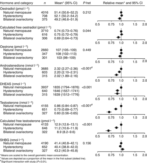
Oestradiol, oestrone and SHBG did not differ significantly by type of menopause (Figure 2). Calculated free oestradiol was 7% lower in women who had undergone a bilateral ovariectomy than in women who had a natural menopause. Compared with women with a natural menopause, concentrations of androstenedione and DHEAS were 5% and 10% lower, respectively, for women who had had a hysterectomy and 13% and 11% lower, respectively, for women who had had a bilateral ovariectomy. Testosterone was 15% lower for women who had had a hysterectomy and 30% lower for those who had had a bilateral ovariectomy than for women who had had a natural menopause, with similar differences in calculated free testosterone. There was significant heterogeneity between studies in the associations of androstenedione and testosterone with type of menopause; for androstenedione, the association with type of menopause was not significant for studies that had used a purification step but was significant for those that had used direct assays, whereas for testosterone the association with type of menopause was significant regardless of whether the assays had used a purification step (results not shown).
Figure 2.
Geometric mean concentration of selected hormones in postmenopausal women by categories of type of menopause, adjusted for study, age at blood collection and BMI.
Body mass index
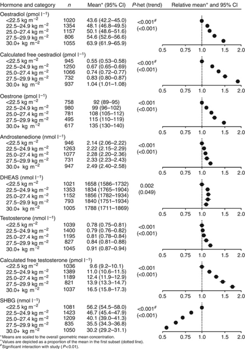
All the hormones and SHBG were significantly associated with BMI (Figure 3). Oestrogen concentrations were much higher in obese (BMI at least 30 kg m−2) than in thin women (BMI <22.5 kg m−2): oestradiol by 47%, calculated free oestradiol by 89% and oestrone by 47%. Concentrations of three androgens were also higher in obese than in thin women: androstenedione by 16%, testosterone by 17% and calculated free testosterone by 72%, whereas DHEAS showed significant heterogeneity by BMI but no clear trend. Sex hormone-binding globulin was 46% lower in obese women than in thin women. The associations of oestrogens, androgens and SHBG with BMI were similar in the subgroup of women who had undergone a bilateral ovariectomy (results not shown). There was significant heterogeneity between studies in the associations of oestradiol, free oestradiol and SHBG with BMI; for oestradiol and free oestradiol, the difference between extreme categories of BMI was significant regardless of the type of assay, but was larger in the studies that had used a purification step than in the studies that had used direct assays (increases from the lowest to the highest BMI category of 82% and 30%, respectively, for oestradiol and 129% and 70%, respectively, for free oestradiol; results not shown).
Figure 3.
Geometric mean concentration of selected hormones in postmenopausal women by categories of BMI, adjusted for study, age at blood collection and type of menopause.
Smoking
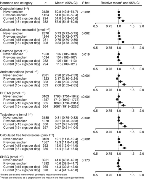
Concentrations of all the sex hormones were higher for women who smoked 15+ cigarettes per day than for never smokers (Figure 4). The largest differences were for androstenedione, testosterone and calculated free testosterone, which were 18%, 20% and 19%, respectively, higher for heavy cigarette smokers than for never smokers. Sex hormone-binding globulin did not differ according to smoking status. There was no significant heterogeneity between studies in the associations of smoking with the hormones or SHBG. The associations of hormones with smoking were less marked before adjustment for BMI, but were almost unaltered after further adjustment for alcohol (results not shown).
Figure 4.
Geometric mean concentration of selected hormones in postmenopausal women by categories of smoking, adjusted for study, age at blood collection, type of menopause and BMI.
Alcohol
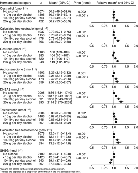
Concentrations of all the sex hormones were higher for women who consumed 20+ g per day of alcohol than for non-drinkers (Figure 5). The largest difference was for DHEAS, which was 25% higher for women consuming 20+ g per day of alcohol than for non-drinkers. Circulating SHBG was 10% lower for women consuming 20+ g per day of alcohol than for non-drinkers. There was no significant heterogeneity between studies in the associations of alcohol with the hormones or SHBG. The associations of hormones with alcohol were less marked before adjustment for BMI, but were almost unaltered after further adjustment for smoking (results not shown).
Figure 5.
Geometric mean concentration of selected hormones in postmenopausal women by categories of usual alcohol consumption, adjusted for study, age at blood collection, type of menopause and BMI.
Reproductive and other factors
Oestradiol was 6% lower in women who had menarche at ages 14 years and above than in women who had menarche before 12 years of age (Table 4). Age at menarche was not associated with the other hormones or SHBG.
Parity was not associated with any of the hormones or with SHBG, with the exception of DHEAS, which varied significantly between categories but with no clear pattern (Table 4). For parous women, none of the hormones or SHBG varied significantly by age at first full-term pregnancy.
Time since menopause was associated with oestradiol, calculated free oestradiol and DHEAS, which were 12%, 17% and 8% higher, respectively, in women who had had their menopause within the previous 5 years than in women who had had their menopause 15 or more years previously (Table 4). The other hormones and SHBG were not associated with time since menopause. Examination of time since menopause of 0–4 years in finer categories (0–1, 2, 3 and 4) showed that the highest concentrations of oestradiol were in the category of 0–1 years since menopause (64.4 pmol l−1), with little difference across the other categories (55.7, 57.2 and 53.7 pmol l−1, respectively); a similar pattern was seen for calculated free oestradiol (means of 0.93, 0.81, 0.83 and 0.80 pmol l−1, respectively).
Family history of breast cancer was not associated with any of the hormones or SHBG (Table 4). Women who had previously used hormonal contraceptives had 5% higher oestradiol and calculated free oestradiol and 6% lower androstenedione than women who had never used hormonal contraceptives. Women who had previously used hormonal therapy for menopause had 9% lower oestrone, 6% lower androstenedione and 7% lower testosterone than women who had not used hormonal therapy for menopause.
Discussion
The results suggest that circulating concentrations of sex hormones are associated with several of the well-established or suspected risk factors for breast cancer. The strongest associations were with BMI: concentrations of all the hormones were higher in obese women than in women with a low BMI, with the largest differences for calculated free oestradiol and free testosterone. Ovariectomy was associated with low levels of androgens, and cigarette smoking and alcohol were associated with moderate increases in all the sex hormones. Age at menarche, parity, age at first full-term pregnancy and family history of breast cancer were not strongly related to any of the hormones examined.
The large sample size provides sufficient power to detect differences in circulating hormone concentrations between categories as small as 10% for most of the analyses presented. Some of the observations replicate well-established associations, whereas others provide novel information of possible relevance to understanding the aetiology of breast cancer, as discussed in more detail below. Many previous publications on this topic are from the studies participating in this collaboration, and these publications are cited together with publications from studies not included in this collaboration. Limitations of this analysis are that the data are cross-sectional, and therefore cannot be used to attribute causality, and that some other potentially important factors such as physical activity and diet were not included in the analyses. Another potential limitation is that, with the exception of the RERF Study, the majority of the women in the contributing studies were of white European ethnic origin.
For some of the analyses, there was significant heterogeneity in the results from different studies. Inspection of the results from individual studies did not show large between-study differences in the variation of hormone levels by categories of risk factors, and suggested that the heterogeneity was largely due to differences in the magnitudes of the associations rather than qualitative differences. It is likely that the accuracy of the assays used varied; therefore, the results should be interpreted simply as an average across assays for the available data. The results suggested that the type of sex hormone assay (with or without a purification step) influenced some of the results. For example, the association of oestradiol with BMI was stronger in the studies that had used an assay with previous purification (see below); in general, hormone assays that include a purification step are both more sensitive and more specific than direct, non-extraction assays (Stanczyk et al, 2003).
For oestradiol and testosterone, we calculated estimates of the concentration of the free hormone using the law of mass action and the concentration of SHBG, and assumed a fixed concentration of albumin. We chose not to report estimates of non-SHBG-bound oestradiol and testosterone, which are often described as ‘bioavailable’ because these hormones dissociate readily from albumin. It should be noted that the calculated non-SHBG-bound oestradiol and testosterone would be perfectly correlated with free oestradiol and testosterone, respectively, and therefore that the associations reported for free oestradiol and free testosterone would be identical for the non-SHBG-bound concentrations of these hormones.
The hormones were all positively correlated with each other, presumably because they are all part of the same metabolic pathway. Whereas oestrogen and progesterone production in premenopausal women is controlled by feedback mechanisms through the anterior pituitary and the hypothalamus, the control of sex hormone production in postmenopausal women is not well understood. The oestrogens are derived from the androgens, which are secreted by the adrenal glands and by the stromal tissue of the ovaries. Adrenal androgen production may be stimulated by stress, as ACTH stimulates the adrenal cortex to synthesise cortisol and, less specifically, the adrenal androgens (Azziz et al, 1990; McKenna et al, 1997). Conversion of androgens to oestrogens is catalysed by the enzyme aromatase, and the major site of aromatase activity in postmenopausal women is in the adipose tissue (Grodin et al, 1973).
Age
The large decrease in DHEAS and androstenedione, the small changes in oestrogens and testosterone and the increase in SHBG with age are broadly consistent with previous reports (Meldrum et al, 1981; Moore et al, 1987; Verkasalo et al, 2001; Spencer et al, 2007). The smaller decreases in oestrogens than in two of the three androgens might suggest that the amount of substrate available only partly determines oestrogen concentrations, and that synthesis by aromatisation may become more active in older women (Misso et al, 2005).
It is likely that the relatively high concentrations of oestradiol in women aged below 55 years are associated with the fact that many of these women have had their menopause within the last 5 years, and exclusion of women who had recently had their menopause reduced this association (see further discussion below in relation to time since menopause).
Hysterectomy and ovariectomy
Previous analyses of data from studies contributing to this collaboration (Laughlin et al, 2000) and others (Judd et al, 1974a; Vermeulen, 1976; Davison et al, 2005; Cappola et al, 2007; Fogle et al, 2007; McTiernan et al, 2008) have shown that postmenopausal women who have had a bilateral ovariectomy have lower circulating androgen concentrations than postmenopausal women with intact ovaries. Furthermore, clinical studies have shown that androgen concentrations are higher in blood samples from the ovarian veins than in the peripheral circulation, demonstrating that the ovaries secrete androgens (Grodin et al, 1973; Judd et al, 1974b). Our analyses replicated these relationships; women who had undergone a bilateral ovariectomy had testosterone concentrations about 30% lower than those of naturally postmenopausal women, together with smaller reductions in androstenedione and DHEAS, but no significant differences for total oestradiol or oestrone. Women who had undergone a hysterectomy without bilateral ovariectomy had circulating androgen concentrations intermediate between those of women who had a natural menopause and women who had undergone a bilateral ovariectomy; this intermediate effect might be due to misclassification of some women who had undergone bilateral ovariectomy into the hysterectomy category and/or damage to the ovarian blood supply during hysterectomy (Laughlin et al, 2000); alternatively, it is possible that long-standing differences in circulating androgens somehow influence the likelihood of hysterectomy.
Body mass index
Levels of the oestrogens were higher in women with higher BMI, probably because women with more adipose tissue have more aromatase (in their adipose tissue) and therefore increased total aromatase activity (Grodin et al, 1973), and the high oestrogen levels of obese postmenopausal women probably explain their increased risk for breast cancer (Endogenous Hormones and Breast Cancer Collaborative Group, 2003a). This relationship has been observed in many previous studies that are not included in this collaboration (Meldrum et al, 1981; Kaye et al, 1991; Madigan et al, 1998; Nagata et al, 2000; Verkasalo et al, 2001; Boyapati et al, 2004; McTiernan et al, 2006; Chavez-MacGregor et al, 2008). Androstenedione and testosterone also increased with increasing BMI, whereas DHEAS was not clearly related to BMI. These associations have been observed previously (Cappola et al, 2007), but the reason why androgens increase with BMI is not clear. Obesity is associated with increases in insulin, which may stimulate androgen production in the ovarian stroma (Barber et al, 2006), whereas adrenal androgen synthesis is not known to be stimulated by insulin; however, our results suggested that androgens also increased with BMI in women who had undergone a bilateral ovariectomy and in whom most androgen synthesis must be in the adrenals. Sex hormone-binding globulin also decreased with increasing BMI, as has been consistently observed in many studies (e.g., Baglietto et al, 2009; Moore et al, 1987; and Newcomb et al, 1995); this decrease may be due to raised insulin concentrations inhibiting SHBG synthesis in the liver (Crave et al, 1995).
Overall, total oestradiol was 47% higher and calculated free oestradiol was 89% higher in obese women than in thin women. There was significant heterogeneity between studies in these estimates, and some of this heterogeneity was explained by the oestradiol assay method, with increases of total oestradiol between thin and obese women of 82% for studies that used assays with previous purification but only 30% for studies that used direct non-extraction assays. A similar difference has been noted previously, and it is likely that the larger estimate of 82% is more accurate because assays with previous purification for oestradiol are more sensitive and more specific than direct assays (Lee et al, 2006).
Smoking
We observed higher concentrations of oestrogens and androgens in current heavy cigarette smokers (at least 15 cigarettes per day) than in non-smokers, whereas SHBG did not vary significantly according to smoking status. Cigarette smokers were on average thinner than non-smokers, and the differences in oestrogens were accentuated by the adjustment for BMI. Previous studies have not shown clear associations of smoking with circulating oestrogens, but have generally observed that at least some of the androgens were higher in smokers than in non-smokers (Khaw et al, 1988; Cassidenti et al, 1992; Baron et al, 1995; Manjer et al, 2005). The mechanism for these associations is unknown but may involve more general effects on the hypothalamic–pituitary–adrenal axis (Kapoor and Jones, 2005).
Most epidemiological studies have suggested that smoking has little or no effect on breast cancer risk (Collaborative Group on Hormonal Factors in Breast Cancer, 2002; International Agency for Research on Cancer, 2004), but some recent large cohort studies have suggested that a long duration of smoking does have a small positive association with breast cancer risk in postmenopausal women, particularly in association with starting to smoke at a young age (Cui et al, 2006; Luo et al, 2011) and perhaps because of the chemical carcinogens in tobacco smoke (Secretan et al, 2009). The moderate differences between heavy smokers and non-smokers in circulating hormones might affect breast cancer risk, although the total effect of smoking on lifetime exposure to hormones also includes the relatively early menopause of smokers (Midgette and Baron, 1990) and possibly changes in oestrogen metabolism (Michnovicz et al, 1986).
Alcohol
Alcohol intake was positively associated with all the sex hormones, with the strongest association for DHEAS, whereas SHBG was inversely associated with alcohol consumption.
High alcohol consumers were on average thinner than non-drinkers, and the differences in oestrogens and in SHBG were accentuated by the adjustment for BMI. These findings are broadly consistent with those of previous observational studies (Onland-Moret et al, 2005). Furthermore, a randomised trial in 51 postmenopausal women showed that DHEAS increased by 5.1% in women consuming 15 g of alcohol per day and by 7.5% in women consuming 30 g of alcohol per day; alcohol consumption also increased circulating concentrations of oestrone sulphate (Dorgan et al, 2001). In another randomised crossover trial in nine postmenopausal women, 30 g of alcohol per day increased circulating DHEAS by 18% but had no detectable effect on oestradiol or testosterone (Sierksma et al, 2004). The mechanism for these associations is unknown.
Alcohol is mostly consumed in the evening, whereas blood samples are usually collected during the day. As the half-life of oestradiol is ∼3 h (Ginsburg et al, 1998), it is possible that there is a marked increase of oestradiol shortly after alcohol consumption in the evening, which cannot be assessed in typical epidemiological studies. In a study of the acute effects of 40 g of alcohol in premenopausal women, circulating oestradiol concentrations increased and reached a peak 25 min after alcohol ingestion (Mendelson et al, 1988). The half-life of DHEAS is longer (∼14 h), and this might explain why this sulphated hormone has the strongest association with alcohol consumption (Dorgan et al, 2001).
The increases in sex hormone concentrations associated with alcohol consumption might contribute to the increase in breast cancer risk with alcohol consumption (Baan et al, 2007), although other factors not related to circulating hormone concentrations might mediate the effect (Seitz and Stickel, 2007).
Other risk factors
Early research suggested that young age at menarche is positively associated with oestrogen levels in young women (MacMahon et al, 1982), but previous studies have not demonstrated clear associations of age at menarche with circulating hormones in postmenopausal women (Verkasalo et al, 2001). We found that, after adjusting for BMI, mean concentrations of oestradiol were 6% lower in postmenopausal women who had had a late menarche than in those who had an early menarche, but age at menarche was not clearly associated with the other hormones or SHBG.
The mean concentration of DHEAS varied significantly with parity, but without a clear pattern. Previous studies have reported that parity was not associated with sex hormones in postmenopausal women (Ness et al, 2000) and that DHEAS was not significantly related to parity in postmenopausal women (Key et al, 1990). Age at first full-term pregnancy in parous women was not associated with any of the sex hormones or SHBG.
Age at menarche, parity and age at first full-term pregnancy are risk factors for breast cancer (Kelsey et al, 1993). Apart from the very weak association of age at menarche with circulating oestradiol, the current findings that these risk factors are not associated with circulating sex hormone concentrations in postmenopausal women suggest that their effects on breast cancer risk are not likely to be mediated through postmenopausal hormone levels. These risk factors may operate through long-term effects on sex hormone levels in premenopausal women (MacMahon et al, 1982), through changes in the duration rather than the level of exposure to premenopausal sex hormones (Key and Pike, 1988), by hormonally mediated long-lasting changes in breast structure (Russo et al, 2005) or by other mechanisms.
Mean concentrations of oestradiol, free oestradiol and DHEAS were significantly higher in women who had had their menopause within 5 years of blood collection than in other women, but the other sex hormones and SHBG did not differ significantly between these groups. The higher levels of oestradiol and free oestradiol were observed primarily up to about 1 year after menopause, and this is compatible with results from a longitudinal study in which oestradiol levels declined from about 2 years before the final menstrual period until ∼2 years after (Sowers et al, 2008). In another report from the same longitudinal study (Crawford et al, 2009), there was a small increase in circulating concentrations of DHEAS during the menopausal transition, followed by a decline (more than 2 years since last menstrual period).
Previous use of exogenous sex hormones was significantly associated with several endogenous hormones, although the magnitude of these associations was small. Previous use of oral contraceptives was associated with higher concentrations of oestradiol and free oestradiol and lower concentrations of androstenedione; previous use of hormonal therapy for menopause was associated with lower concentrations of oestrone, androstenedione, testosterone and free testosterone. In another study (Chan et al, 2008), previous use of oral contraceptives was associated with lower concentrations of oestradiol, oestrone, androstenedione, testosterone and SHBG, and previous use of hormonal therapy for the menopause was associated with lower concentrations of testosterone and 17α-hydroxyprogesterone. It is possible that the associations with hormonal therapy for menopause may be partly because women with relatively low endogenous hormone levels may have more symptoms, and therefore be more likely to be prescribed hormonal therapy.
Conclusions
Circulating sex hormone concentrations in postmenopausal women are strongly associated with several established or suspected risk factors for breast cancer, and may mediate the effects of these factors on breast cancer risk.
Acknowledgments
We thank the women who participated, the research staff and the collaborating laboratories in each of the studies. This work was supported by Cancer Research, UK (C570/A5028) for the central pooling and analysis of the data; details of funding for the original studies are in the relevant publications. The funders of the study had no role in the study design, data collection, data analysis, data interpretation or writing of the report. The authors had full access to all the data and had final responsibility for the decision to submit the manuscript.
Footnotes
The authors declare no conflict of interest.
Contributor Information
Endogenous Hormones and Breast Cancer Collaborative Group:
T J Key, P N Appleby, G K Reeves, A W Roddam, K J Helzlsouer, A J Alberg, D E Rollison, J F Dorgan, L A Brinton, K Overvad, R Kaaks, A Trichopoulou, F Clavel-Chapelon, S Panico, E J Duell, P H M Peeters, S Rinaldi, I S Fentiman, M Dowsett, J Manjer, P Lenner, G Hallmans, L Baglietto, D R English, G G Giles, J L Hopper, G Severi, H A Morris, S E Hankinson, S S Tworoger, K Koenig, A Zeleniuch-Jacquotte, A A Arslan, P Toniolo, R E Shore, V Krogh, A Micheli, F Berrino, E Barrett-Connor, G A Laughlin, M Kabuto, S Akiba, R G Stevens, K Neriishi, C E Land, J A Cauley, Li Yung Lui, Steven R Cummings, M J Gunter, T E Rohan, and H D Strickler
References
- Azziz R, Bradley Jr E, Huth J, Boots LR, Parker Jr CR, Zacur HA (1990) Acute adrenocorticotropin-(1-24) (ACTH) adrenal stimulation in eumenorrheic women: reproducibility and effect of ACTH dose, subject weight, and sampling time. J Clin Endocrinol Metab 70: 1273–1279 [DOI] [PubMed] [Google Scholar]
- Baan R, Straif K, Grosse Y, Secretan B, El Ghissassi F, Bouvard V, Altieri A, Cogliano V (2007) Carcinogenicity of alcoholic beverages. Lancet Oncol 8: 292–293 [DOI] [PubMed] [Google Scholar]
- Baglietto L, English DR, Hopper JL, MacInnis RJ, Morris HA, Tilley WD, Krishnan K, Giles GG (2009) Circulating steroid hormone concentrations in postmenopausal women in relation to body size and composition. Breast Cancer Res Treat 115: 171–179 [DOI] [PubMed] [Google Scholar]
- Barber TM, McCarthy MI, Wass JA, Franks S (2006) Obesity and polycystic ovary syndrome. Clin Endocrinol (Oxf) 65: 137–145 [DOI] [PubMed] [Google Scholar]
- Baron JA, Comi RJ, Cryns V, Brinck-Johnsen T, Mercer NG (1995) The effect of cigarette smoking on adrenal cortical hormones. J Pharmacol Exp Ther 272: 151–155 [PubMed] [Google Scholar]
- Barrett-Connor E, Friedlander NJ, Khaw KT (1990) Dehydroepiandrosterone sulfate and breast cancer risk. Cancer Res 50: 6571–6574 [PubMed] [Google Scholar]
- Berrino F, Muti P, Micheli A, Bolelli G, Krogh V, Sciajno R, Pisani P, Panico S, Secreto G (1996) Serum sex hormone levels after menopause and subsequent breast cancer. J Natl Cancer Inst 88: 291–296 [DOI] [PubMed] [Google Scholar]
- Boyapati SM, Shu XO, Gao YT, Dai Q, Yu H, Cheng JR, Jin F, Zheng W (2004) Correlation of blood sex steroid hormones with body size, body fat distribution, and other known risk factors for breast cancer in post-menopausal Chinese women. Cancer Causes Control 15: 305–311 [DOI] [PubMed] [Google Scholar]
- Cappola AR, Ratcliffe SJ, Bhasin S, Blackman MR, Cauley J, Robbins J, Zmuda JM, Harris T, Fried LP (2007) Determinants of serum total and free testosterone levels in women over the age of 65 years. J Clin Endocrinol Metab 92: 509–516 [DOI] [PubMed] [Google Scholar]
- Cassidenti DL, Pike MC, Vijod AG, Stanczyk FZ, Lobo RA (1992) A reevaluation of estrogen status in postmenopausal women who smoke. Am J Obstet Gynecol 166: 1444–1448 [DOI] [PubMed] [Google Scholar]
- Cauley JA, Lucas FL, Kuller LH, Stone K, Browner W, Cummings SR (1999) Elevated serum estradiol and testosterone concentrations are associated with a high risk for breast cancer. Study of Osteoporotic Fractures Research Group. Ann Intern Med 130: 270–277 [DOI] [PubMed] [Google Scholar]
- Chan MF, Dowsett M, Folkerd E, Wareham N, Luben R, Welch A, Bingham S, Khaw KT (2008) Past oral contraceptive and hormone therapy use and endogenous hormone concentrations in postmenopausal women. Menopause 15: 332–339 [DOI] [PubMed] [Google Scholar]
- Chavez-MacGregor M, van Gils CH, van der Schouw YT, Monninkhof E, van Noord PA, Peeters PH (2008) Lifetime cumulative number of menstrual cycles and serum sex hormone levels in postmenopausal women. Breast Cancer Res Treat 108: 101–112 [DOI] [PMC free article] [PubMed] [Google Scholar]
- Collaborative Group on Hormonal Factors in Breast Cancer (2002) Alcohol, tobacco and breast cancer--collaborative reanalysis of individual data from 53 epidemiological studies, including 58 515 women with breast cancer and 95 067 women without the disease. Br J Cancer 87: 1234–1245 [DOI] [PMC free article] [PubMed] [Google Scholar]
- Crave JC, Lejeune H, Brebant C, Baret C, Pugeat M (1995) Differential effects of insulin and insulin-like growth factor I on the production of plasma steroid-binding globulins by human hepatoblastoma-derived (Hep G2) cells. J Clin Endocrinol Metab 80: 1283–1289 [DOI] [PubMed] [Google Scholar]
- Crawford S, Santoro N, Laughlin GA, Sowers MF, McConnell D, Sutton-Tyrrell K, Weiss G, Vuga M, Randolph J, Lasley B (2009) Circulating dehydroepiandrosterone sulfate concentrations during the menopausal transition. J Clin Endocrinol Metab 94: 2945–2951 [DOI] [PMC free article] [PubMed] [Google Scholar]
- Cui Y, Miller AB, Rohan TE (2006) Cigarette smoking and breast cancer risk: update of a prospective cohort study. Breast Cancer Res Treat 100: 293–299 [DOI] [PubMed] [Google Scholar]
- Davison SL, Bell R, Donath S, Montalto JG, Davis SR (2005) Androgen levels in adult females: changes with age, menopause, and oophorectomy. J Clin Endocrinol Metab 90: 3847–3853 [DOI] [PubMed] [Google Scholar]
- Dorgan JF, Baer DJ, Albert PS, Judd JT, Brown ED, Corle DK, Campbell WS, Hartman TJ, Tejpar AA, Clevidence BA, Giffen CA, Chandler DW, Stanczyk FZ, Taylor PR (2001) Serum hormones and the alcohol-breast cancer association in postmenopausal women. J Natl Cancer Inst 93: 710–715 [DOI] [PubMed] [Google Scholar]
- Dorgan JF, Longcope C, Stephenson Jr HE, Falk RT, Miller R, Franz C, Kahle L, Campbell WS, Tangrea JA, Schatzkin A (1996) Relation of prediagnostic serum estrogen and androgen levels to breast cancer risk. Cancer Epidemiol Biomarkers Prev 5: 533–539 [PubMed] [Google Scholar]
- Dorgan JF, Stanczyk FZ, Longcope C, Stephenson Jr HE, Chang L, Miller R, Franz C, Falk RT, Kahle L (1997) Relationship of serum dehydroepiandrosterone (DHEA), DHEA sulfate, and 5-androstene-3 beta, 17 beta-diol to risk of breast cancer in postmenopausal women. Cancer Epidemiol Biomarkers Prev 6: 177–181 [PubMed] [Google Scholar]
- Endogenous Hormones and Breast Cancer Collaborative Group (2002) Endogenous sex hormones and breast cancer in postmenopausal women: reanalysis of nine prospective studies. J Natl Cancer Inst 94: 606–616 [DOI] [PubMed] [Google Scholar]
- Endogenous Hormones and Breast Cancer Collaborative Group (2003a) Body mass index, serum sex hormones, and breast cancer risk in postmenopausal women. J Natl Cancer Inst 95: 1218–1226 [DOI] [PubMed] [Google Scholar]
- Endogenous Hormones and Breast Cancer Collaborative Group (2003b) Free estradiol and breast cancer risk in postmenopausal women: comparison of measured and calculated values. Cancer Epidemiol Biomarkers Prev 12: 1457–1461 [PubMed] [Google Scholar]
- Fogle RH, Stanczyk FZ, Zhang X, Paulson RJ (2007) Ovarian androgen production in postmenopausal women. J Clin Endocrinol Metab 92: 3040–3043 [DOI] [PubMed] [Google Scholar]
- Garland CF, Friedlander NJ, Barrett-Connor E, Khaw KT (1992) Sex hormones and postmenopausal breast cancer: a prospective study in an adult community. Am J Epidemiol 135: 1220–1230 [DOI] [PubMed] [Google Scholar]
- Ginsburg ES, Gao X, Shea BF, Barbieri RL (1998) Half-life of estradiol in postmenopausal women. Gynecol Obstet Invest 45: 45–48 [DOI] [PubMed] [Google Scholar]
- Gordon GB, Bush TL, Helzlsouer KJ, Miller SR, Comstock GW (1990) Relationship of serum levels of dehydroepiandrosterone and dehydroepiandrosterone sulfate to the risk of developing postmenopausal breast cancer. Cancer Res 50: 3859–3862 [PubMed] [Google Scholar]
- Grodin JM, Siiteri PK, MacDonald PC (1973) Source of estrogen production in postmenopausal women. J Clin Endocrinol Metab 36: 207–214 [DOI] [PubMed] [Google Scholar]
- Gunter MJ, Hoover DR, Yu H, Wassertheil-Smoller S, Rohan TE, Manson JE, Li J, Ho GY, Xue X, Anderson GL, Kaplan RC, Harris TG, Howard BV, Wylie-Rosett J, Burk RD, Strickler HD (2009) Insulin, insulin-like growth factor-I, and risk of breast cancer in postmenopausal women. J Natl Cancer Inst 101: 48–60 [DOI] [PMC free article] [PubMed] [Google Scholar]
- Hankinson SE, Willett WC, Manson JE, Colditz GA, Hunter DJ, Spiegelman D, Barbieri RL, Speizer FE (1998) Plasma sex steroid hormone levels and risk of breast cancer in postmenopausal women. J Natl Cancer Inst 90: 1292–1299 [DOI] [PubMed] [Google Scholar]
- Helzlsouer KJ, Alberg AJ, Bush TL, Longcope C, Gordon GB, Comstock GW (1994) A prospective study of endogenous hormones and breast cancer. Cancer Detect Prev 18: 79–85 [PubMed] [Google Scholar]
- International Agency for Research on Cancer (2004) Tobacco smoke and involuntary smoking. IARC Monogr Eval Carcinog Risks Hum 83: 1–1438 [PMC free article] [PubMed] [Google Scholar]
- Judd HL, Judd GE, Lucas WE, Yen SS (1974b) Endocrine function of the postmenopausal ovary: concentration of androgens and estrogens in ovarian and peripheral vein blood. J Clin Endocrinol Metab 39: 1020–1024 [DOI] [PubMed] [Google Scholar]
- Judd HL, Lucas WE, Yen SS (1974a) Effect of oophorectomy on circulating testosterone and androstenedione levels in patients with endometrial cancer. Am J Obstet Gynecol 118: 793–798 [DOI] [PubMed] [Google Scholar]
- Kaaks R, Rinaldi S, Key TJ, Berrino F, Peeters PH, Biessy C, Dossus L, Lukanova A, Bingham S, Khaw KT, Allen NE, Bueno-de-Mesquita HB, van Gils CH, Grobbee D, Boeing H, Lahmann PH, Nagel G, Chang-Claude J, Clavel-Chapelon F, Fournier A, Thiebaut A, Gonzalez CA, Quiros JR, Tormo MJ, Ardanaz E, Amiano P, Krogh V, Palli D, Panico S, Tumino R, Vineis P, Trichopoulou A, Kalapothaki V, Trichopoulos D, Ferrari P, Norat T, Saracci R, Riboli E (2005) Postmenopausal serum androgens, oestrogens and breast cancer risk: the European prospective investigation into cancer and nutrition. Endocr Relat Cancer 12: 1071–1082 [DOI] [PubMed] [Google Scholar]
- Kabuto M, Akiba S, Stevens RG, Neriishi K, Land CE (2000) A prospective study of estradiol and breast cancer in Japanese women. Cancer Epidemiol Biomarkers Prev 9: 575–579 [PubMed] [Google Scholar]
- Kapoor D, Jones TH (2005) Smoking and hormones in health and endocrine disorders. Eur J Endocrinol 152: 491–499 [DOI] [PubMed] [Google Scholar]
- Kaye SA, Folsom AR, Soler JT, Prineas RJ, Potter JD (1991) Associations of body mass and fat distribution with sex hormone concentrations in postmenopausal women. Int J Epidemiol 20: 151–156 [DOI] [PubMed] [Google Scholar]
- Kelsey JL, Gammon MD, John EM (1993) Reproductive factors and breast cancer. Epidemiol Rev 15: 36–47 [DOI] [PubMed] [Google Scholar]
- Key TJ, Pike MC (1988) The role of oestrogens and progestagens in the epidemiology and prevention of breast cancer. Eur J Cancer Clin Oncol 24: 29–43 [DOI] [PubMed] [Google Scholar]
- Key TJ, Pike MC, Wang DY, Moore JW (1990) Long term effects of a first pregnancy on serum concentrations of dehydroepiandrosterone sulfate and dehydroepiandrosterone. J Clin Endocrinol Metab 70: 1651–1653 [DOI] [PubMed] [Google Scholar]
- Khaw KT, Tazuke S, Barrett-Connor E (1988) Cigarette smoking and levels of adrenal androgens in postmenopausal women. N Engl J Med 318: 1705–1709 [DOI] [PubMed] [Google Scholar]
- Laughlin GA, Barrett-Connor E, Kritz-Silverstein D, von Muhlen D (2000) Hysterectomy, oophorectomy, and endogenous sex hormone levels in older women: the Rancho Bernardo Study. J Clin Endocrinol Metab 85: 645–651 [DOI] [PubMed] [Google Scholar]
- Lee JS, Ettinger B, Stanczyk FZ, Vittinghoff E, Hanes V, Cauley JA, Chandler W, Settlage J, Beattie MS, Folkerd E, Dowsett M, Grady D, Cummings SR (2006) Comparison of methods to measure low serum estradiol levels in postmenopausal women. J Clin Endocrinol Metab 91: 3791–3797 [DOI] [PubMed] [Google Scholar]
- Luo J, Margolis KL, Wactawski-Wende J, Horn K, Messina C, Stefanick ML, Tindle HA, Tong E, Rohan TE (2011) Association of active and passive smoking with risk of breast cancer among postmenopausal women: a prospective cohort study. BMJ 342: d1016. [DOI] [PMC free article] [PubMed] [Google Scholar]
- MacMahon B, Trichopoulos D, Brown J, Andersen AP, Cole P, deWaard F, Kauraniemi T, Polychronopoulou A, Ravnihar B, Stormby N, Westlund K (1982) Age at menarche, urine estrogens and breast cancer risk. Int J Cancer 30: 427–431 [DOI] [PubMed] [Google Scholar]
- Madigan MP, Troisi R, Potischman N, Dorgan JF, Brinton LA, Hoover RN (1998) Serum hormone levels in relation to reproductive and lifestyle factors in postmenopausal women (United States). Cancer Causes Control 9: 199–207 [DOI] [PubMed] [Google Scholar]
- Manjer J, Johansson R, Berglund G, Janzon L, Kaaks R, Agren A, Lenner P (2003) Postmenopausal breast cancer risk in relation to sex steroid hormones, prolactin and SHBG (Sweden). Cancer Causes Control 14: 599–607 [DOI] [PubMed] [Google Scholar]
- Manjer J, Johansson R, Lenner P (2005) Smoking as a determinant for plasma levels of testosterone, androstenedione, and DHEAs in postmenopausal women. Eur J Epidemiol 20: 331–337 [DOI] [PubMed] [Google Scholar]
- McKenna TJ, Fearon U, Clarke D, Cunningham SK (1997) A critical review of the origin and control of adrenal androgens. Baillieres Clin Obstet Gynaecol 11: 229–248 [DOI] [PubMed] [Google Scholar]
- McTiernan A, Wu L, Barnabei VM, Chen C, Hendrix S, Modugno F, Rohan T, Stanczyk FZ, Wang CY (2008) Relation of demographic factors, menstrual history, reproduction and medication use to sex hormone levels in postmenopausal women. Breast Cancer Res Treat 108: 217–231 [DOI] [PubMed] [Google Scholar]
- McTiernan A, Wu L, Chen C, Chlebowski R, Mossavar-Rahmani Y, Modugno F, Perri MG, Stanczyk FZ, Van Horn L, Wang CY (2006) Relation of BMI and physical activity to sex hormones in postmenopausal women. Obesity (Silver Spring) 14: 1662–1677 [DOI] [PubMed] [Google Scholar]
- Meldrum DR, Davidson BJ, Tataryn IV, Judd HL (1981) Changes in circulating steroids with aging in postmenopausal women. Obstet Gynecol 57: 624–628 [PubMed] [Google Scholar]
- Mendelson JH, Lukas SE, Mello NK, Amass L, Ellingboe J, Skupny A (1988) Acute alcohol effects on plasma estradiol levels in women. Psychopharmacology (Berl) 94: 464–467 [DOI] [PubMed] [Google Scholar]
- Michnovicz JJ, Hershcopf RJ, Naganuma H, Bradlow HL, Fishman J (1986) Increased 2-hydroxylation of estradiol as a possible mechanism for the anti-estrogenic effect of cigarette smoking. N Engl J Med 315: 1305–1309 [DOI] [PubMed] [Google Scholar]
- Midgette AS, Baron JA (1990) Cigarette smoking and the risk of natural menopause. Epidemiology 1: 474–480 [DOI] [PubMed] [Google Scholar]
- Missmer SA, Eliassen AH, Barbieri RL, Hankinson SE (2004) Endogenous estrogen, androgen, and progesterone concentrations and breast cancer risk among postmenopausal women. J Natl Cancer Inst 96: 1856–1865 [DOI] [PubMed] [Google Scholar]
- Misso ML, Jang C, Adams J, Tran J, Murata Y, Bell R, Boon WC, Simpson ER, Davis SR (2005) Adipose aromatase gene expression is greater in older women and is unaffected by postmenopausal estrogen therapy. Menopause 12: 210–215 [DOI] [PubMed] [Google Scholar]
- Moore JW, Key TJ, Bulbrook RD, Clark GM, Allen DS, Wang DY, Pike MC (1987) Sex hormone binding globulin and risk factors for breast cancer in a population of normal women who had never used exogenous sex hormones. Br J Cancer 56: 661–666 [DOI] [PMC free article] [PubMed] [Google Scholar]
- Nagata C, Shimizu H, Takami R, Hayashi M, Takeda N, Yasuda K (2000) Relations of insulin resistance and serum concentrations of estradiol and sex hormone-binding globulin to potential breast cancer risk factors. Jpn J Cancer Res 91: 948–953 [DOI] [PMC free article] [PubMed] [Google Scholar]
- Ness RB, Buhari A, Gutai J, Kuller LH (2000) Reproductive history in relation to plasma hormone levels in healthy post-menopausal women. Maturitas 35: 149–157 [DOI] [PubMed] [Google Scholar]
- Newcomb PA, Klein R, Klein BE, Haffner S, Mares-Perlman J, Cruickshanks KJ, Marcus PM (1995) Association of dietary and life-style factors with sex hormones in postmenopausal women. Epidemiology 6: 318–321 [DOI] [PubMed] [Google Scholar]
- Onland-Moret NC, Peeters PH, van der Schouw YT, Grobbee DE, van Gils CH (2005) Alcohol and endogenous sex steroid levels in postmenopausal women: a cross-sectional study. J Clin Endocrinol Metab 90: 1414–1419 [DOI] [PubMed] [Google Scholar]
- Rinaldi S, Key TJ, Peeters PH, Lahmann PH, Lukanova A, Dossus L, Biessy C, Vineis P, Sacerdote C, Berrino F, Panico S, Tumino R, Palli D, Nagel G, Linseisen J, Boeing H, Roddam A, Bingham S, Khaw KT, Chloptios J, Trichopoulou A, Trichopoulos D, Tehard B, Clavel-Chapelon F, Gonzalez CA, Larranaga N, Barricarte A, Quiros JR, Chirlaque MD, Martinez C, Monninkhof E, Grobbee DE, Bueno-de-Mesquita HB, Ferrari P, Slimani N, Riboli E, Kaaks R (2006) Anthropometric measures, endogenous sex steroids and breast cancer risk in postmenopausal women: a study within the EPIC cohort. Int J Cancer 118: 2832–2839 [DOI] [PubMed] [Google Scholar]
- Russo J, Moral R, Balogh GA, Mailo D, Russo IH (2005) The protective role of pregnancy in breast cancer. Breast Cancer Res 7: 131–142 [DOI] [PMC free article] [PubMed] [Google Scholar]
- Secretan B, Straif K, Baan R, Grosse Y, El Ghissassi F, Bouvard V, Benbrahim-Tallaa L, Guha N, Freeman C, Galichet L, Cogliano V (2009) A review of human carcinogens--Part E: tobacco, areca nut, alcohol, coal smoke, and salted fish. Lancet Oncol 10: 1033–1034 [DOI] [PubMed] [Google Scholar]
- Seitz HK, Stickel F (2007) Molecular mechanisms of alcohol-mediated carcinogenesis. Nat Rev Cancer 7: 599–612 [DOI] [PubMed] [Google Scholar]
- Sierksma A, Sarkola T, Eriksson CJ, van der Gaag MS, Grobbee DE, Hendriks HF (2004) Effect of moderate alcohol consumption on plasma dehydroepiandrosterone sulfate, testosterone, and estradiol levels in middle-aged men and postmenopausal women: a diet-controlled intervention study. Alcohol Clin Exp Res 28: 780–785 [DOI] [PubMed] [Google Scholar]
- Sodergard R, Backstrom T, Shanbhag V, Carstensen H (1982) Calculation of free and bound fractions of testosterone and estradiol-17 beta to human plasma proteins at body temperature. J Steroid Biochem 16: 801–810 [DOI] [PubMed] [Google Scholar]
- Sowers MR, Zheng H, McConnell D, Nan B, Harlow SD, Randolph Jr JF (2008) Estradiol rates of change in relation to the final menstrual period in a population-based cohort of women. J Clin Endocrinol Metab 93: 3847–3852 [DOI] [PMC free article] [PubMed] [Google Scholar]
- Spencer JB, Klein M, Kumar A, Azziz R (2007) The age-associated decline of androgens in reproductive age and menopausal Black and White women. J Clin Endocrinol Metab 92: 4730–4733 [DOI] [PubMed] [Google Scholar]
- Stanczyk FZ, Cho MM, Endres DB, Morrison JL, Patel S, Paulson RJ (2003) Limitations of direct estradiol and testosterone immunoassay kits. Steroids 68: 1173–1178 [DOI] [PubMed] [Google Scholar]
- Thomas HV, Key TJ, Allen DS, Moore JW, Dowsett M, Fentiman IS, Wang DY (1997) A prospective study of endogenous serum hormone concentrations and breast cancer risk in post-menopausal women on the island of Guernsey. Br J Cancer 76: 401–405 [DOI] [PMC free article] [PubMed] [Google Scholar]
- Toniolo PG, Levitz M, Zeleniuch-Jacquotte A, Banerjee S, Koenig KL, Shore RE, Strax P, Pasternack BS (1995) A prospective study of endogenous estrogens and breast cancer in postmenopausal women. J Natl Cancer Inst 87: 190–197 [DOI] [PubMed] [Google Scholar]
- Verkasalo PK, Thomas HV, Appleby PN, Davey GK, Key TJ (2001) Circulating levels of sex hormones and their relation to risk factors for breast cancer: a cross-sectional study in 1092 pre- and postmenopausal women (United Kingdom). Cancer Causes Control 12: 47–59 [DOI] [PubMed] [Google Scholar]
- Vermeulen A (1976) The hormonal activity of the postmenopausal ovary. J Clin Endocrinol Metab 42: 247–253 [DOI] [PubMed] [Google Scholar]
- Wysowski DK, Comstock GW, Helsing KJ, Lau HL (1987) Sex hormone levels in serum in relation to the development of breast cancer. Am J Epidemiol 125: 791–799 [DOI] [PubMed] [Google Scholar]
- Zeleniuch-Jacquotte A, Bruning PF, Bonfrer JM, Koenig KL, Shore RE, Kim MY, Pasternack BS, Toniolo P (1997) Relation of serum levels of testosterone and dehydroepiandrosterone sulfate to risk of breast cancer in postmenopausal women. Am J Epidemiol 145: 1030–1038 [DOI] [PubMed] [Google Scholar]
- Zeleniuch-Jacquotte A, Shore RE, Koenig KL, Akhmedkhanov A, Afanasyeva Y, Kato I, Kim MY, Rinaldi S, Kaaks R, Toniolo P (2004) Postmenopausal levels of oestrogen, androgen, and SHBG and breast cancer: long-term results of a prospective study. Br J Cancer 90: 153–159 [DOI] [PMC free article] [PubMed] [Google Scholar]


