Abstract
The Alexander Technique (AT) is used to improve postural and movement coordination and has been reported to be clinically beneficial, however its effect on movement coordination is not well-characterized. In this study we examined the sit-to-stand (STS) movement by comparing coordination (phasing, weight-shift and spinal movement) between AT teachers (n=15) and matched control subjects (n=14). We found AT teachers had a longer weight-shift (p<0.001) and shorter momentum transfer phase (p=0.01), than control subjects. AT teachers also increased vertical foot force monotonically, rather than unweighting the feet prior to seat-off, suggesting they generate less forward momentum with hip flexors. The prolonged weight-shift of AT teachers occurred over a greater range of trunk inclination, such that their weight shifted continuously onto the feet while bringing the body mass forward. Finally, AT teachers had greatly reduced spinal bending during STS (cervical, p<0.001; thoracic, p<0.001; lumbar, p<0.05). We hypothesize that the low hip joint stiffness and adaptive axial postural tone previously reported in AT teachers underlies this novel “continuous” STS strategy by facilitating eccentric contractions during weight-shift.
Keywords: Muscle Tone, Posture, Sit-to-Stand, Alexander Technique, Motor Processes
1. Introduction
The Alexander Technique (AT) is a method to improve habitual postural and movement coordination commonly used by performing artists [1]. It is offered in the music and theatre departments at major colleges for the purpose of improving performance and preventing injury. Recent reports indicate AT is clinically beneficial for back pain [2], Parkinson’s disease [3] and balance in the elderly [4]. However, the mechanisms underlying its clinical and claimed performance improvements are poorly understood. A greater understanding of improved coordination could have broad implications for rehabilitation.
The emphasis of AT is on axial behavior, the positional and tensional relationships within the neck and trunk, during posture and movement [5]. In particular, AT aims to reduce unnecessary tension and maintain elongation along the spine, referred to as the head-neck-back relationship. Proponents consider this relationship fundamental to any clinical or performance benefit from AT [1, 5].
Recently, AT has been found to alter postural tone. This was observed as a reduction of stiffness along the spine and hips in response to slowly applied torsion during unsupported stance [6]. Interestingly, this stiffness reduction resulted from an increase in the extent muscle tone dynamically adapted to yield to the applied movement. It is unclear, however, how such altered axial and proximal postural behavior may influence movement coordination. In general, the relationship between postural tone and movement coordination is not understood. In particular, the importance of regulating postural tone dynamically throughout movement has been hypothesized previously by Bernstein and others [7, 8], but has not been studied to date. Populations with atypical axial postural tone, such as AT, might help elucidate how tone affects movement coordination.
AT is taught by bringing attention to one’s head-neck-back relationship and specific features of movement, such as preparation and smoothness, in various postures and movements. A primary aim is to minimize abrupt shifts in tension and position along the body axis at movement onset [1, 5, 9]. AT instruction uses manual guidance to increase one’s awareness of these features and facilitate the desired head-neck-back relationship. The resulting coordination is claimed to be more efficient [1, 5, 9]. Movements typically performed in AT include sit-to-stand (STS), stand-to-sit, knee bends, lunges and squats. Of these, only STS has been studied with AT. Jones and colleagues found that, for this movement, horizontal head velocity, vertical acceleration and cervical extension decreased following AT training [10]. This group also found the movement was perceived as smoother and lower in effort [11]. Although AT may alter STS coordination, its effect is not well characterized and the significance of the resulting coordination differences is not clear.
In the present study, we aimed to better characterize STS coordination following AT training by examining 1) the overall phasing of the STS movement, 2) features of weight-shift, because it is perceived as smoother with AT, and 3) spinal coordination, because AT emphasizes axial behavior. A preliminary version of this work has appeared previously in abstract form [12].
2. Methods
2.1 Subjects
A total of 15 AT teachers (4 male, 11 female) participated in the study. AT teachers were selected as they are highly trained, spending 80% of their 1600-hour training improving their own proficiency in AT. All teachers were certified by affiliates of the Society for Teachers of the Alexander Technique and had a mean age of 42.7±9.1 years, height of 169.3±8.5 cm, and weight of 74.5±11.3 kg. The gender bias reflected that of US teachers. AT teachers had an average of 10.4±9.3 years experience post-certification.
Fourteen control subjects (4 male, 10 female) were recruited to match the age, height, and weight of AT teachers: 38.1±10.0 years (F1,27=1.15, p=0.29), 164.7±9.7 cm (F1,27=2.29, p=0.14), and 70.8±10.8 kg (F1,27=0.56, p=0.46). All subjects were free of pain and orthopedic conditions and provided informed consent in accordance with the Oregon Health & Science University Institutional Review Board.
2.2 Experimental Procedure
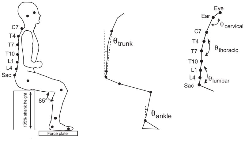
Subjects sat on an adjustable height backless chair with feet resting on a custom-built force plate. The chair height was adjusted to 105% of each subject’s shank height (from floor to lateral knee epicondyle). Initial foot position was adjusted so the knee angle was 85° (Fig. 1). In pilot data, we observed AT teachers to have a prolonged, monotonic weight-shift when given no specific instruction regarding how to stand up. In the present study, we instructed participants to stand up “as smoothly as possible, without using momentum”. This instruction aimed to encourage controls to mimic the gradual AT weight-shift, in order to understand whether it is simply a ‘choice’ of how to move or is difficult to perform, perhaps reflecting a more fundamental aspect of coordination. Subjects were told to start from sitting upright and to keep their arms crossed in front of their body. Subjects stood up at a self-selected speed 5 times.
Figure 1.
Initial position and kinematic quantification. Left panel: Subjects began the movement from a standardized initial position with marker placements as shown. Middle panel: Trunk angle was calculated as the sagittal plane angle between C7 and the sacrum, relative to vertical. Right panel. The total sagittal thoracic angle (θthoracic) was obtained by adding the individual joint angles at T4, T7 and T10 computed between adjacent markers. Similarly, θlumbar was obtained by summing L1 and L4 joint angles.
2.3 Data Collection
2.3.1 Kinematics
Kinematics were collected at 60Hz using a 7-camera passive marker system (Falcon, Motion Analysis) and low-pass filtered at 6Hz. Markers were placed bilaterally on the lateral orbital margin, tragus of the ear, posterior superior iliac spine, greater trochanter, lateral knee epicondyle, 3 cm proximal to the ankle joint along the fibula, lateral posterior calcaneus, and the head of the first metatarsal, as well as the spinal processes of C7, T4, T7, T10, L1, L4 and the midpoint of the sacral crest.
Trunk-tilt (θtrunk) was defined as the sagittal plane segment angle from C7 to sacrum relative to vertical. Ankle angle (θankle) was computed between ankle and knee markers relative to vertical averaged across both legs. Positive indicates dorsiflexion.
2.3.2 Forces
Forces were anti-alias filtered, sampled at 480 Hz and low-pass filtered at 15Hz.
2.4 Data Analysis
2.4.1 Movement phases
STS movement phases were calculated according to Schenkman [13] as follows. Flexion-momentum phase (Phase I) began when θtrunk exceeded 5° of the seated value and ended when foot Fz > 100% bodyweight (tso). The momentum-transfer phase (Phase II) occurred between tso and the occurrence of max(θankle). The extension phase (Phase III) occurred between max(θankle) and when θtrunk reached 5° of its value during stance. We did not examine the subsequent stabilization phase. To better quantify weight-transfer, we subdivided Phase I into flexion-only (Phase Ia) and weight-transfer (Phase Ib). Phase Ia ended and Phase Ib began when foot Fz exceeded 30% of bodyweight (BW). Phase durations were normalized by movement time (Phase I onset to Phase III cessation).
2.4.2 Monotonicity of weight-shift
Smoothness of weight transfer was examined by quantifying the monotoniticy of Fz before and after weight-shift as: undershoot = 100*(Fz (t0) – min Fz)/BW; and overshoot = 100*(max Fz - BW)/BW. This undershoot likely results from a hip flexor moment, which acts to accelerate the trunk forward and simultaneously lift the feet from the floor. Overshoot reflects the maximal leg extensor moments following seatoff.
2.4.3 Spinal angles
Spinal angles were computed between adjacent markers in the sagittal plane (e.g. θT4 was computed between C7, T4 and T7) relative to the initial seated value. Positive indicates extension. Our pilot data revealed thoracic segments typically extended earlier than lumbar segments [14, 15]. To decrease noise due to marker proximity, thoracic and lumbar angles were separately summed to obtain overall “joint” angles (θthoracic=θT4+θT7+θT10, θlumbar=θL1+θL4). Neck angle was computed between C7, ear and orbit markers, averaged bilaterally to exclude head rotation.
2.4.4 Statistics
Measures were computed for each trial and averaged across repetitions. Significant differences between groups were determined using a one-way ANOVA with α=0.05.
3. Results
The mean duration to stand up was similar for control subjects (2.2±0.9 s) and AT teachers (2.3±0.5 s); (F1,27=0.087, p=0.77). These STS durations are slightly longer than typical self-selected speeds for young adults (1.5-2 s [16, 17]), likely due to the instruction to stand up as smoothly as possible without momentum.
3.1 Movement Phases
Durations of STS phases are shown in Fig. 2. The mean duration of Phase Ia, prior to weight-shift onset, was smaller in AT teachers (15.8±7.9%) than control subjects (20.0±4.8%), but this was not statistically different (F1,27=3.01, p=0.09). AT teachers spent a significantly (F1,27=25.3, p<0.001) longer percentage in Phase Ib (19.4±5.5%), transferring weight to the feet, compared to control subjects (11.3±2.5%). AT teachers also spent a significantly (F1,27=7.15, p=0.01) shorter percentage of time (6.1±15.7%) in Phase II, the momentum transfer phase, than control subjects (18.1±6.3%). Phase III was similar between groups (AT=59.0±18.8%; CTL=50.0±10.6%; F1,27=2.53, p=0.12).
Figure 2.
STS movement phasing for AT teachers and matched control subjects. Phase durations for the flexion only (Ia), weight-shift (Ib), momentum transfer (II) and extension (III) phases as a percentage of total movement time. ** indicates p=0.01, *** indicates p<0.001.
3.2 Weight-shift
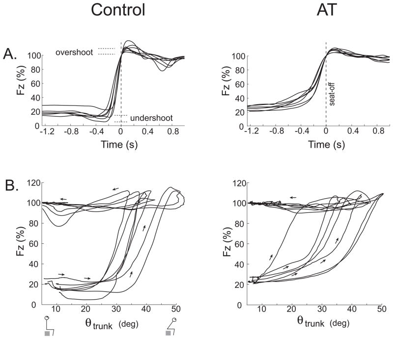
Fig. 3A shows the transfer of weight onto the feet. Most (12/14) control subjects had a distinct decrease in Fz before weight-shift compared to the minority (3/15) of AT teachers. This undershoot differed significantly between groups (CTL=3.7±2.8% vs. AT=1.1±1.3%, F1,27=10.4, p<0.01). In most AT teachers Fz increased monotonically and more gradually than in control subjects, corresponding to the longer Phase Ib. Following weight-shift, Fz overshoot was similar for AT teachers and controls 9.4±2.6% vs. 9.1±4.3%, respectively (F1,27=0.046, p=0.83).
Figure 3.
Weight-shift in AT teachers and control subjects. Panels show single trials from seven subjects in each population. A: Combined vertical foot force normalized to BW over time. All traces have been aligned at seatoff (time=0, vertical dotted line). B: Vertical combined-foot force relative to trunk inclination.
Fig. 3B shows Fz relative to θtrunk. AT subjects reached Fz=30% BW at a lower θtrunk (16.4±6.3°) than control subjects (20.9±2.5°, F1,27=6.19, p<0.05). During weight-shift (Phase Ib), θtrunk changed by 19.4±4.2° in AT teachers, with only around half that (10.6±3.9°) occurring in control subjects (F1,27=34.1, p<0.001). Thus, AT teachers began to weight the feet earlier (relative to θtrunk) and over a greater range of trunk inclination than controls.
3.3 Momentum transfer phase
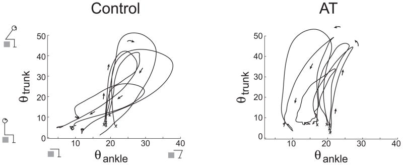
Fig. 4 shows the relation between θtrunk and θankle. While controls tended to sequentially increase θtrunk and then θankle (11/14 controls), AT teachers tended to increase these together (14/15 teachers). Additionally, following maximum θtrunk, 12/14 controls increased θankle, while 13/15 teachers reduced it. Thus, Phase II was shortened in AT teachers as θankle decreased, rather than increased, shortly following seat-off.
Figure 4.
Relationship between trunk tilt and shank movement. Data from single trials of five subjects are overlaid. The initial position is marked with an “x” for each trial.
3.4 Spinal angles
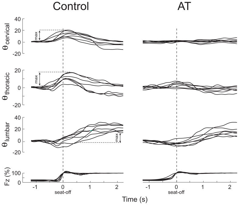
Spinal angles during STS are presented in Fig. 5. The cervical and thoracic spine tended to extend prior to weight-shift during Phase I and reached maximum shortly after. In contrast maximal lumbar extension occurred during Phase III. AT teachers had less cervical movement than controls (AT max(θneck)=2.2±1.0°; CTL max(θneck)=11.7±5.4°; F1,26=40.88, p<0.001) as well as less thoracic movement (AT max(θthoracic)=3.8±1.7°; CTL max(θthoracic)=12.6±4.6°; F1,27=82.03, p<0.001). AT teachers also had less change in lumbar angle (max(θlumbar)=17.0±6.3) than controls (max(θ lumbar)=25.2±5.9; F1,27=13.00, p<0.05). Some subjects in both groups flexed the lumbar spine around seatoff prior to extending it [15].
Figure 5.
Sagittal head, thoracic and lumbar spinal angles during STS. Data are shown for individual trials from seven subjects in each group. All traces are aligned to seatoff (time=0, dashed vertical line) and represent deviations from the initial seated angles. Combined foot vertical force is given at bottom.
4. Discussion
4.1 STS coordination in AT and controls
The aim of this study was to compare the coordination of the STS movement between AT teachers and matched healthy control subjects. AT teachers had altered STS phasing, weight-shift and spinal coordination, suggesting they employ a novel strategy for this task.
4.1.1 Continuous and sequential STS strategies
AT teachers shifted their weight continuously as the trunk inclined forward, rather than at a more specific trunk angle. To our knowledge this “continuous strategy” for transferring weight has not been reported previously in adults for STS. The continuous strategy indicates that AT teachers simultaneously generate anti-gravity leg-extensor moments while solving the balance problem — bringing the center-of-mass (COM) forward over the feet. In contrast, control subjects have two distinct actions prior to weight-shift: bringing the trunk forward and then shifting weight (Fig. 3B), which we refer to as the “sequential strategy”. Continuous vs. sequential movement features are also apparent in the relationship between trunk and ankle angles (Fig 4), which likely underlies the inter-group difference in momentum transfer duration. Because our control subjects had foot unloading and STS phase durations similar to previous reports (see section 4.1.3), we expect that untrained adults typically employ the sequential strategy when rising from a chair. The continuous strategy theoretically requires sustained eccentric contractions in the legs and trunk (flexing the joints to bring the body forward while generating extensor moments to weight the feet), which may be a fundamental aspect of this strategy.
4.1.2 Comparison with previous STS strategy classifications
Several categorizations of STS coordination have been made to date. Such categorizations include momentum transfer vs. stabilization strategies [18], distinguished by the COM-base of support distance at seat-off, or knee vs. hip strategies [19, 20], based on the maximal joint moments. Other classification schemes also exist. However, to date all reported classification schemes relate to hip angle, trunk lean and COM position at an instant in time and do not correspond to the relationship between forward trunk movement and weight-shift across time [21]. Also, because continuous and sequential strategies each occurred across a similar range of maximal trunk angles (Fig. 3B), the distinction between continuous and sequential strategies is complementary to existing classification schemes.
4.1.3 Previous reports of weight-shift in untrained subjects
The weight-shift (Phase Ib) of AT teachers occurred over roughly twice the proportion of the STS action (19.4%) compared with either our matched controls (11.3%) or healthy untrained adults reported previously. In previous reports the weight-shift duration was measured to be 9.5% ([22], Fig. 6) and 9.6% [19] of the total STS duration, and can be calculated from other available data to be 8.5% (c.f. Fig. 2 in [23]) and 12.0% (c.f. Fig. 6 in [24]). In other studies that did not report this phase duration, weight-shift typically occurs over a short time (e.g. [13, 25]), in contrast to the prolonged weight transfer in AT teachers.
The foot unloading prior to weight-shift in our control subjects is also typically observed in other untrained healthy adults [13, 22–29]. Thus, control subjects appear to use greater hip flexor moments to stand. A smooth monotonic weight shift, as occurred in AT teachers, has also been reported in 4–5 year old children [30]. Interestingly, proponents of AT consider children of this age to be especially well-coordinated, based on their head-neck-back relationship [1].
4.2 Axial STS coordination and axial postural tone
It is interesting that control subjects reported difficulty weighting their feet smoothly whilst standing up. We suspect this difficulty arises from the conflict between leg extensor torques that weight the feet and leg flexion required to move the COM forward. AT teachers’ near-isometric spinal behavior would act mechanically to maximize power transmission through the spine and pelvis during weight-shift. This would facilitate transferring forward trunk momentum to overcome (i.e., perform positive work on) hip and knee extensors while they generate moments to weight the feet, thus maintaining forward COM movement to solve the balance constraint. This could help AT teachers to perform the continuous STS strategy.
It is possible that the atypical postural tone previously observed in AT teachers [6] could help to solve these tasks simultaneously. For the hip, teachers’ low stiffness would reduce the energy absorbed eccentrically in this joint during weight-shift. Low hip stiffness could also reduce the forward trunk velocity used to stand up, explaining the prolonged AT weight-shift. For controls, greater hip stiffness could explain their Fz undershoot, as this group might need more energy to move the body forward.
The relationship between AT teachers’ minimal spinal bending and low stiffness in the neck and trunk [6] is less obvious. Other studies have reported spinal movement during STS similar to our control group [14, 15], suggesting that AT teachers’ lack of spinal movement is atypical. This seems paradoxical as we would expect lower axial stiffness to result in greater spinal movement. One possibility is that teachers’ axial resistance to flexion and extension (i.e. during STS) differs from that previously measured in torsion. However, this would be surprising as many muscles stretched during torsion would also be stretched during flexion and extension. Another possibility is that AT teachers’ heightened dynamic tone modulation acts to precisely counteract the changing axial loads during STS. This would require a change in sign so that it resists spinal movement rather than yielding to it. In support of this possibility, AT procedures practice both resistance and compliance [1].
4.3 AT STS coordination and AT theory
The lower spinal bending we observed and that was reported previously for the neck [10] are consistent with the elongated head-neck-back relationship proposed by AT [1, 5]. Because minimizing spinal bending would facilitate power transfer through the complex trunk structure, the AT head-neck-back relationship might generally serve to transfer trunk momentum and work performed on the trunk to the limbs. It is notable that the primary movements in AT (knee bends, squats, lunges, stand-to-sit, as well as STS — the so-called procedures of mechanical advantage [1]) all involve flexing leg joints while they provide antigravity support. Thus, transferring mechanical energy to these joints to drive eccentric contractions may be a fundamental principle of AT.
For AT, the smaller force undershoot, momentum-transfer duration, head velocity and acceleration [10] as well as perceived effort [11] are consistent with AT claims of lower energy expenditure [1, 5]. However, this is not yet clear. In addition, the early force undershoot observed in controls, to generate forward momentum, may correspond to the undesired movement preparation within AT [1, 5].
It is possible that the continuous S2S strategy could help impaired populations rise from a chair, such as the elderly or those with Parkinson’s disease. More generally, however, the features that we hypothesize to underlie AT coordination, i.e. increased ability to drive eccentric contractions and transfer power through the trunk, may constitute basic motor skills that are not specific to STS. In this case, AT coordination could plausibly provide functional benefit for other tasks such as stair climbing or skilled performance.
5. Conclusions
We found that AT teachers raise themselves from a chair with altered movement coordination compared to matched control subjects. In particular, AT teachers had a prolonged weight-shift duration, shorter momentum transfer phase and reduced spinal movement resulting in continuous weight shift onto the feet as they inclined the trunk forwards. We hypothesize that decreased leg stiffness and increased power transmission through the spine enable this continuous STS strategy. Future studies are necessary to understand whether features of AT STS coordination are beneficial, from a performance or clinical standpoint, and whether they generalize to other motor behaviors, particularly those not explicitly practiced in AT.
Acknowledgments
We are grateful to Shoshana Kaminitz for sharing her valuable insight into the Alexander Technique, to Charley Russell and Andrew Owings for technical support, and to Kevi Ames for her assistance with data collection. We also thank Omar Mian for discussion. TWC was supported by NIH F32 HD-008520. We are grateful to the FM Alexander Trust and the Medical Research Council for their financial support to write up the manuscript.
Footnotes
Publisher's Disclaimer: This is a PDF file of an unedited manuscript that has been accepted for publication. As a service to our customers we are providing this early version of the manuscript. The manuscript will undergo copyediting, typesetting, and review of the resulting proof before it is published in its final citable form. Please note that during the production process errors may be discovered which could affect the content, and all legal disclaimers that apply to the journal pertain.
References
- 1.de Alcantara P. Indirect procedures: a musician’s guide to the Alexander Technique. Oxford; New York: Clarendon Press; 1996. [Google Scholar]
- 2.Little P, Lewith G, Webley F, Evans M, Beattie A, Middleton K, et al. Randomised controlled trial of Alexander technique lessons, exercise, and massage (ATEAM) for chronic and recurrent back pain. BMJ. 2008;337:a884. doi: 10.1136/bmj.a884. [DOI] [PMC free article] [PubMed] [Google Scholar]
- 3.Stallibrass C, Sissons P, Chalmers C. Randomized controlled trial of the Alexander technique for idiopathic Parkinson’s disease. Clin Rehabil. 2002;16(7):695–708. doi: 10.1191/0269215502cr544oa. [DOI] [PubMed] [Google Scholar]
- 4.Dennis RJ. Functional reach improvement in normal older women after Alexander Technique instruction. J Gerontol A. 1999;54(1):M8–11. doi: 10.1093/gerona/54.1.m8. [DOI] [PubMed] [Google Scholar]
- 5.Alexander FM. The use of the self. New York: E. P. Dutton and co. inc; 1932. [Google Scholar]
- 6.Cacciatore TW, Gurfinkel VS, Horak FB, Cordo PJ, Ames KE. Increased dynamic regulation of postural tone through Alexander Technique training. Hum Mov Sci. 2011;30(1):74–89. doi: 10.1016/j.humov.2010.10.002. [DOI] [PMC free article] [PubMed] [Google Scholar]
- 7.Sherrington C. Postural activity of muscle and nerve. Brain. 1915;38:191–234. [Google Scholar]
- 8.Angel RW, Lewitt PA. Unloading and shortening reactions in Parkinson’s disease. J Neurol Neurosurg Psychiatry. 1978;41(10):919–23. doi: 10.1136/jnnp.41.10.919. [DOI] [PMC free article] [PubMed] [Google Scholar]
- 9.Cacciatore TW, Horak FB, Henry SM. Improvement in automatic postural coordination following alexander technique lessons in a person with low back pain. Phys Ther. 2005;85(6):565–78. [PMC free article] [PubMed] [Google Scholar]
- 10.Jones FP, Gray FE, Hanson JA, Oconnell DN. An experimental study of the effect of head balance on patterns of posture and movement in man. Journal of Psychology. 1959;47:247–58. [Google Scholar]
- 11.Jones FP. Method for changing stereotyped response patterns by the inhibition of certain postural sets. Psychological Review. 1965;72:196–214. doi: 10.1037/h0021752. [DOI] [PubMed] [Google Scholar]
- 12.Cacciatore TW, Horak FB, Gurfinkel VS. Differences in the coordination of sit-to-stand in teachers of the Alexander Technique. Gait & Posture. 2005;21:S128. doi: 10.1016/j.gaitpost.2011.06.026. [DOI] [PMC free article] [PubMed] [Google Scholar]
- 13.Schenkman M, Berger RA, Riley PO, Mann RW, Hodge WA. Whole-body movements during rising to standing from sitting. Phys Ther. 1990;70(10):638–48. doi: 10.1093/ptj/70.10.638. discussion 48–51. [DOI] [PubMed] [Google Scholar]
- 14.Tully EA, Fotoohabadi MR, Galea MP. Sagittal spine and lower limb movement during sit-to-stand in healthy young subjects. Gait Posture. 2005;22(4):338–45. doi: 10.1016/j.gaitpost.2004.11.007. [DOI] [PubMed] [Google Scholar]
- 15.Johnson MB, Cacciatore TW, Hamill J, Van Emmerik RE. Multi-segmental torso coordination during the transition from sitting to standing. Clin Biomech (Bristol, Avon) 2010;25(3):199–205. doi: 10.1016/j.clinbiomech.2009.11.009. [DOI] [PubMed] [Google Scholar]
- 16.Hanke TA, Pai YC, Rogers MW. Reliability of measurements of body center-of-mass momentum during sit-to-stand in healthy adults. Phys Ther. 1995;75(2):105–13. doi: 10.1093/ptj/75.2.105. discussion 13–8. [DOI] [PubMed] [Google Scholar]
- 17.Pai YC, Rogers MW. Control of body mass transfer as a function of speed of ascent in sit-to-stand. Med Sci Sports Exerc. 1990;22(3):378–84. [PubMed] [Google Scholar]
- 18.Hughes M, Weiner D, Schenkman M, Long R, Studenski S. Chair rise strategies in the elderly. Clin Biomech. 1994;9:187–92. doi: 10.1016/0268-0033(94)90020-5. [DOI] [PubMed] [Google Scholar]
- 19.Coghlin SS, McFadyen BJ. Transfer strategies used to rise from a chair in normal and low back pain subjects. Clin Biomech. 1994;9:85–92. doi: 10.1016/0268-0033(94)90029-9. [DOI] [PubMed] [Google Scholar]
- 20.Sibella F, Galli M, Romei M, Montesano A, Crivellini M. Biomechanical analysis of sit-to-stand movement in normal and obese subjects. Clin Biomech (Bristol, Avon) 2003;18(8):745–50. doi: 10.1016/s0268-0033(03)00144-x. [DOI] [PubMed] [Google Scholar]
- 21.Scarborough DM, McGibbon CA, Krebs DE. Chair rise strategies in older adults with functional limitations. J Rehabil Res Dev. 2007;44(1):33–42. doi: 10.1682/jrrd.2005.08.0134. [DOI] [PubMed] [Google Scholar]
- 22.Kralj A, Jaeger RJ, Munih M. Analysis of standing up and sitting down in humans: definations and normative data presentation. J Biomech. 1990;23:1123–38. doi: 10.1016/0021-9290(90)90005-n. [DOI] [PubMed] [Google Scholar]
- 23.Gioftsos G, Grieve DW. The use of artificial neural networks to identify patients with chronic low-back pain conditions from patterns of sit-to-stand manoeuvres. Clin Biomech (Bristol, Avon) 1996;11(5):275–80. doi: 10.1016/0268-0033(96)00013-7. [DOI] [PubMed] [Google Scholar]
- 24.Millington PJ, Myklebust BM, Shambes GM. Biomechanical analysis of the sit-to-stand motion in elderly persons. Arch Phys Med Rehabil. 1992;73(7):609–17. [PubMed] [Google Scholar]
- 25.Hirschfeld H, Thorsteinsdottir M, Olsson E. Coordinated ground forces exerted by buttocks and feet are adequately programmed for weight transfer during sit-to-stand. J Neurophysiol. 1999;82(6):3021–9. doi: 10.1152/jn.1999.82.6.3021. [DOI] [PubMed] [Google Scholar]
- 26.Brunt D, Greenberg B, Wankadia S, Trimble MA, Shechtman O. The effect of foot placement on sit to stand in healthy young subjects and patients with hemiplegia. Arch Phys Med Rehabil. 2002;83(7):924–9. doi: 10.1053/apmr.2002.3324. [DOI] [PubMed] [Google Scholar]
- 27.Kawagoe S, Tajima N, Chosa E. Biomechanical analysis of effects of foot placement with varying chair height on the motion of standing up. J Orthop Sci. 2000;5(2):124–33. doi: 10.1007/s007760050139. [DOI] [PubMed] [Google Scholar]
- 28.Mak MK, Levin O, Mizrahi J, Hui-Chan CW. Joint torques during sit-to-stand in healthy subjects and people with Parkinson’s disease. Clin Biomech (Bristol, Avon) 2003;18(3):197–206. doi: 10.1016/s0268-0033(02)00191-2. [DOI] [PubMed] [Google Scholar]
- 29.Yamada T, Demura S. Relationships between ground reaction force parameters during a sit-to-stand movement and physical activity and falling risk of the elderly and a comparison of the movement characteristics between the young and the elderly. Arch Gerontol Geriatr. 2009;48(1):73–7. doi: 10.1016/j.archger.2007.10.006. [DOI] [PubMed] [Google Scholar]
- 30.Cahill BM, Carr JH, Adams R. Inter-segmental co-ordination in sit-to-stand: an age cross-sectional study. Physiother Res Int. 1999;4(1):12–27. doi: 10.1002/pri.1999.4.1.12. [DOI] [PubMed] [Google Scholar]


