

Abstract
Introduction:
The estimation of the age of a person has been an archaic exercise, and since decades even dentists have contributed to this science with several methods through radiography. The tooth with its developmental stages provides us with a non-invasive modality to determine the age of the person.
Aim:
To evaluate the reliability of age estimation using Demirjian's 8 teeth method following the French maturity scores and India specific formula.
Materials and Methods:
The study was conducted on 121 archived digital orthopantamographs which were predominantly pre-treatment orthodontic radiographs from patients without any obvious developmental anomalies. The radiographs were divided into two gender specific groups and further sub-divided into two smaller groups of 7–16 years and 16.1–23 years. The radiographs were evaluated as per Demirjian's criteria and age was calculated using the formula developed for the Indian population.
Results:
The results showed that the mean absolute error for the study sample was 1.18 years; in 57.9% of cases the error rate was within ±1 year. The mean absolute error in males (7-16 years) was 1.2 years; in males (16.1-23 years) was 1.3 years; in females (7-16 years) was 0.95 years and in females (16.1-23 years) was 1.16 years.
Conclusion:
The age estimation using this method narrows down the error rate to just over one year making this method reliable. However the inclusion of third molar increases the error rates in the older individuals within the sample.
Keywords: Age prediction, children and adolescents, Demirjian's method, dental maturity, forensic dentistry
Introduction
The broadening frontiers of dentistry have taken the dentist as an expert witness in legal room proceedings and in the field of forensic sciences. But forensic odontology for long had been a less explored area of dentistry. Age estimation forms one of the most important sub-disciplines of forensic sciences and is of paramount importance in medico-legal issues. The age estimation process has to be highly accurate in predicting the individual's age and easy to use. In the current scenario, most of the age estimation modalities are invasive, requiring lengthy processing times, use of expensive instruments and the services of an experienced pathologist to deduce the age of the person. But the biggest pitfall had been the lack of the usability of these methods in-vivo.[1] It is in this juncture, that the branch of radiology comes handy as it offers an insight into the developmental stages of the teeth, which provides a baseline data for age estimation in children and adolescents.
It was in this background that Demirjian et al.[2] classified the development of teeth into 8 stages and arrived at an age estimation method. The original method used only the seven mandibular teeth on the left side and assigned a gender specific maturity score to each tooth. The scores were summed up and compared with the centile charts to arrive at the age.[2] However, the method has now been cited as having a lack of reliability in several studies.[3] The aforementioned method has gone in for several modifications. One of the commonest changes carried out was the replacement of centile maturity curves with regression formulas and the incorporation of the third molar to expand the scope and duration of age prediction using this method.[4] In this scenario, Acharya had carried out a regression analysis and worked out a formula incorporating the third molar as well, into the age estimation process in an Indian population.[3]
The aim of this study was to evaluate the reliability of age estimation using the modified Demirjian's 8 teeth method following the gender specific French maturity scores, and the regression formula derived in the Indian population by Acharya in a population of Chennai city, South-eastern India. The objectives included: 1) The prediction of accuracy of age estimation using the India-specific formula and 2) to estimate the reliability of age estimation after incorporating the third molar.
Materials and Methods
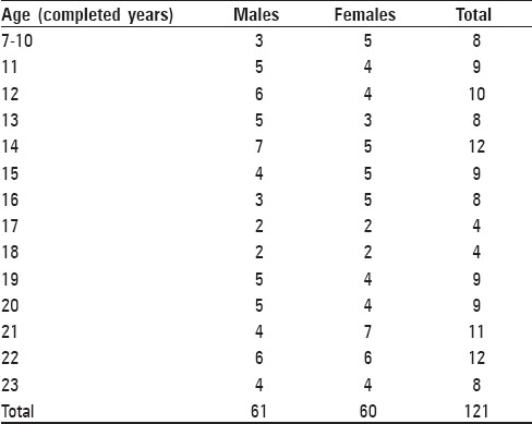
The study was carried out on 121 digital orthopantamograms (OPG) of patients in the age group of 7–23 years (males = 61; females = 60) [Table 1] which were predominantly pre-treatment orthodontic radiographs from patients without any obvious developmental anomalies. The radiographs were taken from the archives of patients visiting our college during the years 2008–2010. The soft copy of these radiographs were retrieved from the computer attached to the digital OPG machine (Orthophos XG5, Sirona Dental Systems, Inc., Long Island, NY, USA).
Table 1.
Sample distribution across age-groups and sexes during the study
The inclusion criteria of the radiographs were:
Patients free of obvious developmental anomalies
OPGs without any distortions
Radiographs of patients with the full complement of teeth in the mandibular left or right side
The exclusion criteria were:
Radiographs of patients with developmental anomalies
Distortion and crowding of teeth where the root structures of the teeth were not clearly discernible
Radiographs of patients with bilaterally missing teeth in the mandible
The radiographs collected were divided into the following groups:
Group A: Males in the age group of 7–16 years
Group B: Males in the age group of 16.1–23 years
Group C: Females in the age group of 7–16 years
Group D: Females in the age group of 16.1–23 years.
The rationale for dividing the sample based on sex was that the maturity scores assigned to each tooth based on its developmental stages was gender specific due to the differing rates in the development of the teeth in either sex.[4] Within each sex, the sample was divided into two subgroups to assess the reliability of the third molar in age estimation, since after 16 years, it is only the third molar, which is still developing under normal conditions. As the images were from the digital OPG machine, they were exported to JPEG format using the Sidexis Next generation imaging software, ver 2.4®, integrated with the Orthophos XG5 Sirona OPG machine (Sirona Dental Systems, Inc., Long Island, NY, USA)®. The digital images were then analysed with Adobe Photoshop 7.0 (San Jose, CA, USA). During the analysis, ‘Magnify’ and ‘Ruler’ tools were used.
The teeth on the mandibular left side - from the mandibular central incisor to the mandibular third molar - were scored based on the Demirjian's modified criteria, which included ten stages of tooth development.[4] A representation of the stages based on the development of mandibular third molar from our own radiographs used for the study are depicted in Figure 1. The stages were entered into a separate scoring pro-forma following which the sex-specific maturity score for each tooth was entered depending on the scoring grade. Since, at present, no India-specific maturity scores exists, the French maturity scores, as modified by Challiet and Demirjian[4] and used by Acharya[3] himself in his study to establish the formula, were used. If a tooth in the mandibular left quadrant was missing the contralateral tooth was included in the study.
Figure 1.
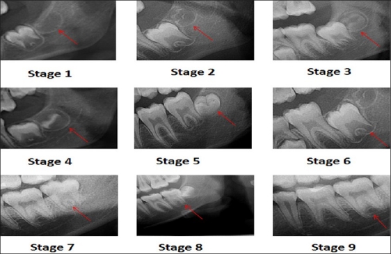
Representative stages of Demirjian's method based on the development of mandibular third molar as seen on the radiographs used during the study
The scores were summed up and substituted in the regression formula shown below:[3]
Males: Age = 27.4351 – (0.0097×S2) + (0.000089×S3)
Females: Age = 23.7288 – (0.0088×S2) + (0.000085×S3)
The value so obtained was designated as the age calculated. The chronologic age of the patient was obtained from the date of birth recorded into the Sidexis software, which is mandatory to obtain a radiograph in the digital OPG machine, and the date of exposure as retrieved from the patient database on the software. To rule out intra-observer difference, 30 randomly selected radiographs were re-evaluated. The inter-observer agreeability was arrived at by scoring 30 randomly selected radiographs between the two authors and the results obtained were compared. The intra-observer difference and inter observer agreeability were evaluated using the Wilcoxon signed rank test. The calculation of results and statistical analysis were carried out using Microsoft Excel (MS Office 2010 Microsoft Corp., Redmond, WA, USA) and Statistical package for the social sciences (SPSS) statistical software Version 10 (SPSS Inc, Chicago, IL, USA). The effectiveness of age prediction is usually represented by the mean absolute error (MAE), which is calculated as the difference between the estimated age and the actual age at the time of exposure, and the number of estimates that fell in the error group of <±1 year, within 1.1-2 years, and >±2 years. In age estimation studies, generally MAE is considered as a standard measure to estimate the effectiveness of the methods.[3,5,6] Error of <±1 year is considered a good result and error rate of >±2 years is considered as inaccurate.[3]
Results
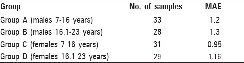
Test for inter-observer difference and intra-observer bias did not yield any significant statistical variation (P>0.05). The results of the study were calculated based on the grouping as mentioned above. It was found that the overall MAE was 1.18 years. The most accurate estimation as denoted by a smaller MAE was obtained in Group C (females, 7 to 16 years) at 0.95 years followed by Group D (females, 16.1 to 25 years) at 1.16 years. In case of males, the error rates were slightly higher at 1.2 years in Group A (7 to 16 years) and 1.3 years in Group B (16.1 to 23 years) [Table 2]. 70 out of the 121 test subjects (approximately 57.85%) were estimated to be within ±1 year while 38 (approximately 31.40%) age estimates fell within 1.1 to 2 years from the actualage; in 13 samples (approximately 10.75%), age estimates fell outside the ±2 year range. It was noted that in case of 20 samples the actual age of the person was greater than 21 years.
Table 2.
Error of age estimation in all the groups during the study
Discussion
The estimation of age is an important exercise in medico-legal practice. The need for age estimation has certain important reasons at certain specific age groups in the Indian context: 1) 12 years: children below this age are not liable for certain offences; 2) 14 years: a child cannot be employed below 14 years; 3) 18 years: determines the status of majority and the legally permissible age for marriage in females; 4) 21 years: the legally permissible age of marriage in males.[3,7] Teeth have contributed to an array of methods for age estimation which include both - methods requiring the extraction of teeth and histopathological examination,and in-vivo methods which merely require radiographs.[8]
Demirjian introduced a method for age estimation in children, adolescents and young adults which relies on the developmental stages of teeth.[2] The original method relied on the mandibular left side teeth from the central incisor to the second molar and had assigned eight stages of development which were categorised as A to H. A score was assigned as a function of age and the predictive interval was given for the maturity score and computed to obtain the age.[2] This original method of age estimation enabled the clinicians to know the deviation of the dental maturity for one individual, however was inappropriate for age determination.[4,9] Furthermore, when the method was applied to the Indian population, it resulted in an average overestimation of about 3 years.[10] Hence to overcome the shortcomings, polynomial or multiple regression analysis was used to obtain an age with a confidence interval as a function of score and also to limit the problems of missing data.[11]
The original Demirjian's method using the seven mandibular teeth had a high accuracy but poor reliability. For an age estimation method to be accurate and reliable is a tedious task as they are contrarian parameters. Hence the goal is to achieve the best of these two factors using the appropriate method and the most adopted biological indicators.[4] The original Demirjian's method excluded the third molar due to the variability in its development, eruption and anatomy.[5] However, the pitfall of its exclusion was that the age prediction by the original Demirjian's method is not feasible after about 16 years of age, which coincides with the completion of the root development of the second molar.[12] The third molar offers the only reliable radiological parameter for age determination in the age group of 16–23 years.[13] In a study carried out by Mincer et al., it was concluded that the third molar may provide reasonable accuracy for the likelihood that a person is atleast 18 years of age, rather than giving the exact chronological age, due to the absence of any other marker in the late adolescence.[13]
Hence subsequently Chaillet and Demirjian[4] modified their method to incorporate the third molar and developed a new maturity score based on a French population. Another major modification made in this study was that the stages of teeth were modified to include two additional stages of non-formation of tooth (Stage “0”) and crypt development (Stage “1”); furthermore, the stage of development were assigned numerals which were designated as 0–9 for easier calculation and develop a multiple regression formula based on cubic functions which gave better reliability when the third molar was incorporated into the study.[4]
Previous studies have found no observer errors using the original method, and Acharya[3] found the same even for the modified method, which our results confirm. Acharya's study, which arrived at the formula used in the present assessment, had deduced an MAE of 1.43 years, with 44% of samples within ± 1 year, 36% within ± 1.1 to ± 2 years, and 20% beyond ± 2 years.[3] In the present study, the MAE (1.18 years) for the overall sample was slightly better than the original study. One difference between the previous study and our study was the use of digital OPG's for analysis. The study resulted in better age prediction in females than in males. The MAEs were lower in the younger age group in both males and females, indicating that the presence of third molar only in 16.1 to 23 year old individuals results in greater inaccuracy in age estimation, which was also noted recently by Acharya.[3] Moreover, beyond the age of 21 years, the MAE had risen considerably to 1.62 years where the number of samples where only 18. The reason attributed is due to the completion of third molar development and the paucity of samples.[3]
Conclusion
In conclusion, the reliability of age estimation using the Demirjian's 8 teeth method following the French maturity scores and India specific formula provides fairly reliable results. This has resulted in the error of age prediction narrowing down to just over 1 year, which is a slight improvement compared to the original method carried out in the Indian population.[3] We also noted that incorporation of the third molar results in slightly greater errors in age estimates, as was also noted previously.[3] As in-vivo non extraction method of age estimation would be the most sought after method to determine the age in case of medico-legal issues, and this method may prove to be useful. However, the pitfalls of this method is the lack of India weighted maturity scores which, if developed, can go further to increase the accuracy of this method. Although this method is more of a subjective method of determining the stage of tooth development, no significant variations were noted in the inter observer agreeability and intra observer bias during our study.
Acknowledgments
We wish to thank Prof. Dr. P. Jayakumar, Principal and Head of Department of Orthodontics, Meenakshi Ammal Dental College and Hospital, for giving permission to access their archives of pre-treatment radiographs.
Footnotes
Source of Support: Nil
Conflict of Interest: None declared
References
- 1.Willems G, Moulin-Romsee C, Solheim T. Non-destructive dental-age calculation methods in adults: Intra and inter observer effects. Forensic Sci Int. 2002;126:221–6. doi: 10.1016/s0379-0738(02)00081-6. [DOI] [PubMed] [Google Scholar]
- 2.Demirjian A, Goldstein H, Tanner JM. A new system of dental age assessment. Hum Biol. 1973;45:211–27. [PubMed] [Google Scholar]
- 3.Acharya AB. Age estimation in Indians using Demirjian's 8-teeth method. J Forensic sci. 2011;56:124–7. doi: 10.1111/j.1556-4029.2010.01566.x. [DOI] [PubMed] [Google Scholar]
- 4.Chaillet N, Demirjian A. Dental maturity in South France: A comparison between Demirjian's method and polynomial functions. J Forensic Sci. 2004;49:1059–66. [PubMed] [Google Scholar]
- 5.Solari AC, Abramovitch K. The accuracy of age and precision of third molar development as an indicator of chronologic age in Hispanics. J Forensic Sci. 2002;47:531–5. [PubMed] [Google Scholar]
- 6.Arany S, Lino M, Yoshioka N. Radiographic survey of third molar development in relation to chronologic age among Japanese juveniles. J Forensic Sci. 2004;49:534–8. [PubMed] [Google Scholar]
- 7.Reddy KSN. The essential of forensic medicine andtoxicology. 3rd ed. Hyderabad: K Suguna Devi publication; 2009. pp. 73–4. [Google Scholar]
- 8.Soomer H, Ranta H, Lincoln MJ, Pentillä A, Leibur E. Reliability and validity of eight dental age estimation methods of adults. J Forensic Sci. 2003;48:149–52. [PubMed] [Google Scholar]
- 9.Sherrer B. In: Biostastique. Morin G, editor. Montrea: Quebec publishers; 1984. [Google Scholar]
- 10.Koshy S, Tandon S. Dental age assessment: the applicability of the assessment in south Indian children. Forensic SciInt. 1998;94:73–85. doi: 10.1016/s0379-0738(98)00034-6. [DOI] [PubMed] [Google Scholar]
- 11.Teivens A, Mörnstad H. A modification of the Demirjian method for age estimation in children. J Forensic Odontostomatol. 2001;19:26–30. [PubMed] [Google Scholar]
- 12.Ash M, Nelson SJ. Wheeler's dental anatomy, physiology and occlusion. 8th ed. London: W B Saunders company Ltd; 2006. p. 316. [Google Scholar]
- 13.Mincer HH, Harris EF, Berryman HE. The ABFO study of third molar development and its use as an estimator of chronological age. J Forensic Sci. 1993;38:379–90. [PubMed] [Google Scholar]