

Abstract
Monopolar mitotic apparatus can be produced in sea urchin eggs by a manoeuvre that distributes the four poles of the second mitosis into four separate blastomeres. The pole of the monopolar mitotic apparatus generates a half-spindle that is similar in structural details to the half-spindle of a normal bipolar mitotic apparatus, although the chromosomes are not as well aligned as in a normal metaphase plate. The chromosomes are oriented; one kinetochore faces the pole while its sister kinetochore faces away from the pole. The poleward kinetochore is connected to the pole by bundles of microtubules. No microtubules are seen on the sister kinetochore that faces away from the pole. Therefore, a single pole can direct most of the events in the establishment of a mitotic apparatus. Our interpretation examines the cooperation of kinetochores and poles in the formation of microtubules between them, stressing the half-spindle as the medium of cooperation and leaving open the question whether the kinetochores are origins or terminations of microtubules.
Full text
PDF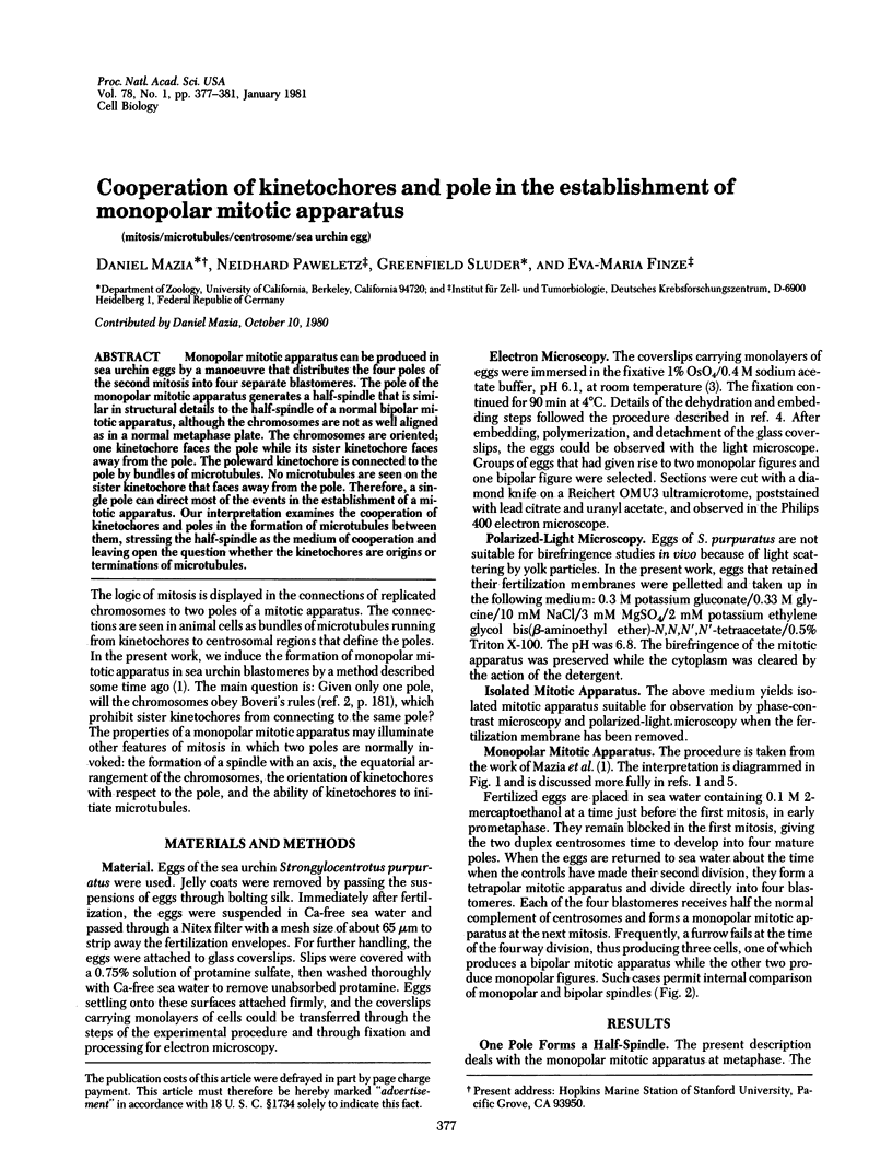
Images in this article
Selected References
These references are in PubMed. This may not be the complete list of references from this article.
- Andersen B., Osborn M., Weber K. Specific visualization of the distribution of the calcium dependent regulatory protein of cyclic nucleotide phosphodiesterase (modulator protein) in tissue culture cells by immunofluorescence microscopy: mitosis and intercellular bridge. Cytobiologie. 1978 Aug;17(2):354–364. [PubMed] [Google Scholar]
- Bajer A. Chromosome movement and fine structure of the mitotic spindle. Symp Soc Exp Biol. 1968;22:285–310. [PubMed] [Google Scholar]
- HARRIS P. Some structural and functional aspects of the mitotic apparatus in sea urchin embryos. J Cell Biol. 1962 Sep;14:475–487. doi: 10.1083/jcb.14.3.475. [DOI] [PMC free article] [PubMed] [Google Scholar]
- Paweletz N., Mazia D. Fine structure of the mitotic cycle of unfertilized sea urchin eggs activated by ammoniacal sea water. Eur J Cell Biol. 1979 Oct;20(1):37–44. [PubMed] [Google Scholar]
- Telzer B. R., Moses M. J., Rosenbaum J. L. Assembly of microtubules onto kinetochores of isolated mitotic chromosomes of HeLa cells. Proc Natl Acad Sci U S A. 1975 Oct;72(10):4023–4027. doi: 10.1073/pnas.72.10.4023. [DOI] [PMC free article] [PubMed] [Google Scholar]
- Tippit D. H., Pickett-Heaps J. D., Leslie R. Cell division in two large pennate diatoms Hantzschia and Nitzschia III. A new proposal for kinetochore function during prometaphase. J Cell Biol. 1980 Aug;86(2):402–416. doi: 10.1083/jcb.86.2.402. [DOI] [PMC free article] [PubMed] [Google Scholar]
- Welsh M. J., Dedman J. R., Brinkley B. R., Means A. R. Calcium-dependent regulator protein: localization in mitotic apparatus of eukaryotic cells. Proc Natl Acad Sci U S A. 1978 Apr;75(4):1867–1871. doi: 10.1073/pnas.75.4.1867. [DOI] [PMC free article] [PubMed] [Google Scholar]