Abstract
The immunopathogenesis of Chlamydia trachomatis-induced oviduct pathological sequelae is not well understood. Mice genetically deficient in perforin (perforin−/− mice) or tumor necrosis factor alpha (TNF-α) production (TNF-α−/− mice) displayed comparable vaginal chlamydial clearance rates but significantly reduced oviduct pathology (hydrosalpinx) compared to that of wild-type mice. Since both perforin and TNF-α are effector mechanisms of CD8+ T cells, we evaluated the role of CD8+ T cells during genital Chlamydia muridarum infection and oviduct sequelae. Following vaginal chlamydial challenge, (i) mice deficient in TAP I (and therefore the major histocompatibility complex [MHC] I pathway and CD8+ T cells), (ii) wild-type mice depleted of CD8+ T cells, and (iii) mice genetically deficient in CD8 (CD8−/− mice) all displayed similar levels of vaginal chlamydial clearance but significantly reduced hydrosalpinx, compared to those of wild-type C57BL/6 mice, suggesting a role for CD8+ T cells in chlamydial pathogenesis. Repletion of CD8−/− mice with wild-type or perforin−/−, but not TNF-α−/−, CD8+ T cells at the time of challenge restored hydrosalpinx to levels observed in wild-type C57BL/6 mice, suggesting that TNF-α production from CD8+ T cells is important for pathogenesis. Additionally, repletion of TNF-α−/− mice with TNF-α+/+ CD8+ T cells significantly enhanced the incidence of hydrosalpinx and oviduct dilatation compared to those of TNF-α−/− mice but not to the levels found in wild-type mice, suggesting that TNF-α production from CD8+ T cells and non-CD8+ cells cooperates to induce optimal oviduct pathology following genital chlamydial infection. These results provide compelling new evidence supporting the contribution of CD8+ T cells and TNF-α production to Chlamydia-induced reproductive tract sequelae.
INTRODUCTION
Genital Chlamydia trachomatis infection in humans leads to pathological sequelae in the upper genital tract (UGT), including pelvic inflammatory disease, ectopic pregnancy, and infertility (3, 13, 23). Studies from humans and the mouse model of Chlamydia muridarum genital infection have provided important clues toward the mechanisms of pathogenesis and protective immunity against genital chlamydial infection. Cell-mediated immunity, particularly T helper (Th) 1 type CD4+ T cells and gamma interferon (IFN-γ) production, has been shown to be important for chlamydial clearance after a primary genital infection (3, 18, 23, 25, 32). In contrast, the pathogenic mechanisms of chlamydial UGT sequelae are less well understood.
It is generally accepted that the host immune response to infection leads to the pathological sequelae in the UGT (3, 12, 13). The inciting event for Chlamydia-induced sequelae appears to be the cellular damage in infected tissues, which is followed by fibrotic reparative mechanisms in the reproductive mucosa. Cellular damage may occur via perforin-mediated cytolysis of infected cells (34). In this regard, Perry et al. (33) reported that perforin-deficient mice display a reduced incidence of hydrosalpinx compared to that of wild-type control mice following genital C. muridarum challenge. Another mechanism for potent induction of apoptosis and/or fibrosis in infected cells is the effect of tumor necrosis factor alpha (TNF-α) (1, 11, 14, 30, 31). For example, chlamydial scarring trachoma has been shown to be associated with polymorphisms in TNF-α gene promoter and elevated levels of TNF-α in the tear fluid (10). Thus, it would seem that perforin-mediated cytolysis and TNF-α production may be important mediators of chlamydial pathogenesis leading to the severe UGT disease sequelae.
CD8+ T cells are an important cell type that utilizes both perforin-mediated cytolysis and TNF-α production as effector mechanisms for killing of target cells. Resolution of genital C. muridarum infections induced by some Chlamydia-specific CD8+ T cell clones has been shown to correlate with the levels of TNF-α production (17). However, CD8+ T cells, TNF-α production and perforin-mediated cytolysis have been suggested to play a minor role in the clearance of genital C. muridarum infections (12, 24, 33). Despite a minimal role in chlamydial clearance, a contribution of CD8+ T cells to chlamydial pathogenesis has been suggested by correlative evidence in different models, including the induction of infertility in the mouse model of C. muridarum genital infection (16), salpingitis in nonhuman primates (41, 42), and trachoma in human individuals (19).
In this study, we directly evaluated the contribution of CD8+ T cells to chlamydial clearance and oviduct pathology following primary genital chlamydial challenge, using three different models of CD8+ T cell deficiency: (i) mice that are genetically deficient in the major histocompatibility complex (MHC) I processing pathway and lacking CD8+ T cells (transport-associated protein I [C57BL/6 TAP I]−/− mice) (40), (ii) C57BL/6 mice treated with neutralizing antibody to deplete the CD8+ T cell compartment, and (iii) CD8 gene-deficient (C57BL/6 CD8−/−) mice. We found that CD8+ T cells contribute significantly to the oviduct pathological sequelae, but not bacterial clearance, following primary genital chlamydial challenge. Importantly, the reduction in pathology in CD8−/− mice could be reversed by repletion of the CD8+ T cell compartment. Moreover, we found that TNF-α production, not perforin, from CD8+ T cells is primarily responsible for mediating this effect.
MATERIALS AND METHODS
Bacteria.
Chlamydia muridarum subtype Nigg was grown on confluent HeLa cell monolayers as described previously (29, 44). Cells were lysed by sonication, and elementary bodies (EBs) were purified on Renografin gradients. Aliquots of bacteria were stored at −70°C in sucrose-phosphate-glutamine (SPG) buffer.
Mice.
Four- to six-week-old female mice were used for all experiments. Mice deficient in (i) transport-associated protein 1 (TAP I; TAP I−/− mice) and therefore incapable of MHC class I pathway processing and lacking CD8+ T cells, (ii) CD8 (CD8−/− mice), (iii) TNF-α production (TNF-α−/− mice), and (iv) perforin (perforin−/− mice) and (v) wild-type C57BL/6 mice were purchased from Jackson Laboratory (Bar Harbor, ME) and bred at the University of Texas at San Antonio (UTSA). Animal care and experimental procedures were performed at the UTSA in compliance with the Institutional Animal Care and Use Committee (IACUC) guidelines.
Vaginal infection, determination of chlamydial shedding, and UGT pathology.
At 10 and 3 days prior to challenge, mice were treated with 2.5 mg of Depo-Provera (Upjohn, Kalamazoo, MI) and subsequently infected intravaginally (i.vag.) on day 0 with 5 × 10⁴ inclusion-forming units (IFU) of C. muridarum. Vaginal swab material was collected every third day after challenge, and chlamydial enumeration was conducted by plating swab material on HeLa cell monolayers followed by immunofluorescent staining (9). On day 80 after challenge, mice were euthanized; the genital tracts were removed, placed next to a standard metric ruler, and photographed, gross oviduct diameter was measured for each oviduct, and the results were reported individually and as mean ± standard error of the mean (SEM) in a group, as described previously (27). Additionally, the number of normal oviducts (numerator) and the total number of oviducts evaluated (denominator) have been indicated in the parentheses of each respective figure. The enumeration of chlamydial counts and oviduct measurements was conducted in a blinded fashion.
Estimation of granzyme B and TNF-α production in the genital tract.
On the day of i.vag. C. muridarum (5 × 10⁴ IFU) challenge, and every week thereafter, groups of three mice were euthanized, the upper genital tracts (uterine horns and oviducts but not ovaries) were homogenized and centrifuged to remove debris, and the supernatants were collected. The total protein content was evaluated using the Bradford assay. The supernatants were then analyzed for granzyme B production and for TNF-α production using enzyme-linked immunosorbent assay (ELISA) kits (eBioscience, San Diego, CA), according to the manufacturer's instructions. The cytokine values were normalized to total protein content and mean ± SEM of the values per group shown for each analyzed time point.
In vivo CD8+ T cell depletion.
The hybridoma cell line producing anti-CD8 neutralizing antibody (clone 2.43) (26) was purchased from ATCC and grown according to the manufacturer's instructions. Antibodies were purified using ammonium sulfate precipitation. On days −6, −5, −4, and −3, and on the day of challenge and every third day afterwards, animals were injected intraperitoneally (i.p.) with 0.5 mg of purified anti-CD8 monoclonal antibody or control rat IgG. The last injection was given on day 27 after challenge. In another group of mice, the injections were given between days 30 and 78 on every third day. Cellular depletion was monitored by flow cytometry using a PE-Cy7-labeled anti-CD8 antibody (BD Biosciences).
Adoptive transfer of CD8+ T cells.
Mice were euthanized, and their spleens were removed. Single cell suspensions were made and layered over a Ficoll density gradient (Cedarlane Laboratories, Canada) to obtain mononuclear cells. CD8+ T cell populations were enriched by negative selection using magnetic particles (Stem Cell Technologies). The purity of the CD8+ T cell population was determined to be at least >95% of total splenocytes by flow cytometry using a PE-Cy7-labeled anti-CD8 antibody (BD Biosciences). The CD8+ T cells were identified within the lymphocytic population (forward and side scatter parameters), and >99% of these cells were stained positively with a PerCP-labeled anti-CD3 antibody (BD Biosciences). Two hours before transfer, female C57BL/6 mice (4 to 8 weeks old) were challenged i.vag. with 5 × 10⁴ IFU of C. muridarum. Adoptive transfer was accomplished with 106 CD8+ T cells transferred intravenously into the mice.
Statistics.
Sigma Stat (Systat Software Inc., San Jose, CA) was used to perform all tests of significance. Student's t test was used for comparisons between two groups and analysis of variance (ANOVA) between multiple groups for vaginal chlamydial shedding and dilatation of hydrosalpinx. A chi-square test was used to compare the incidence of hydrosalpinx. P ≤ 0.05 was considered statistically significant. All experiments were repeated at least twice, and each experiment was analyzed independently. In some experiments, where oviduct diameter data are shown as a composite of two experiments, the indicated significant difference holds true when the experiments are analyzed individually.
RESULTS
Deficits in cytolytic activity or TNF-α production do not alter genital chlamydial clearance but result in reduced oviduct pathology.
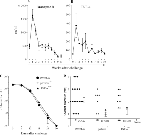
Based on suggestions from previous studies that cytolytic activity (33) as well as TNF-α induced cellular apoptosis (12) may be important in genital chlamydial infection and sequelae, we evaluated the levels of granzyme B and TNF-α, respectively, in the upper genital tracts of female C57BL/6 mice at weekly intervals for 10 weeks following i.vag. C. muridarum (5 × 10⁴ IFU) challenge. As shown in Fig. 1A, granzyme B levels were elevated at the end of weeks 1 (1,631 ± 213 pg/ml) and 2 (1,045 ± 185 pg/ml) compared to those of naïve mice (week 0, 478 ± 87 pg/ml) and reduced to prechallenge levels during subsequent time periods. Additionally, TNF-α levels (Fig. 1B) were elevated at the end of weeks 1 (372 ± 132 pg/ml), 2 (174 ± 28 pg/ml), 3 (157 ± 78 pg/ml), and 4 (199 ± 86 pg/ml), compared to those of naïve mice (week 0, 57 ± 4 pg/ml) and reduced to prechallenge levels during subsequent time periods. We then evaluated the course of chlamydial clearance and development of oviduct pathology in perforin−/− (deficient in cytolytic activity) and TNF-α−/− mice. As shown in Fig. 1C, wild-type C57BL/6 mice shed high numbers of chlamydial organisms from days 3 to 6 with progressive reduction thereafter and completely cleared the vaginal infection by day 30 after challenge. The kinetics of chlamydial shedding in perforin−/− and TNF-α−/− mice were comparable to those in the C57BL/6 mice. On day 80 after challenge, the number of oviducts developing hydrosalpinx and the gross diameter of respective dilated oviducts were evaluated. As shown in Fig. 1D, the incidence of hydrosalpinx and oviduct dilatation were significantly (P ≤ 0.05) reduced in both perforin−/− (29% positive oviducts, 0.95 ± 0.17 mm) and TNF-α−/− mice (37% positive oviducts, 1.14 ± 0.2 mm) compared to those in C57BL/6 animals (87.5% positive oviducts, 2.1 ± 0.22 mm). These results suggest that both perforin-mediated cytolytic activity and TNF-α are involved in inducing oviduct pathology, but not bacterial clearance, following primary genital chlamydial challenge. Perforin-mediated cytolytic activity is an important effector mechanism by which CD8+ T cells induce apoptosis in infected cells (34). Moreover, antigen-specific TNF-α production by C57BL/6 Chlamydia-specific CD8+ T cells (1.4 ± 0.07 ng/ml) was significantly (P ≤ 0.05) greater than from equal numbers of CD4+ T cells (0.8 ± 0.12 ng/ml) (data not shown), suggesting that CD8+ T cells may be an important source of both perforin and TNF-α production involved in chlamydial pathogenesis.
Fig. 1.
Role of perforin and TNF-α in genital chlamydial infection and pathology. (A and B) Groups (n = 33) of C57BL/6 mice were challenged i.vag. with C. muridarum, and upper genital tracts (n = 3) were collected on day 0 before challenge and at weekly intervals after challenge for 10 consecutive weeks. Tissue homogenates were examined for levels of granzyme B (A) and TNF-α (B). Results are expressed as mean ± SEM of cytokine level at each time point per group and are representative of two independent experiments. (C and D) Groups (n = 6) of perforin−/−, TNF-α−/−, and C57BL/6 mice were challenged i.vag. with C. muridarum. (C) The vaginal chlamydial shedding was measured over a period of 1 month following challenge. (D) On day 80 after challenge, the gross oviduct diameter was measured. Each individual marker represents one oviduct, and the mean ± SEM of oviduct diameter per group is also shown. The number of normal oviducts (numerator) and the total number of oviducts evaluated (denominator) per respective group of mice have been indicated in parentheses. *, significant (P ≤ 0.05) difference between indicated group and C57BL/6 mice in incidence of hydrosalpinx (chi-square test) and the oviduct diameter (ANOVA). Results are a composite from two independent experiments.
Deficiency of CD8+ T cells does not affect bacterial clearance following genital C. muridarum challenge.
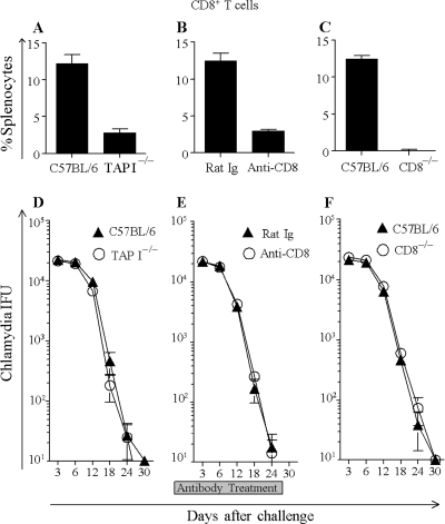
CD8+ T cells were present in minimal numbers in the uninfected mouse genital tract, but following C. muridarum challenge, they progressively infiltrated the genital tract up to the end of week 2, followed by a progressive reduction in numbers by 5 weeks after challenge to the levels seen in naïve mice (data not shown). We also evaluated the role of CD8+ T cells in clearance of infection following i.vag. C. muridarum challenge using different mouse models of CD8+ T cell deficiency. As shown in Fig. 2A, the frequency of splenic CD8+ T cells in TAP I−/− mice (2.8% ± 0.5%) was significantly reduced compared to that in wild-type animals (12.2% ± 1.2%), as also has been shown previously (20). As shown in Fig. 2D, C57BL/6 mice shed high numbers of chlamydial organisms from days 3 to 9, with progressive reduction and complete clearance of the vaginal infection by day 27 after challenge. The kinetics of chlamydial shedding in TAP I−/− mice was comparable to that in the wild-type mice (Fig. 2D). Since TAP I−/− mice have a residual CD8+ T cell population, we also evaluated C57BL/6 mice depleted of the CD8+ T cell compartment using neutralizing anti-CD8 antibody injections. As shown in Fig. 2B, anti-CD8 antibody treatment significantly depleted CD8+ T cells (3% ± 0.2% remaining), whereas mice receiving control rat Ig (12.5% ± 1% remaining) did not exhibit appreciable alterations in splenic CD8+ T cell frequency compared to untreated wild-type animals (12.2% ± 1.2%). Both anti-CD8 antibody- and control Ig-treated mice cleared the vaginal infection by day 27 after challenge (Fig. 2E), similar to untreated C57BL/6 animals. Since neutralizing antibody depletion also did not result in total depletion of the CD8+ T cell compartment, we also evaluated CD8−/− mice that display a total deficiency of CD8+ T cells (Fig. 2C). As shown in Fig. 2F, CD8−/− mice displayed clearance of the genital chlamydial infection by day 30 after challenge, which was comparable to the result for C57BL/6 mice. Collectively, these results from three different mouse models demonstrate that a deficiency of CD8+ T cells does not significantly affect chlamydial clearance following primary i.vag. C. muridarum challenge.
Fig. 2.
Role of CD8+ T cells in genital chlamydial infection. (A to C) Groups (n = 3) of mice (C57BL/6 and TAP I−/− [A], control rat Ig and anti-CD8 antibody treated [B], and C57BL/6 and CD8−/− [C]) were euthanized, and splenic populations of CD8+ T cells were analyzed by flow cytometry. Results are shown as mean ± SEM of the percentage of CD8+ T cells in total splenocytes and are representative of two independent experiments. (D to F) Groups (n = 6) of mice (C57BL/6 and TAP I−/− [D], control rat Ig and anti-CD8 antibody treated [E], and C57BL/6 and CD8−/− [F]) were challenged with 5 × 10⁴ IFU of C. muridarum on day 0, and chlamydial shedding was enumerated every third day for a period of 1 month. The mean ± SEM of vaginal chlamydial shedding per group at each time point is shown. Results are representative of three independent experiments.
Deficiency of CD8+ T cells leads to reduced oviduct pathology following genital C. muridarum challenge.
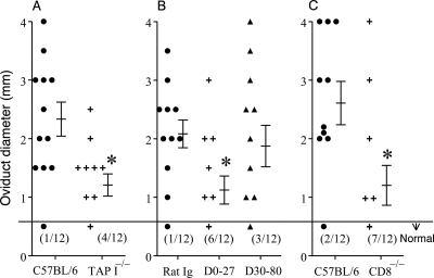
The development of hydrosalpinx, a characteristic marker of reproductive tract pathological sequelae, was characterized at day 80 following genital chlamydial challenge in the three different mouse models of CD8+ T cell deficiency. Our extensive analyses conducted previously have demonstrated that oviduct sequelae are fully developed by day 80 after genital chlamydial challenge in mice (28, 31). As shown in Fig. 3A, TAP I−/− mice displayed a reduction in the incidence of hydrosalpinx (67% oviducts) and a significant (P ≤ 0.05) reduction in oviduct dilatation (1.21 ± 0.2 mm), compared to those in C57BL/6 mice (92% and 2.3 ± 0.3 mm, respectively) following primary genital chlamydial challenge. Significant (P ≤ 0.05) reductions in the incidence of hydrosalpinx and oviduct dilatation also were observed in mice treated with anti-CD8 antibody (Fig. 3B) during the infection between days 0 and 27 (50% and 1.1 ± 0.24 mm, respectively) but not afterwards between days 30 and 80 (75% and 1.9 ± 0.4 mm, respectively), compared to results for control rat Ig-treated animals (92% and 2.1 ± 0.23 mm, respectively). Additionally, CD8−/− mice (Fig. 3C) displayed significantly (P ≤ 0.05) reduced incidences of hydrosalpinx (42%) and oviduct dilatation (1.2 ± 0.34 mm) following primary chlamydial challenge, compared to results for infected wild-type C57BL/6 mice (83% and 2.6 ± 0.4 mm, respectively). Collectively, these results from three different mouse models suggest that a deficiency of CD8+ T cells leads to significant reductions in upper genital tract pathological sequelae following primary chlamydial challenge.
Fig. 3.
Role of CD8+ T cells in oviduct sequelae after vaginal C. muridarum infection. Groups (n = 6) of mice (C57BL/6 and TAP I−/− [A], control rat Ig and anti-CD8 antibody treated [B], and C57BL/6 and CD8−/− [C]) were challenged with 5 × 10⁴ IFU of C. muridarum on day 0, and the macroscopic oviduct diameter was measured on day 80 after challenge. Each individual marker represents one oviduct, and the mean ± SEM of oviduct diameter per group of mice also is shown. The number of normal oviducts (numerator) and the total number of oviducts evaluated (denominator) per respective group of mice have been indicated in parentheses. *, significant (P ≤ 0.05, Student's t test) difference in oviduct diameter between the indicated group and C57BL/6 mice in panels A and C or control rat Ig-treated mice in panel B). In panels B and C, * indicates significant (P ≤ 0.05, chi-square test) difference in oviduct diameter between the indicated group and control rat Ig-treated mice or C57BL/6 mice, respectively. Results are representative of three independent experiments.
TNF-α production from CD8+ T cells mediates Chlamydia-induced oviduct pathology.
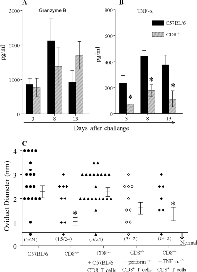
The production of granzyme B and TNF-α in the upper genital tracts of C57BL/6 and CD8−/− mice was compared on days 3, 8, and 13 following genital C. muridarum challenge. As shown in Fig. 4A, granzyme B production in the upper genital tract of CD8−/− mice on days 3, 8, and 13 following challenge (756 ± 265 pg/ml, 1,386 ± 851 pg/ml, and 1,695 ± 402 pg/ml, respectively) was not significantly different from that in C57BL/6 mice (857 ± 174 pg/ml, 2,123 ± 636 pg/ml, and 927 ± 323 pg/ml, respectively), although the CD8−/− mice had reduced levels of granzyme B on day 8 after challenge compared to levels in C57BL/6 mice. TNF-α production (Fig. 4B) was significantly (P ≤ 0.05) reduced on days 3, 8, and 13 following challenge in the upper genital tract of CD8−/− mice (72 ± 15 pg/ml, 179 ± 43 pg/ml, and 113 ± 63 pg/ml, respectively) compared to that for C57BL/6 mice (235 ± 57 pg/ml, 444 ± 43 pg/ml, and 379 ± 72 pg/ml, respectively). These results suggested that cytolytic activity and TNF-α production may be potential mechanisms by which CD8+ T cells mediate pathogenesis during genital chlamydial infections. To directly examine the causation of oviduct pathology by CD8+ T cells and associated mechanisms, we repleted CD8−/− mice with wild-type, perforin−/−, or TNF-α−/− CD8+ T cells at the time of challenge. Specifically, CD8+ T cells were purified from donor mice and 1 × 10⁶ cells (>95% CD8+ T cells) were injected intravenously 2 h before i.vag. chlamydial challenge into recipient CD8−/− mice. Following challenge, we found that the adoptively transferred CD8+ T cells infiltrated into the genital tract of the CD8−/− mice (2.1% ± 1.2% of upper genital tract cells at the end of week 1 and 3.3% ± 1.7% at the end of week 2) with kinetics and frequencies comparable to those of the CD8+ T cell infiltration in wild-type C57BL/6 mice (2.6% ± 0.5% at week 1 and 4.4% ± 1% at week 2). As expected, the course of chlamydial clearance was not significantly different between the groups, with all groups of mice displaying complete clearance of the vaginal infection by day 30 following challenge (data not shown). The mice were evaluated further for hydrosalpinx development on day 80 after challenge. As shown in Fig. 4, CD8−/− mice displayed significant (P ≤ 0.05) reduction in the incidence of hydrosalpinx (37%) and oviduct dilatation (1 ± 0.2 mm) compared to those of C57BL/6 mice (79% and 2.27 ± 0.24 mm). Importantly, oviducts of CD8−/− mice repleted with C57BL/6 CD8+ T cells displayed a significantly greater incidence of hydrosalpinx (88%) and oviduct dilatation (2.28 ± 0.18 mm) than those of CD8−/− mice and comparable to incidences in wild-type C57BL/6 mice, suggesting that CD8+ T cells mediate Chlamydia-induced oviduct pathology. Furthermore, oviducts of CD8−/− mice repleted with perforin−/− CD8+ T cells displayed an increase in the incidence of hydrosalpinx (75%) and oviduct dilatation (1.6 ± 0.3 mm) but not to the extent of those of CD8−/− mice repleted with C57BL/6 CD8+ T cells. However, oviducts of CD8−/− mice repleted with TNF-α−/− CD8+ T cells displayed a significant reduction in the incidence of hydrosalpinx (50%) and oviduct dilatation (1.3 ± 0.3 mm) compared to those in CD8−/− mice repleted with C57BL/6 CD8+ T cells. Collectively, these results suggest that TNF-α production from CD8+ T cells may be more important than perforin in mediating the oviduct pathological sequelae following primary genital chlamydial challenge.
Fig. 4.
Role of TNF-α production from CD8+ T cells in oviduct pathological sequelae following vaginal C. muridarum infection. (A and B) Groups (n = 3) of C57BL/6 and CD8−/− mice were challenged i.vag. with 5 × 10⁴ IFU of C. muridarum on day 0, and granzyme B (A) and TNF-α (B) production was evaluated in the upper genital tract homogenates on days 3, 8, and 13 following challenge. *, significant (P ≤ 0.05, Student's t test) difference between the CD8−/− and C57BL/6 mice. Results are representative of two independent experiments. (C) Groups (n = 3 to 6) of mice as indicated were challenged i.vag. with 5 × 10⁴ IFU of C. muridarum on day 0, and macroscopic oviduct dilatation was measured on day 80 after challenge. Each individual marker represents one oviduct, and the mean ± SEM of oviduct diameter per group of mice also is shown. The number of normal oviducts (numerator) and the total number of oviducts evaluated (denominator) per respective group of mice have been indicated in parentheses. *, significant (P ≤ 0.05, chi-square test and ANOVA) difference in incidence and dilatation, respectively, of hydrosalpinx between CD8−/− and C57BL/6 mice and between CD8−/− mice repleted with TNF-α−/− CD8+ T cells and CD8−/− mice repleted with C57BL/6 CD8+ T cells. Results are a composite from two independent experiments.
TNF-α production from both CD8+ T cells and non-CD8+ T cells may mediate chlamydial pathological sequelae.
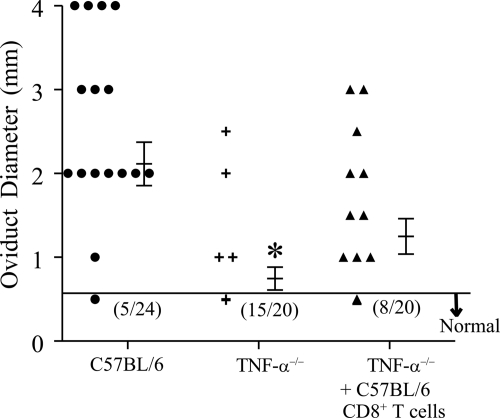
Since TNF-α production from CD8+ T cells was important for induction of hydrosalpinx, we also determined if CD8+ T cells were a sufficient source of TNF-α, in the absence of all other cellular sources of TNF-α production, to mediate the pathology. Specifically, purified (>95%) splenic CD8+ T cells from C57BL/6 mice were transferred (1 × 10⁶ cells/mouse) into TNF-α−/− mice 2 h before i.vag. C. muridarum challenge. The vaginal chlamydial clearance in the TNF-α−/− mice repleted with C57BL/6 CD8+ T cells was comparable to that in TNF-α−/− mice and C57BL/6 mice, with all mice clearing the vaginal infection by day 30 after challenge (data not shown). Additionally, as shown in Fig. 5, TNF-α−/− mice repleted with C57BL/6 CD8+ T cells displayed a significantly (P ≤ 0.05) greater incidence of hydrosalpinx (60%) than TNF-α−/− mice (25%) but a significantly (P ≤ 0.05) lesser incidence than C57BL/6 mice (79%). TNF-α−/− mice repleted with C57BL/6 CD8+ T cells also displayed oviduct dilatation (1.25 ± 0.2 mm) which was intermediate between levels for TNF-α−/− (0.8 ± 0.1 mm) and C57BL/6 mice (2.1 ± 0.35 mm) not receiving cellular transfers. These results suggest that following genital chlamydial challenge, TNF-α production from CD8+ T cells may be sufficient to mediate a certain degree of oviduct pathology, but the induction of optimal pathology also may require TNF-α production from non-CD8+ cells.
Fig. 5.
Role of TNF-α production from CD8+ T cells in oviduct pathological sequelae following vaginal C. muridarum infection. Groups (n = 3) of mice as indicated were challenged with 5 × 10⁴ IFU of C. muridarum on day 0, and macroscopic oviduct dilatation was measured on day 80 after challenge. Each individual marker represents one oviduct, and the mean ± SEM of oviduct diameter per group of mice also is shown. The number of normal oviducts (numerator) and the total number of oviducts evaluated (denominator) per respective group of mice have been indicated in parentheses. *, significant (P ≤ 0.05, chi-square test and ANOVA) difference in incidence and dilatation, respectively, of hydrosalpinx between TNF-α−/− and C57BL/6 mice and in incidence of hydrosalpinx between TNF-α−/− and TNF-α−/− receiving C57BL/6 CD8+ T cells. Results are a composite from two independent experiments.
DISCUSSION
We provide evidence for a role of CD8+ T cells in the induction of oviduct pathology following primary genital C. muridarum challenge in C57BL/6 mice. Specifically, the incidence and severity of hydrosalpinx were reduced significantly by deficiency of CD8+ T cells and could be reversed to wild-type levels by repletion of the CD8+ T cell compartment at the time of bacterial challenge in CD8-deficient mice. We also confirm and extend previous reports that perforin and TNF-α contribute to oviduct pathological sequelae following primary genital C. muridarum challenge. However, the production of TNF-α from CD8+ T cells was found to be more important than perforin for induction of the oviduct pathology. Vaginal chlamydial clearance in mice was not affected significantly by the absence of CD8+ T cells, TNF-α, or perforin.
It has been known that CD8+ T cells infiltrate the murine genital tract in measurable numbers and that chlamydial antigen-specific CD8+ T cell responses are induced following C. muridarum challenge (36). However, barring a few clones that produce high levels of IFN-γ (17, 38), CD8+ T cells in general have been thought to contribute minimally to genital chlamydial clearance in mice (23), leading one to question the role of these infiltrating CD8+ T cell populations during mouse genital chlamydial infections. In this study, the deficiency of CD8+ T cells led to reduction in Chlamydia-induced oviduct pathology, whereas the pathology could be induced by adoptively transferred CD8+ T cells, providing clear evidence that CD8+ T cells mediate Chlamydia-induced oviduct pathology. These results confirm and extend previous correlative evidence on the role of CD8+ T cells in different models, including the induction of infertility in the mouse model of C. muridarum genital infection (16), salpingitis in nonhuman primates (41, 42), and trachoma in human individuals (19). While these results suggest that CD8+ T cells do respond to the genital infection and contribute to pathogenesis, it is counterintuitive that these cells do not contribute to clearance of C. muridarum, an obligate intracellular pathogen. Perhaps the majority of the CD8+ T cell responses in mice may be targeted toward the pathogenic, as opposed to protective, chlamydial antigens. It is to be noted that this study did not directly address whether the pathology was induced by Chlamydia-specific CD8+ T cells, but that remains a strong possibility, albeit pending experimental confirmation. Moreover, given that (i) the length of the developmental cycle and IFN-γ sensitivity are different between C. trachomatis and C. muridarum and that (ii) the CD4+ and CD8+ T cell ratios in the infected genital tract differ in larger animals and humans compared to those in mice (1, 35, 45), the implications of these findings to human genital C. trachomatis infections remain to be determined in future studies.
This study also provides evidence that the production of TNF-α may be more important for the induction of pathogenesis than perforin (and subsequent lysis of infected cells) from CD8+ T cells. However, a gene deficiency of perforin per se was sufficient to significantly reduce oviduct pathology compared to that in wild-type mice. These results suggest that perforin production from non-CD8+ T cells may be more important in inducing oviduct pathology following genital chlamydial challenge. To this end, natural killer (NK) cells and/or NKT cells may be important players in perforin-mediated induction of oviduct damage (8). Bilenki et al. (2) demonstrated that CD1 knockout mice, deficient in NKT cells, display reduced chlamydial growth and histopathology following intranasal C. muridarum challenge, whereas α-galactosylceramide activation of NKT cells had the opposite effect. These authors further suggested that the NKT cells enhance Th2 type interleukin 4 (IL-4) responses leading to enhanced in vivo chlamydial growth. Our results do not directly exclude that possibility, but they suggest that the cytolytic activity of NKT cells may be involved in chlamydial pathogenesis. It also is to be noted that Bilkeni et al. studied pulmonary chlamydial infection, which per se may induce immune responses different from those of the genital chlamydial infection used in the current study.
The absence of TNF-α resulted in significantly reduced oviduct pathology, and TNF-α production was found to be an important mechanism by which CD8+ T cells mediate chlamydial pathogenesis. Additionally, in TNF-α-deficient mice, transferred TNF-α+/+ CD8+ T cells were able to induce oviduct pathology that was intermediate between those of wild-type C57BL/6 mice and TNF-α−/− mice, suggesting that TNF-α production from both CD8+ T cells and non-CD8+ T cells may mediate chlamydial pathogenesis. TNF-α can be produced by a broad variety of cell types, including fibroblasts, macrophages, mast cells, and lymphocytes (4). TNF-α binds to two receptors, TNF-R1 and TNF-R2, and is known to activate at least three different signaling cascades (5, 43): (i) NF-κB signaling leading to very strong anti-apoptotic effects, (ii) JNK pathway leading to generally proapoptotic effects, and (iii) induction of death signaling by TNF-R1. Since TNF-α can induce both proapoptotic and anti-apoptotic signals, the ability of TNF-α to induce cell death may depend on the local microenvironment, such as the presence of reactive oxygen species (15). Such cell death may be followed by reparative fibrosis (39) that leads to the oviduct luminal obstruction and hydrosalpinx, and eventually infertility, as sequelae to genital chlamydial infections. TNF-α has been shown to induce the development of fibrotic tissue in several disease models. For example, TNF-α has been shown to mediate upper urinary tract obstruction-induced renal fibrosis in rats (22), granulomatous experimental autoimmune thyroiditis in mice (6), and spontaneous pulmonary fibrosis in transgenic mice constitutively expressing TNF-α (21). This pathology may result from a variety of mechanisms, in addition to proapoptotic effects, including the ability of TNF-α to stimulate production of other proinflammatory cytokines, such as IL-1β, IL-17, and MCP-1 (6), and the inhibition of the collagen phagocytic pathway (7), which have been suggested to lead to fibrosis. TNF-α production from CD8+ T cells specifically also has been shown to contribute to the immunopathology in respiratory syncytial virus infections (37).
In summary, we have shown that CD8+ T cells, TNF-α, and perforin contribute to Chlamydia-induced oviduct pathology but not to bacterial clearance. Additionally, TNF-α, not perforin, production may be the primary mechanism by which CD8+ T cells mediate chlamydial pathogenesis. Perforin and TNF-α production from non-CD8+ T cells also seems to contribute to the process. Collectively, our results suggest that chlamydial pathogenesis may be the result of inflammatory processes elicited by both the infected epithelial cells (cellular hypothesis) and the responding lymphocytes (immunological hypothesis) (3), and that the two hypotheses may not be mutually exclusive. Importantly, the results of this study provide new avenues for exploration to enhance our understanding of the pathogenesis of genital chlamydial infections in humans.
ACKNOWLEDGMENTS
This work was supported by San Antonio Area Foundation grant and NIH grant 1R03AI088342 to A.K.M. and by NIH grant 1RO1AI074860 to B.P.A.
Footnotes
Published ahead of print on 2 May 2011.
REFERENCES
- 1. Alikhani M., Alikhani Z., Raptis M., Graves D. T. 2004. TNF-alpha in vivo stimulates apoptosis in fibroblasts through caspase-8 activation and modulates the expression of pro-apoptotic genes. J. Cell. Physiol. 201:341–348 [DOI] [PubMed] [Google Scholar]
- 2. Bilenki L., et al. 2005. NK T cell activation promotes Chlamydia trachomatis infection in vivo. J. Immunol. 175:3197–3206 [DOI] [PubMed] [Google Scholar]
- 3. Brunham R. C., Rey-Ladino J. 2005. Immunology of Chlamydia infection: implications for a Chlamydia trachomatis vaccine. Nat. Rev. Immunol. 5:149–161 [DOI] [PubMed] [Google Scholar]
- 4. Chatzidakis I., Mamalaki C. 2010. T cells as sources and targets of TNF: implications for immunity and autoimmunity. Curr. Dir. Autoimmun. 11:105–118 [DOI] [PubMed] [Google Scholar]
- 5. Chen G., Goeddel D. V. 2002. TNF-R1 signaling: a beautiful pathway. Science 296:1634–1635 [DOI] [PubMed] [Google Scholar]
- 6. Chen K., Wei Y., Sharp G. C., Braley-Mullen H. 2007. Decreasing TNF-alpha results in less fibrosis and earlier resolution of granulomatous experimental autoimmune thyroiditis. J. Leukoc. Biol. 81:306–314 [DOI] [PMC free article] [PubMed] [Google Scholar]
- 7. Chou D. H., Lee W., McCulloch C. A. 1996. TNF-alpha inactivation of collagen receptors: implications for fibroblast function and fibrosis. J. Immunol. 156:4354–4362 [PubMed] [Google Scholar]
- 8. Clement M. V., et al. 1990. Involvement of granzyme B and perforin gene expression in the lytic potential of human natural killer cells. Res. Immunol. 141:477–489 [DOI] [PubMed] [Google Scholar]
- 9. Cong Y., et al. 2007. Intranasal immunization with chlamydial protease-like activity factor and CpG deoxynucleotides enhances protective immunity against genital Chlamydia muridarum infection. Vaccine 25:3773–3780 [DOI] [PMC free article] [PubMed] [Google Scholar]
- 10. Conway D. J., et al. 1997. Scarring trachoma is associated with polymorphism in the tumor necrosis factor alpha (TNF-alpha) gene promoter and with elevated TNF-alpha levels in tear fluid. Infect. Immun. 65:1003–1006 [DOI] [PMC free article] [PubMed] [Google Scholar]
- 11. Cooker L. A., et al. 2007. TNF-alpha, but not IFN-gamma, regulates CCN2 (CTGF), collagen type I, and proliferation in mesangial cells: possible roles in the progression of renal fibrosis. Am. J. Physiol. Renal Physiol. 293:F157–F165 [DOI] [PubMed] [Google Scholar]
- 12. Darville T., Andrews C. W., Jr., Rank R. G. 2000. Does inhibition of tumor necrosis factor alpha affect chlamydial genital tract infection in mice and guinea pigs? Infect. Immun. 68:5299–5305 [DOI] [PMC free article] [PubMed] [Google Scholar]
- 13. Debattista J., Timms P., Allan J., Allan J. 2003. Immunopathogenesis of Chlamydia trachomatis infections in women. Fertil. Steril. 79:1273–1287 [DOI] [PubMed] [Google Scholar]
- 14. D'Incalci M. 2002. One of the most interesting pro-apoptotic proteins recently identified is the TNF-related apoptosis-inducing ligand (TRAIL). Eur. J. Cancer 38:17. [DOI] [PubMed] [Google Scholar]
- 15. Gaur U., Aggarwal B. B. 2003. Regulation of proliferation, survival and apoptosis by members of the TNF superfamily. Biochem. Pharmacol. 66:1403–1408 [DOI] [PubMed] [Google Scholar]
- 16. Igietseme J. U., et al. 2009. Role of T lymphocytes in the pathogenesis of Chlamydia disease. J. Infect. Dis. 200:926–934 [DOI] [PMC free article] [PubMed] [Google Scholar]
- 17. Igietseme J. U., Magee D. M., Williams D. M., Rank R. G. 1994. Role for CD8+ T cells in antichlamydial immunity defined by Chlamydia-specific T-lymphocyte clones. Infect. Immun. 62:5195–5197 [DOI] [PMC free article] [PubMed] [Google Scholar]
- 18. Kelly K. A., Rank R. G. 1997. Identification of homing receptors that mediate the recruitment of CD4+ T cells to the genital tract following intravaginal infection with Chlamydia trachomatis. Infect. Immun. 65:5198–5208 [DOI] [PMC free article] [PubMed] [Google Scholar]
- 19. Kimani J., et al. 1996. Risk factors for Chlamydia trachomatis pelvic inflammatory disease among sex workers in Nairobi, Kenya. J. Infect. Dis. 173:1437–1444 [DOI] [PubMed] [Google Scholar]
- 20. Ljunggren H. G., Van Kaer L., Ashton-Rickardt P. G., Tonegawa S., Ploegh H. L. 1995. Differential reactivity of residual CD8+ T lymphocytes in TAP1 and beta 2-microglobulin mutant mice. Eur. J. Immunol. 25:174–178 [DOI] [PubMed] [Google Scholar]
- 21. Lundblad L. K., et al. 2005. Tumor necrosis factor-alpha overexpression in lung disease: a single cause behind a complex phenotype. Am. J. Respir. Crit. Care Med. 171:1363–1370 [DOI] [PMC free article] [PubMed] [Google Scholar]
- 22. Meldrum K. K., et al. 2007. TNF-alpha neutralization ameliorates obstruction-induced renal fibrosis and dysfunction. Am. J. Physiol. Regul. Integr. Comp. Physiol. 292:R1456–R1464 [DOI] [PubMed] [Google Scholar]
- 23. Morrison R. P., Caldwell H. D. 2002. Immunity to murine chlamydial genital infection. Infect. Immun. 70:2741–2751 [DOI] [PMC free article] [PubMed] [Google Scholar]
- 24. Morrison R. P., Feilzer K., Tumas D. B. 1995. Gene knockout mice establish a primary protective role for major histocompatibility complex class II-restricted responses in Chlamydia trachomatis genital tract infection. Infect. Immun. 63:4661–4668 [DOI] [PMC free article] [PubMed] [Google Scholar]
- 25. Morrison S. G., Morrison R. P. 2000. In situ analysis of the evolution of the primary immune response in murine Chlamydia trachomatis genital tract infection. Infect. Immun. 68:2870–2879 [DOI] [PMC free article] [PubMed] [Google Scholar]
- 26. Morrison S. G., Su H., Caldwell H. D., Morrison R. P. 2000. Immunity to murine Chlamydia trachomatis genital tract reinfection involves B cells and CD4+ T cells but not CD8+ T cells. Infect. Immun. 68:6979–6987 [DOI] [PMC free article] [PubMed] [Google Scholar]
- 27. Murthy A. K., et al. 2009. A limited role for antibody in protective immunity induced by rCPAF and CpG vaccination against primary genital Chlamydia muridarum challenge. FEMS Immunol. Med. Microbiol. 55:271–279 [DOI] [PMC free article] [PubMed] [Google Scholar]
- 28. Murthy A. K., et al. 2006. Chlamydial protease-like activity factor induces protective immunity against genital chlamydial infection in transgenic mice that express the human HLA-DR4 allele. Infect. Immun. 74:6722–6729 [DOI] [PMC free article] [PubMed] [Google Scholar]
- 29. Murthy A. K., Sharma J., Coalson J. J., Zhong G., Arulanandam B. P. 2004. Chlamydia trachomatis pulmonary infection induces greater inflammatory pathology in immunoglobulin A deficient mice. Cell Immunol. 230:56–64 [DOI] [PubMed] [Google Scholar]
- 30. Oikonomou N., et al. 2006. Soluble TNF mediates the transition from pulmonary inflammation to fibrosis. PLoS One 1:e108. [DOI] [PMC free article] [PubMed] [Google Scholar]
- 31. Ortis F., et al. 2008. Induction of nuclear factor-kappaB and its downstream genes by TNF-alpha and IL-1beta has a pro-apoptotic role in pancreatic beta cells. Diabetologia 51:1213–1225 [DOI] [PubMed] [Google Scholar]
- 32. Perry L. L., Feilzer K., Caldwell H. D. 1997. Immunity to Chlamydia trachomatis is mediated by T helper 1 cells through IFN-gamma-dependent and -independent pathways. J. Immunol. 158:3344–3352 [PubMed] [Google Scholar]
- 33. Perry L. L., Feilzer K., Hughes S., Caldwell H. D. 1999. Clearance of Chlamydia trachomatis from the murine genital mucosa does not require perforin-mediated cytolysis or Fas-mediated apoptosis. Infect. Immun. 67:1379–1385 [DOI] [PMC free article] [PubMed] [Google Scholar]
- 34. Podack E. R., Hengartner H. 1989. Structure of perforin and its role in cytolysis. Year Immunol. 6:245–261 [PubMed] [Google Scholar]
- 35. Rank R. G., Bowlin A. K., Reed R. L., Darville T. 2003. Characterization of chlamydial genital infection resulting from sexual transmission from male to female guinea pigs and determination of infectious dose. Infect. Immun. 71:6148–6154 [DOI] [PMC free article] [PubMed] [Google Scholar]
- 36. Roan N. R., Starnbach M. N. 2006. Antigen-specific CD8+ T cells respond to Chlamydia trachomatis in the genital mucosa. J. Immunol. 177:7974–7979 [DOI] [PubMed] [Google Scholar]
- 37. Rutigliano J. A., Graham B. S. 2004. Prolonged production of TNF-alpha exacerbates illness during respiratory syncytial virus infection. J. Immunol. 173:3408–3417 [DOI] [PubMed] [Google Scholar]
- 38. Starnbach M. N., Bevan M. J., Lampe M. F. 1994. Protective cytotoxic T lymphocytes are induced during murine infection with Chlamydia trachomatis. J. Immunol. 153:5183–5189 [PubMed] [Google Scholar]
- 39. Swynghedauw B., Baillard C. 2000. Biology of hypertensive cardiopathy. Curr. Opin. Cardiol. 15:247–253 [DOI] [PubMed] [Google Scholar]
- 40. Van Kaer L., Ashton-Rickardt P. G., Ploegh H. L., Tonegawa S. 1992. TAP1 mutant mice are deficient in antigen presentation, surface class I molecules, and CD4−8+ T cells. Cell 71:1205–1214 [DOI] [PubMed] [Google Scholar]
- 41. Van Voorhis W. C., Barrett L. K., Sweeney Y. T., Kuo C. C., Patton D. L. 1996. Analysis of lymphocyte phenotype and cytokine activity in the inflammatory infiltrates of the upper genital tract of female macaques infected with Chlamydia trachomatis. J. Infect. Dis. 174:647–650 [DOI] [PubMed] [Google Scholar]
- 42. Van Voorhis W. C., Barrett L. K., Sweeney Y. T., Kuo C. C., Patton D. L. 1997. Repeated Chlamydia trachomatis infection of Macaca nemestrina fallopian tubes produces a Th1-like cytokine response associated with fibrosis and scarring. Infect. Immun. 65:2175–2182 [DOI] [PMC free article] [PubMed] [Google Scholar]
- 43. Wajant H., Pfizenmaier K., Scheurich P. 2003. Tumor necrosis factor signaling. Cell Death Differ. 10:45–65 [DOI] [PubMed] [Google Scholar]
- 44. Wang J., et al. 2009. A chlamydial type III-secreted effector protein (Tarp) is predominantly recognized by antibodies from humans infected with Chlamydia trachomatis and induces protective immunity against upper genital tract pathologies in mice. Vaccine 27:2967–2980 [DOI] [PMC free article] [PubMed] [Google Scholar]
- 45. Whittum-Hudson J. A., Taylor H. R., Farazdaghi M., Prendergast R. A. 1986. Immunohistochemical study of the local inflammatory response to chlamydial ocular infection. Invest. Ophthalmol. Vis. Sci. 27:64–69 [PubMed] [Google Scholar]


