1. Introduction
Protein lipidation is the covalent attachment of a lipid group to protein. Lipids modify large numbers of eukaryotic proteins and regulate protein function and localization. The hydrophobic character of lipid modifications makes the study of protein lipidation challenging. Chemical biology has played an increasingly important role in advancing the field of protein lipidation through novel methods of detection and isolation, design and synthesis of inhibitors, strategies to monitor the behavior of lipidated proteins in cells, and methods to produce lipidated proteins for structural and biophysical analyses. In this review, we discuss the chemical tools that have been developed to facilitate discovery in the field of protein lipidation and provide examples of how this has created new understanding of the scope of protein lipidation and its biological consequences.
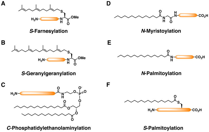
The content of this review is limited to the major posttranslational modifications that occur in the cytoplasm or on the cytoplasmic face of membranes: S-prenylation, N-myristoylation, and S-palmitoylation (Fig. 1). Protein lipidation of molecules destined for secretion occurs in the lumen of organelles within the secretory pathway. Glycosylphosphatidylinositol (GPI) anchors attached to proteins in the endoplasmic reticulum tether proteins to the extracellular face of the plasma membrane. Other secreted proteins that are lipidated include morphogens, cytokines, and hormones. Examples include the octanoylated peptide hormone ghrelin and the Hedgehog signaling protein, which is modified with both cholesterol and palmitate. Lipid modifications that occur in the lumen of secretory organelles are reviewed elsewhere. 1
FIGURE 1.
Survey of cytosolic protein lipidation in eukaryotes. A) S-Farnesylation. B) S-Geranylgeranylation. C) C-phosphatidylethanolaminylation. D) N-myristoylation. E) N-palmitoylation. F) S-palmitoylation.
2. Lipid Modifications on the Cytoplasmic Face of Membranes
Fatty acids and isoprenoids anchor proteins to the cytoplasmic leaflet of cell membranes (Fig. 1). Protein S-prenylation and N-myristoylation are mediated by cytoplasmic enzymes, whereas S-palmitoylation occurs at the membrane surface. Two other lipid modifications that occur in the cytoplasm are more rare. Amide-linked palmitate (N-palmitoylation) is found on the amino terminus of the signal transducing protein Gsα (Fig. 1E)2; phosphatidylethanolamine is linked to the C-terminal glycine residue of LC3/Atg8, a protein involved in autophagy (Fig. 1C) 3. In this section we briefly review the structure and mechanism of the major lipid modifications.
S-Prenylation refers to the irreversible addition of an isoprenoid, either C15 farnesyl (Fig. 1A) or C20 geranylgeranyl (Fig. 1B), to protein through a thioether linkage.4 S-Prenylated proteins are recognized by their characteristic C-terminal motifs. Farnesyl transferase (FTase) and geranylgeranyltransferase type I (GGTase-I) modify proteins at a Cys residue that is four amino acids from the C terminus (the -CaaX box, where ‘a’ is a small, typically aliphatic amino acid and the identify of X contributes to the specificity of the prenyl group added). S-prenylation is followed by the proteolytic removal of the three terminal amino acids (–aaX) by Ras converting enzyme 1 and carboxylmethylation of the S-prenylated cysteine by isoprenylcysteine methyltransferase.5 Members of the Rab family of GTPases are prenylated by a dedicated enzyme, Rab geranylgeranyltransferase (RGGT).6 Rab proteins terminate in –CC, –CCXX, or –CXC motifs and are typically modified with two geranylgeranyl groups. Proteins modified with prenyl groups require the modification for membrane association and consequently to exert their functions.4 The realization that S-prenylation is essential for proper protein function has stimulated the development of small molecule S-prenyltransferase inhibitors to block the activity of oncogenic Ras and other S-prenylated proteins that malfunction in various disease states.7
Protein N-myristoylation refers to the irreversible addition of myristic acid to eukaryotic and viral proteins through an amide linkage to an N-terminal glycine residue (Fig. 1D).8 Modification of the protein typically occurs cotranslationally as the nascent polypeptide chain emerges from the ribosome and following removal of the initiator methionine by methionylpeptidase. Alternatively, during apoptosis, N-myristoylation can occur posttranslationally on proteins where caspase cleavage exposes a cryptic N-terminal glycine residue.9 The enzyme responsible for myristoylation is N-myristoyltransferase (NMT).8 Loss of NMT function in S. cereviseae is lethal10 and Drosophila lacking NMT have multiple developmental defects11. Thus, like S-prenylation, N-myristoylation is an essential process. Humans and mice have two N-myristoyltransferase genes, NMT1 and NMT2, that encode enzymes with overlapping functions. NMT1 is required for early embryonic development in mice.12
Proteins are fatty acylated at cysteine residues through reversible thioester linkages in a process commonly referred to as S-palmitoylation (Fig. 1F).13 Thioacylation or S-acylation is a more appropriate term for this modification given that other long chain fatty acids besides palmitate are incorporated into proteins through this mechanism. Both integral and peripheral membrane proteins can be modified with palmitate and this occurs both on intracellular membranes and at the plasma membrane. Promoting membrane association of soluble proteins is an obvious function for S-palmitoylation, but this modification also regulates protein trafficking, stability, and activity.14
The reversibility of protein S-palmitoylation distinguishes it from other lipid modifications and predicts the existence of enzymes that palmitoylate and depalmitoylate proteins. Protein S-acyltransferases (PATs) that modify proteins on the cytoplasmic face of membranes were first characterized molecularly in 2002.15 These are a family of integral membrane proteins that share a protein domain referred to as the DHHC domain, a cysteine-rich domain with a conserved Asp-His-His-Cys signature motif that is required for enzymatic activity. Yeast have seven DHHC-PATs; genetic and biochemical analyses have revealed that these enzymes can account for nearly all palmitoylation events in yeast.16 A larger family of 23 DHHC enzymes is found in mammals.17
The process of depalmitoylation is less well characterized. The lysosomal enzyme responsible for degradation of S-palmitoylated proteins may be protein palmitoylthioesterase 1 (PPT1).18 A cytoplasmic enzyme, acylprotein thioesterase 1 (APT1) removes palmitate from G-protein alpha subunits and Ras proteins in vitro.19 In vivo, APT1 has been linked to G-protein depalmitoylation in yeast20 and to Ras depalmitoylation in mammalian cells.21 The development of small molecule APT1 inhibitors has provided an important new tool to investigate the functional consequences of reversible palmitoylation of Ras.21 These are discussed in section 6.
S-palmitoylation is often found in combination with other lipid modifications.14 Proteins with Cys residues adjacent to or near to an N-myristoylated Gly are often S-palmitoylated. Signal-transducing proteins such as G-protein α subunits22 and non-receptor tyrosine kinases23 are examples of proteins that are modified in this way. Similarly, S-prenylated proteins with cysteine residues immediately upstream of the S-prenylated cysteine are also substrates for S-palmitoylation. N-Ras and H-Ras are modified with palmitate at one or two cysteines, respectively. Early studies revealed that N-myristoylation or S-farnesylation was a requirement for subsequent S-palmitoylation and that both the myristoyl or farnesyl modification and S-palmitoylation were necessary for stable membrane association and plasma membrane localization. As discussed in the following sections, the development of synthetic lipopepides and semisynthesis of lipidated proteins was key to understanding the physical basis of the membrane interactions and the functional significance of reversible S-palmitoylation.
3. Synthetic Lipopeptides
Short amino acid sequences that encode the recognition motifs for modification are sufficient for lipidation to occur in cells. For example, the last ten amino acids of H-Ras are sufficient for CaaX processing and S-palmitoylation in cells when transplanted onto a soluble protein.24 Genetically encoded fusions of a lipidation sequence and a fluorescent protein have been exploited to study the cell biology of lipidated proteins. Similarly, synthetic lipidated peptides have also been widely used as surrogates to study the behavior of their cognate lipid-modified proteins.25 A variety of synthetic methods have been developed to overcome the challenges associated with the synthesis of cysteinyl-containing peptides modified with lipids. The reader is referred to several excellent reviews on this topic.26
Biophysical studies monitoring the association of fluorescent lipopeptides with model membranes provided an important framework to understand how dual lipid modifications mediate interactions with membranes. The kinetic membrane-trapping model emerged from measurements of the rates of interbilayer transfer of singly and dually lipid-modified peptides. Proteins modified with myristate or a farnesyl group undergo rapid association and reassociation with membranes, whereas proteins with dual lipid modifications displayed greatly diminished exchange between membranes.27 The model provided a rationale for how lipid modifications might target proteins to a specific membrane through restricted localization of a “membrane-targeting receptor” that facilitates addition of the second lipid modification. It also had implications for modes of protein trafficking. S-Farnesylated or N-myristoylated proteins could potentially move about the cell through the cytoplasm independent of vesicular transport, sampling different membrane compartments. Proteins modified with two or more lipids would transit on vesicles because of their stable membrane assocation. As discussed in section 7, live cell imaging studies monitoring the trafficking of lipidated proteins provide further support for the kinetic membrane-trapping model.
4. Semisynthetic Lipidated Proteins
In cells, lipopeptides appear to replicate the localization and dynamics of their full-length counterparts in many respects. However, these studies are naturally limited by the absence of other protein determinants that influence protein-protein and protein-membrane interactions. Semisynthetic proteins in which synthetic peptides modified with lipids are conjugated to recombinant proteinsto afford native or near-native homogenous preparations of lipidated proteins for structural, biophysical, and cell biological applications.
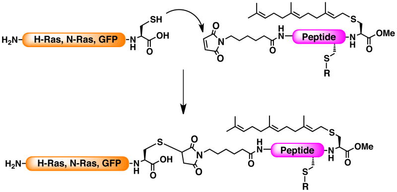
Two strategies have been used to produce semisynthetic lipid-modified proteins. In the first, a lipopeptide activated at the amino-terminal end with a maleimido modification is linked to the carboxyl-terminal cysteine residue of a recombinant protein (Fig. 2).28 Semisynthetic proteins prepared using this method were used to study the trafficking of S-palmitoylated isoforms of Ras proteins (Section 7).25b,29 The maleimido caproic acid (MIC) coupling of truncated Ras proteins to lipopetides was efficient at neutral pH in a buffered detergent solution, conditions compatible with preservation of native protein structure. The semisynthetic protein was active as assessed by its ability to bind guanine nucleotides and to the Ras-binding domain (RBD) of its effector Raf. An oncogenic form induced neurite extension when microinjected into PC12 cells, an indicator that semisynthetic lipidated Ras could transform cells. These experiments suggest that the MIC linkage does not grossly perturb the function of semisynthetic H- and N-Ras28. MIC coupling has also been used successfully to link GFP modified with a C- terminal cysteine with peptides derived from proteins that are modified at the N-terminus by N-myristoylation and S-palmitoylation.29 Key to the success with MIC coupling is the accessibility of the C-terminal cysteine residue and inaccessibility of other cysteines in the protein. The MIC approach was not feasible for K-Ras4B due to the presence of multiple C-terminal lysine residues, complications with separation of products, and low yield of lipopeptide.30
FIGURE 2.
Schematic of maleimidocaproyl (MIC)-controlled ligation of S-farnesylated peptide to Ras GTPases or GFP.
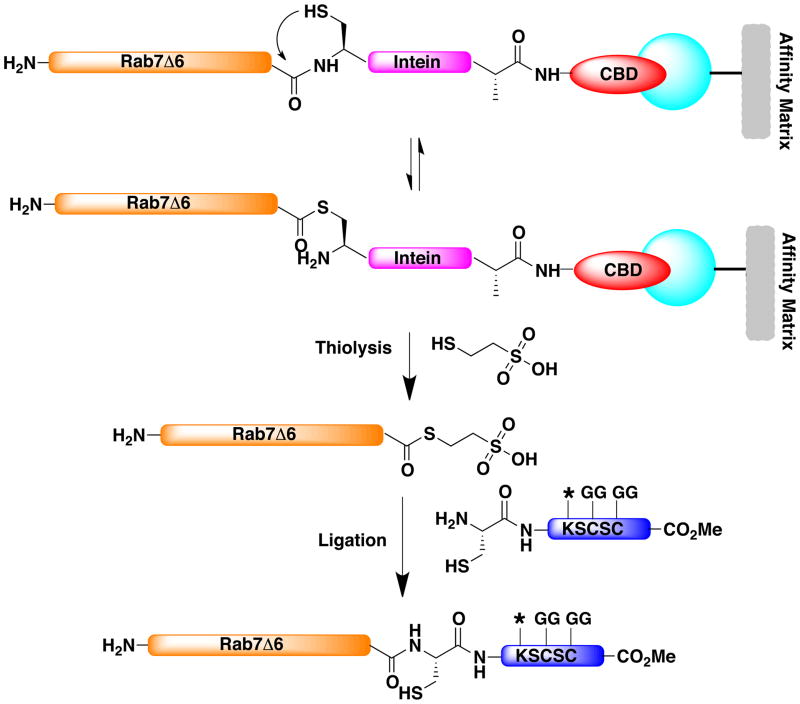
The second approach uses expressed protein ligation (EPL) in which a recombinant protein thioester is reacted with a lipidated peptide containing an N-terminal cysteine residue to yield a native amide bond (Fig. 3). The EPL method exploits intein-based autocleavage to produce a protein thioester.31 The protein of interest is expressed in bacteria as a fusion protein with an intein and chitin-binding domain, permitting affinity purification on a chitin column. On-column cleavage initiated by the addition of mercaptoethanesulfonate (MESNA) yields the protein thioester, which is then reacted with a lipidated peptide. In contrast to MIC coupling, the linkage results in a native peptide bond. EPL has been used to produce S-prenylated Rab proteins for structural and biophysical analysis32, and more recently to generate S-farnesylated K-Ras and Rheb30. One disadvantage of EPL is that it leaves a cysteine residue at the junction of the protein and peptide. If the semisynthetic protein is introduced into cells, a non-native cysteine is well positioned for S-palmitoylation when in proximity to the S-prenylated C-terminus, introducing an additional lipid modification and its associated consequences on function and localization.
FIGURE 3.
Schematic of expressed protein ligation of Rab7 ligation to a fluorescent digeranylgeranylated hexapeptide. Chitin binding domain (CBD). * represents a dansyl group. Geranylgeranyl group (GG).
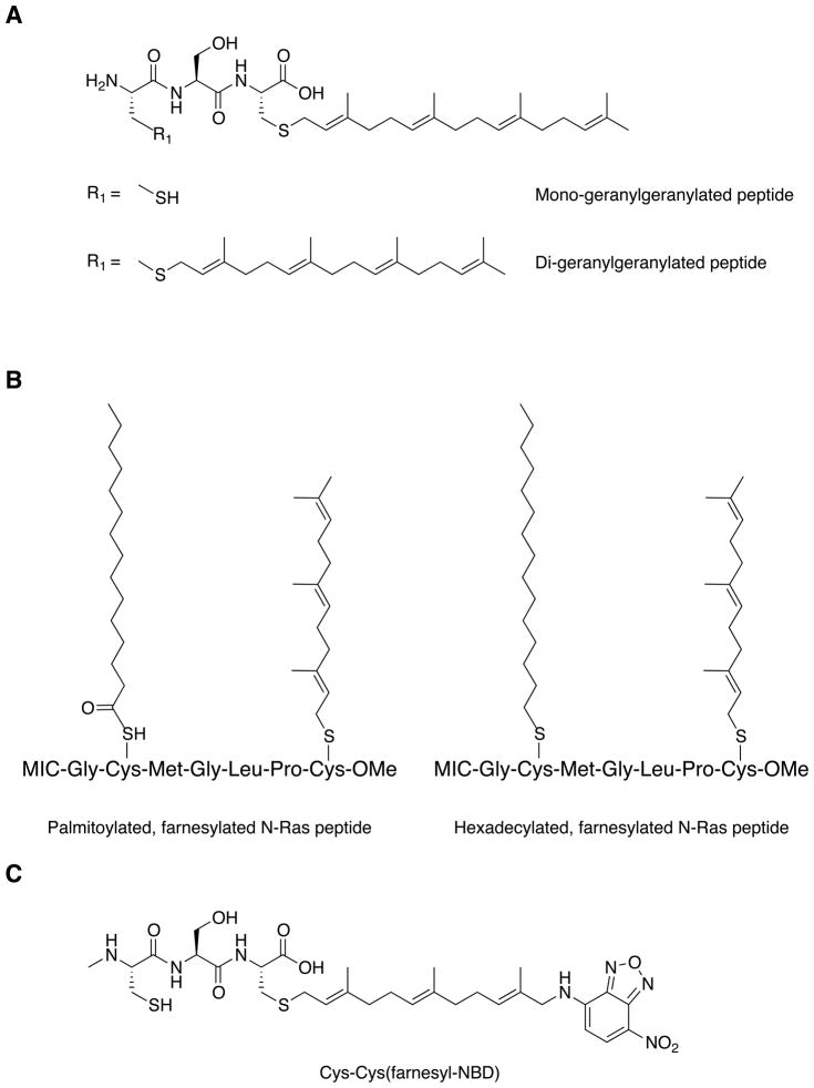
A significant advantage of semisynthetic proteins is the versatility of the synthetic peptides that can be added. The lipid or combination of lipids modifying the protein can be varied, for example, mono- versus digeranylgeranylation (Fig. 4A).26b The importance of the reversibility of S-palmitoylation was demonstrated by comparing the behavior of proteins modified with palmitate through a reversible thioester linkage versus stable thioether attachment (Fig. 4B).25b The significance of reversible S-palmitoylation could not be addressed using genetically encoded fluorescent proteins that rely on cellular enzymes to incorporate the lipids into protein. Finally, fluorescent reporters or unnatural amino acids are easily introduced into the protein through chemical modification of the synthetic peptide (Fig. 4C).26b,29
FIGURE 4.
Schematics of lipopeptides used in EPL to generate lipidated semisynthetic proteins. A) Mono- and digeranylgeranylated peptides. B) S-farnesylated peptides modified with a cleavable thioester-linked palmitate (left) or noncleavable thioether-linked hexadecanoate (right) for maleimidocaproyl (MIC) ligation. C) Cys-Ser-Cys peptide modified with farnesyl-NBD (nitrobenzoxadiazole).
A clever application of the semisynthetic protein technology was the creation of recombinant Rab7 linked to a peptide modified with NBD-farnesyl (Fig. 4C).33 The advantages of this configuration are two fold. First, the farnesyl-NBD moiety mimics the native geranylgeranyl (GG) group but has reduced hydrophobicity, rendering a soluble protein that can be analyzed in the absence of detergent, which perturbs protein-protein and membrane interactions. Second, the introduction of the fluorescent moiety allowed time-resolved measurement of prenylated Rab interactions with GDI or REP proteins.
To date, monomeric GTPases and lipidated Green Flurorescent Protein (GFP) are the only lipidated proteins synthesized using MIC linkage or EPL.28–29,31b,34 Successful application of the method requires identifying conditions in which coupling of the peptide occurs only at the desired residue and the semisynthetic protein can be recovered in an active form. Semisynthetic Ras proteins were purified under native conditions, separated on the basis of their newly acquired hydrophobicity using detergent partitioning28, whereas the introduction of the polybasic, S-farnesylated C-terminal peptide into truncated K-Ras resulted in an increase in the isoelectric point of the protein, enabling separation by ion exchange chromatography.30 Semisynthetic Rab and Rheb proteins were precipitated following ligation and refolded.30,34 Recovery of active mono- and di-geranylgeranylated proteins required refolding in the presence of a chaperone to prevent aggregation.34,33 Extension of MIC coupling or EPL to other lipidated proteins, particularly larger proteins with multiple domains will be technically challenging.
5. Structural and Biophysical Characterization of Rab GTPases Using Semisynthetic Proteins
Semisynthetic Rab proteins have been key to illuminating how S-prenylation interfaces with the protein-protein interactions that are necessary for reversible interactions with membranes.33,35 Rab proteins constitute the largest family of monomeric GTPases and regulate organelle biogenesis and vesicular transport.36 Like all GTPases, they undergo cycles of nucleotide exchange and hydrolysis. Superimposed on the GTP-GDP molecular switch is a cycle of membrane association and dissociation that is dependent upon S-prenylation and regulatory proteins. In this section we discuss a current model of membrane targeting based on recent structural and biophysical studies.33
Most Rab proteins are digeranylgeranylated (Rab-diGG) at the C-terminus, but a few are monogeranylgeranylated (Rab-mG).6 Rab proteins are S-prenylated by RGGT, a S-prenyltransferase that like FTase and GGTaseI is an α/β heterodimer. However, RGGT also requires an accessory protein, Rab Escort Protein (REP), for function. REP binds to unprenylated Rab and positions it for modification by RGGT. REP also binds to mono- and di-prenylated Rabs, shielding the hydrophobic prenyl groups from the cytoplasm and facilitating initial membrane targeting. REP shares sequence homology with RabGDI, a protein that binds to S-prenylated Rab proteins in the inactive GDP-bound form. The function of RabGDI is to mediate the transfer of Rab proteins between membrane compartments. The structures of Rab GDI and REP in complex with S-prenylated Rab proteins reveal a shared two-domain architecture.32,37 Domain I provides most of the binding surface for the core GTPase domain, whereas Domain II contains a hydrophobic pocket that harbors one or two geranylgeranyl groups.
Fluorescent semisynthetic proteins have permitted real-time quantitative assessment of the interactions between S-prenylated Rab7 and its regulators, providing insight into how RabGDI and REP carry out their distinct functions in the cell.33,35 REP has multiple roles, first to “escort” unprenylated Rab to RGGT; second, to present monoprenylated Rab to RGGT for the addition of the second isoprenoid; and third, to facilitate the initial membrane targeting of the Rab protein.6 REP binds to unmodified and diGG Rab7 with low nM affinity.35 Interestingly, REP displays pM affinity for monoprenylated Rab7. The tight association of REP with monoGG Rab7 facilitates its retention in the complex for the addition of the second geranylgeranyl moiety. The reduced affinity of REP for the di-GG modified protein facilitates the release of Rab7 from REP and deposition on membranes.35
Although REP and RabGDI share a function of facilitating the insertion of Rab proteins into membranes, only GDI is able to efficiently extract S-prenylated proteins from membranes. Quantitative comparison of the impact of lipidation on Rab7 interactions with REP and GDI provides insight into this distinction.35 In contrast to the nearly equivalent binding affinities of unprenylated Rab7 and Rab7-diGG for REP, unprenylated Rab7 and Rab7-diGG displayed a difference in affinities for GDI of at least 3 orders of magnitude. The large change in affinity when Rab is docked to GDI through the C-terminus and tandem isoprenoids provides the thermodynamic driving force required for membrane extraction. In contrast, the binding energy in REP:Rab7 complexes is associated primarily with the core GTPase domain and there is no additional “benefit” to binding the di-GG moiety to drive extraction of the protein from membranes.35
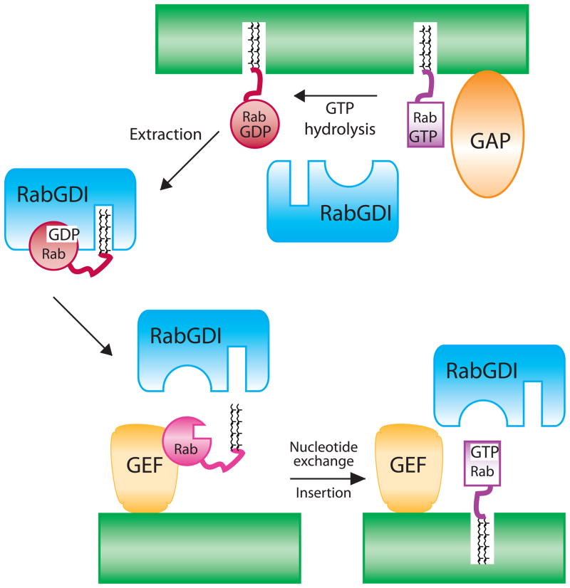
A recent study expanded this type of analysis to examine the impact of the nucleotide bound to Rab7 on its interactions with GDI and proposed a model of how nucleotide exchange is integrated with membrane extraction and insertion of Rab proteins (Fig. 5).33 Lipidated semisynthetic proteins were refolded in the presence of GDP or GppNHp, a nonhydrolyzable analog of GTP, to permit comparison of affinities of S-prenylated Rab proteins in the GDP- and GTP-bound states. The large, three-order of magnitude difference between GDI’s affinity for Rab in the GTP- versus the GDP-form is much larger than previous estimates and provides a strong rationale for GDI’s ability to selectively extract GDP-bound Rab from membranes.33
FIGURE 5.
Model of the membrane association and GTPase cycles of Rab proteins.
An outstanding problem in the biology of Rab proteins is how they are targeted to specific membrane compartments from a common pool of cytoplasmic Rab:RabGDI complexes. A current model proposes that interactions of the Rab:GDI complex with a GDI displacement factors (GDF) dislodges S-prenylated Rab from GDI, facilitating insertion of Rab into membranes.38 The first GDF characterized, YIP3, is one of three polytopic proteins that bind S-prenylated Rab proteins. However, only YIP3 has been demonstrated to catalyze GDI displacement, leaving open the possibility that S-prenylated Rab interactions with YIP proteins have functions beyond or in addition to membrane targeting.38–39
Evidence is accumulating that guanine nucleotide exchange factors (GEFs) for Rab proteins play the key role in targeting Rab proteins to membranes. DrrA is a soluble bacterial effector from Legionella pneumophila that was proposed to have separable GEF and GDF activities for human Rab1.40 The structure of the Drr/Rab1 complex and biochemical characterization revealed that displacement of RabGDI from S-prenylated Rab1 was a consequence of the GEF activity of Drr, demonstrating that GDF and GEF activities of DrrA were one and the same.41 These studies were extended using a fluorescent analogue of S-prenylated Rab produced using the chemoenzymatic CaaX tag method (see section 8.1).33 GDI displacement was assayed by the decrease in fluorescence when Rab1 dissociated from GDI. In the presence of the GEF domain of DrrA, Rab1 was dissociated from GDI. Dissociation was dependent on the addition of GTP and reversed when excess GDP was added to the reaction, suggesting that the GTP/GDP switch controls GDI dissociation and insertion of Rab into membranes.
The analysis of protein interactions between Rab and RabGDI provides a thermodynamic basis for the mechanisms that regulate Rab membrane targeting and dissociation (Fig 5).33 Recently, the interaction of the Rho family GTPase, Cdc42 with RhoGDI was studied in the presence of model membranes.42 In contrast to the pronounced difference in affinities observed with S-prenylated Rab proteins and RabGDI in the presence of GTP or GDP33, Cdc42 displayed the same affinity for RhoGDI in solution whether it was GDP- or GTP-bound. In the presence of a membrane surface, however, the affinity of RhoGDI for Cdc42-GDP was much higher than for Cdc42-GTP. Cdc42 dissociation from membrane vesicles occurred at similar rates alone or when bound to RhoGDI, suggesting that the primary function of RhoGDI in extracting Cdc42 from membranes is to prevent its rebinding to the membrane surface.42 It will be of interest to measure the impact of model membranes on the dynamics of Rab and RabGDI interactions. Rab proteins modified with two prenyl groups will dissociate from membranes with much slower kinetics than monoprenylated Cdc4227, suggesting differences in how RabGDI will interact with its cognate Rab to extract it from membranes. Both studies support the importance of GEF-catalyzed nucleotide exchange to free the GTPases from the action of their corresponding GDIs, and suggest that GEF localization plays a prominent role in imparting specific membrane localization.33,42
6. Small Molecule Inhibitors of Reversible S-Palmitoylation
Identification of the enzymes that modify proteins with lipids enabled small molecule discovery programs to find inhibitors of enzyme activity. These reagents are key to elucidating the biology and enzymology of protein lipidation. NMT43 and S-prenyltransferases7,44 have been the subjects of intensive programs to discover inhibitors and determine their utility in treating disease. In contrast, small molecules that specifically inhibit S-palmitoylation in vitro and in vivo are noticeably lacking. The discovery and characterization of DHHC-PATs makes molecular characterization of molecules that inhibit S-palmitoylation feasible, as well as expanding strategies for high throughput screening. Development of inhibitors of protein depalmitoylation has progressed with the advent of small molecules that inhibit the thioesterase APT1.21 In this section, we review the status of small molecules that affect reversible protein S-palmitoylation.
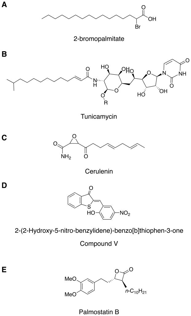
Pharmacological inhibitors of S-palmitoylation currently in use block modification of proteins with palmitate, but their cellular effects are not limited to protein S-palmitoylation.45 2-bromopalmitate (2-BP) is most commonly used to block incorporation of palmitate into proteins (Fig. 6A).45 A non-metabolizable fatty acid analogue, 2-BP also inhibits enzymes involved in lipid synthesis, as well as NADPH cytochrome-c reductase and glucose-6-phosphatase.46 Accordingly, 2-BP has pleiotropic effects in cells and its inhibition of S-palmitoylation may be due as much to an effect on lipid metabolism as it is on direct inhibition of S-palmitoyltransferases. Other reagents used to inhibit cellular S-palmitoylation are tunicamycin (Fig. 6B)47 and cerulenin (Fig. 6C), which like 2-BP are likely to exert their effects through inhibition of fatty acid or acyl-CoA synthesis.45 Of these reagents, 2-BP is the only inhibitor that has been demonstrated to inhibit DHHC-PATs in vitro. Four DHHC-PATs, two from yeast and two from mammalian cells, were tested and displayed irreversible inhibition with 2-BP.48
FIGURE 6.
Inhibitors of protein palmitoylation (A–D) and depalmitoylation (E). A) 2-bromopalmitate. B) Tunicamycin. C) Cerulenin. D) Compound V, 2-(2-hydroxy-5-nitro-benzylidene)-benzo[b]thiophen-3-one. E) Palmostatin B.
Screening of a compound library using several cell-based assays that report processes associated with S-palmitoylation identified five classes of compounds as potential S-palmitoylation inhibitors.49 Representative compounds from the five classes were shown to inhibit endogenous PAT activity in membranes using S-farnesylated or N-myristoylated peptides as substrates.49 Subsequent testing of four of the five compounds with four DHHC-PATs showed only Compound V (2-(2-Hydroxy-5-nitro-benzylidene)-benzo[b]thiophen-3-one) inhibited the purified enzymes (Fig. 6D). Inhibition was reversible, time-dependent, and was less potent than 2-BP (Fig. 6A).48
Inhibitor development to block protein depalmitoylation has been more successful. APT1 is a soluble enzyme that acts as an acylprotein thioesterase19 and as a lysophospholipase50 in vitro. Kinetic characterization of the enzyme suggested that S-palmitoylated proteins were better substrates for the enzyme19, supported by studies in mammalian cells and yeast in which gene silencing of APT1 expression resulted in a steady state increase of S-palmitoylated Ras proteins21 and G-protein alpha subunits20. The structure of APT1 shows that the enzyme is an α/β hydrolase with a classic Ser-His-Asp catalytic triad.51 Two related serine hydrolases, APT2 (LYPLA2) and APT3 (LYPLAL1) are predicted to have acylprotein thioesterase activity.52 Although APT2 has not been purified and tested in vitro, exogenous expression of APT2 in cells accelerated deacylation of the neuronal S-palmitoylation substrate GAP-43, suggesting that it is a bona fide acylprotein thioesterase.53
Early efforts to design inhibitors of APT1 focused on synthesis of peptidomimetics. A number of compounds that inhibited APT1 activity in vitro were identified but were not active in cells.54 Recently, a β-lactone-containing compound named Palmostatin B was shown to inhibit APT1 enzyme activity in vitro and appears to target APT1 in cells (Fig. 6E). Kinetic characterization revealed that Palmostatin B acts as a competitive inhibitor with an IC50 of 670 nM.21 Based on the mechanism of gastric lipase inhibition by β-lactones 55, Palmostatin B inhibits activity by covalently modifying the serine residue in the active site. Pre-steady state kinetics indicated that the initial interaction with the enzyme is fast, followed by a slow reactivation of the enzyme upon hydrolysis of the compound.21
In cells treated with Palmostatin B, steady state S-palmitoylation of Ras was increased modestly, similar to the increase that was observed when APT1 expression was reduced by siRNA.21 These data are the first evidence that Ras is a substrate for APT1 in cells and are consistent with APT1 as a target of Palmostatin B in cells. Various phospholipases were ruled out as cellular targets for Palmostatin B. Phospholipases A1, A2, D, and Cβ activities were unaffected by Palmostatin B concentrations that inhibited Ras depalmitoylation in cells, arguing for inhibitor specificity.21 However, the effects of Palmostatin B on other members of the large family of serine hydrolases52, including the related APT2 and APT3 enzymes and the lysosomal palmitoyl protein thioesterases, awaits further study. Palmostatin B perturbed the steady state localization of Ras proteins and induced a partial phenotypic reversion in oncogenic Ras-transformed fibroblasts. These results suggest that inhibition of Ras depalmitoylation may represent a novel approach to therapeutically intervene with oncogenic Ras signaling.21
7. The S-Palmitoylation Cycle and Protein Trafficking of Lipidated Peripheral Membrane Proteins
Lipidated signaling proteins are present both at the plasma membrane and on intracellular organelles.56 Advances in live cell imaging enabled the discovery that signaling proteins are actively cycling between endomembranes and the plasma membrane through a process regulated by reversible S-palmitoylation.25b,57 Fluorescent lipopeptides and semisynthetic proteins in combination with small molecule inhibitors were essential reagents for developing a model for S-palmitoylation-dependent cycling of peripheral membrane proteins that originated with studies of H- and N-Ras trafficking.
Early studies revealed that S-palmitoylation and an intact secretory pathway were required for plasma membrane localization of H- and N-Ras.24,58 In the absence of S-palmitoylation, S-farnesylated GFP-H-Ras and GFP-N-Ras localized on endomembranes and underwent rapid exchange between the cytoplasm and membranes,57 analogous to the rapid interbilayer transfer that occurred with S-farnesylated peptides and model membranes.27 In the Golgi, a stable pool of Ras could be observed in addition to the rapidly exchanging pool, suggesting that S-palmitoylation was occurring at the Golgi.57 The dependence of plasma membrane localization of H- and N-Ras on an intact secretory pathway indicated that anterograde movement to the plasma membrane was mediated by vesicular transport.24,58 Fluorescence recovery after photobleaching (FRAP) provided evidence for retrograde transport of H- and N-Ras from the plasma membrane to the Golgi that was observed only when Ras could be S-palmitoylated. Experiments with semisynthetic fluorescent Ras proteins provided the key evidence that the steady state localization of H- and N-Ras proteins at the Golgi and plasma membrane was dependent on constitutive trafficking regulated by a cycle of S-palmitoylation and depalmitoylation.25b Semisynthetic N-Ras linked to a S-farnesylated and S-palmitoylated peptide recapitulated the Golgi and plasma membrane localization seen for GFP-N-Ras when microinjected into cells. However, when thioether-linked hexadecanoate was substituted for palmitate on cysteine, the modified Ras protein distributed non-specifically on membranes throughout the cell. This suggested that depalmitoylation was also a requirement for localization at the Golgi and plasma membrane.25b Treatment of cells with Palmostatin B or gene silencing of APT1 also resulted in non-specific association of Ras with endomembranes.21 Hence, conditions that result in permanent S-palmitoylation or the absence of S-palmitoylation yielded the same non-specific membrane localization of H- and N-Ras, indicating that the cycle of S-palmitoylation and depalmitoylation was required for the steady state Golgi and plasma membrane distribution.
To determine whether other peripheral membrane proteins modified with palmitate traffic similarly to Ras, fluorescent lipopeptides microinjected directly or as semisynthetic C-terminal fusion proteins were tested.25b,29 N-Myristoylated and S-palmitoylated peptides with sequences derived from known myristoyl-palmitoyl proteins (Gαi1, endothelial nitric oxide synthase, the nonreceptor tyrosine kinase Fyn) as well as a synthetic sequence, displayed Golgi and plasma membrane distributions and rapid kinetics of retrograde transport from the plasma membrane to the Golgi. A GAP-43 peptide dipalmitoylated near the amino terminus displayed similar properties, suggesting that the S-palmitoylation cycle is not confined to S-farnesylated or N-myristoylated substrates for S-palmitoylation. Steady state enrichment of the proteins at the plasma membrane versus Golgi and slower kinetics of retrograde transport to the Golgi correlated with the number of S-palmitoylation sites in the protein, consistent with depalmitoylation controlling the residency time on membranes. The similar kinetics and localization of lipidated peripheral membrane proteins with diverse sequence contexts suggested that the core S-palmitoylation machinery lacks substrate specificity. This was further supported by the finding that semisynthetic N-Ras with a heptapeptide that is a stereoisomer or composed of β amino acids that contain an additional carbon atom between the amino and carboxy groups accumulated at the Golgi with kinetics similar to the native counterpart.
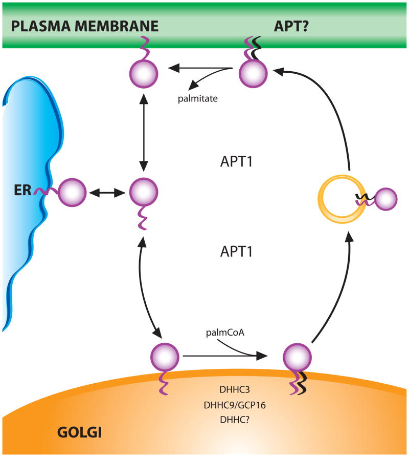
The model that emerged from these studies (Fig. 7) describes a constitutive cycle of protein trafficking between endomembranes and the plasma membrane that is regulated by reversible S-palmitoylation.25b,29 S-palmitoylation of proteins is concentrated at the Golgi apparatus and the longer residence times of S-palmitoylated proteins on membranes allows them to feed into the secretory pathway for transport to the plasma membrane. This unidirectional transport opposes entropy-driven redistribution of S-palmitoylated proteins throughout cellular membranes. The model proposes that thioesterase activity is distributed throughout the cell and returns proteins to a state where they rapidly dissociate and reassociate with all cellular membranes until trapped again at the Golgi by S-palmitoylation and subsequent vesicular transport to the plasma membrane.
FIGURE 7.
Model of Acylation-dependent Ras Trafficking. Endoplasmic reticulum (ER). Acylprotein thioesterase 1(APT1).
Independent studies have demonstrated constitutive trafficking of heterotrimeric G proteins between endomembranes and the plasma membrane that is dependent upon S-palmitoylation. Gqα is an exclusively S-palmitoylated G-protein alpha subunit that is modified by the Golgi-localized DHHC3 and DHHC7 PATs. These enzymes were required for continuous shuttling of Gqα between the plasma membrane and Golgi.59 Similarly, N-myristoylated and S-palmitoylated Goα displayed similar trafficking that was inhibited by treatment of cells with 2-BP.60 FRAP analysis of GAD65, an isoform of glutamic acid decarboxylase, suggests that a S-palmitoylation/depalmitoylation cycle regulates transport between ER-Golgi and post-Golgi membranes. 61
Concentration of palmitoylating activity at the Golgi apparatus and widespread distribution of acylprotein thioesterase activity are central tenets of the model depicted in Figure 7. In mammalian cells, most DHHC proteins are localized at the endoplasmic reticulum and Golgi apparatus when ectopically expressed.62 Golgi localization has been verified for endogenous DHHC363, a PAT for both integral and peripheral membrane proteins, and GCP1664, an obligate binding partner of DHHC9 which has PAT activity for Ras proteins in vitro.65 A variety of protein substrates with diverse sequence contexts have been assigned to Golgi-localized PATs 66, consistent with the Golgi as a central hub for S-palmitoylation. However, S-palmitoylation of peripheral membrane proteins is not exclusive to the Golgi apparatus. The synaptic scaffold, PSD-95 and R7BP, a S-palmitoylated membrane anchor for G-protein regulators, are S-palmitoylated by DHHC267, an enzyme that is localized at the plasma membrane and on endosomal vesicles.68
The cytoplasmic distribution of the acylprotein thioesterase APT1 is consistent with its ability to come in contact with S-palmitoylated proteins on membranes throughout the cell.19 The localization of APT2 and APT3 is unknown. Evidence for the presence of Ras depalmitoylating activity in crude membrane fractions from yeast20 and from mammalian cells69 suggests that additional protein thioesterases remain to be identified. APT1 was recently detected in a S-palmitoylproteome screen, suggesting a possible mechanism for its membrane association.70
The kinetics of membrane association and spatial organization of fluorescent lipopetides and semisynthetic lipidated proteins in cells supports a mechanism of S-palmitoylation that is rapid, not stereoselective, and that does not require specific amino acid sequences.29 Whether the properties of DHHC-PATs can be reconciled with the proposed mechanism is unresolved. DHHC autoacylation could facilitate rapid transfer of palmitate to substrates in cells. Recent kinetic analysis of the yeast Ras PAT (Erf2/Erf4) strongly supports that autoacylation represents an acyl-intermediate.71 Autoacylation of the enzyme occurs in seconds and in the absence of Ras, the acyl group undergoes rapid turnover. The rapid regeneration of acylated DHHC-PAT suggests that in cells in the presence of palmitoyl-CoA, the enzyme is charged with palmitate and ready to deliver it to a protein substrate.71 DHHC proteins are S-palmitoylated in vivo, but to date the only S-palmitoylation sites that have been mapped are distinct from the cysteine in the DHHC motif, the presumed catalytic cysteine of the enzyme.70 The susceptibility of the autoacylated enzyme to hydrolysis71 may make trapping the acyl-enzyme intermediate difficult.
The question of whether there is any substrate specificity to S-palmitoylation at the Golgi is provocative. With respect to the absence of stereoselectivity29, this can be directly tested by determining whether peptides with unnatural amino acid sequences are substrates for DHHC PATs. The presence of multiple DHHC proteins at the Golgi may contribute to the recognition of a group of substrates with diverse sequence contexts. However, evidence in yeast suggests substantial functional overlap of DHHC proteins for different substrates, particularly for peripheral membrane proteins.16 Because it is not possible to achieve the complete absence of DHHC proteins in yeast and maintain viability of the organism, the possibility of non-enzymatic S-palmitoylation or other enzymes cannot be excluded.16 There is evidence to suggest that determinants outside the sequence surrounding the S-palmitoylation site are important for specific recognition of a substrate by the enzyme72 and that regions of the enzymes outside the catalytic DHHC domain contribute to substrate recognition.73 The issue of substrate specificity of DHHC enzymes is an area of active investigation and is discussed in more detail in a recent review.66
The process of sorting of S-palmitoylated proteins into vesicular carriers and their post-Golgi trafficking is also an area of obvious interest. A recent study reports that S-palmitoylation directs H-Ras to recycling endosomes on its exocytic route to the plasma membrane74 and there is evidence to suggest that recycling endosomes may operate as a sorting hub for other S-palmitoylated substrates and DHHC proteins.68
The model of constitutive palmitate turnover and cycling of peripheral membrane proteins represents a core pathway that can accommodate regulatory inputs. To date, there is little evidence for regulation of the enzymes that S-palmitoylate and depalmitoylate proteins, but the field is in its early days. Protein interactions that influence the accessibility of a substrate have been documented as a mechanism to regulate the S-palmitoylation status of a protein. A recent example is the promotion of Ras depalmitoylation by the prolyl isomerase FKBP12.69 Discussion of how signaling pathways are integrated with the acylation cycle is beyond the scope of this review, but has been discussed elsewhere.75
8. Chemical and Enzymatic Methods for Analyzing Protein Lipidation
Biochemical analysis of covalent lipid modifications is essential for determining the function and regulatory mechanisms of many proteins. While bioinformatic programs based on primary amino acid sequences for protein lipidation have provided convenient means of predicting lipid-modified proteins from genomic data, direct biochemical detection is required for demonstrating lipid modifications on proteins in specific cell types and cellular states. Radiolabeled lipids have historically been used to monitor lipid modification of proteins,45 but the long exposure times for detection (weeks to months) and hazards associated with radioactivity have inspired many researchers to develop more sensitive and convenient methods for analyzing protein lipidation. In this section, we highlight chemical and enzymatic methods as well as bioorthogonal lipid chemical reporters that have enabled more rapid visualization of protein lipidation and also facilitated the discovery of new lipid-modified proteins.76
8.1 Chemoenzymatic Detection of S-Prenylated Proteins
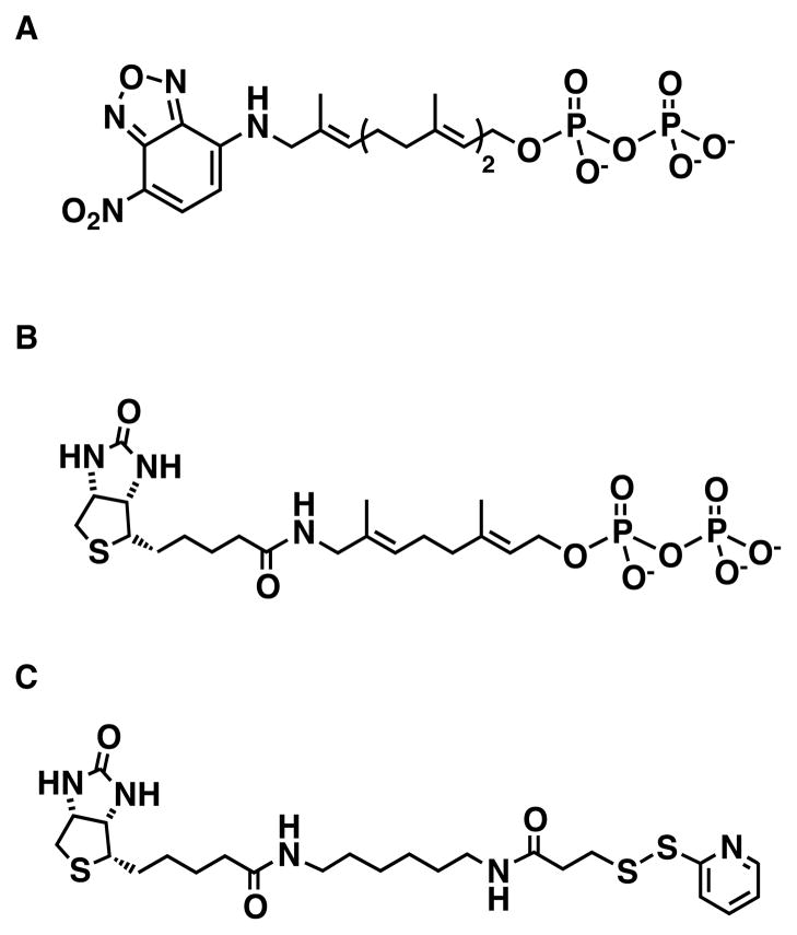
Protein S-prenylation can be predicted based on C-terminal CaaX/CC-motifs using PrenBase (http://mendel.imp.ac.at/PrePS/PRENbase/) and biochemically validated by radioactive isoprenoid pyrophosphate labeling with S-prenyltransferases in vitro or by analysis of target proteins following metabolic labeling of cells with 3H-melavonic acid. Alternatively, the substrate promiscuity of S-prenyltransferases allows utilization of modified isoprenoid substrates that circumvents the limitations of radioactivity and enables the non-radioactive analysis of protein S-prenylation. Notably, the synthesis of 7-nitro-benzo[1,2,5]oxadiazol-4-ylamino (NBD)-modified isoprenoid pyrophosphates have afforded fluorescent substrates for monitoring the activity of all three classes of S-prenyltransferases (Fig. 8A).77 These fluorescent S-prenyltransferase substrates have facilitated the analysis of specific protein substrates in vitro as well as discovery and characterization of small molecular inhibitors.77 For affinity purification of S-prenylated proteins, biotinylated isoprenoid pyrophosphates were synthesized and evaluated with S-prenyltransferases in vitro.78 These biotinylated analogs are not utilized by wild-type enzymes, but biotinylgeranylpyrophosphate (BGPP) is compatible with S-prenyltransferase mutants that accommodate a more sterically demanding isoprenoid substrate and allow enzyme-specific profiling of S-geranylgeranylated proteins (Fig. 8B).78 The development of fluorescent and biotinylated isoprenoids that are utilized by wild-type or mutant S-prenyltransferases have provided more efficient tools to monitor protein S-prenylation and facilitated the characterization of protein substrates as well as inhibitor discovery for therapeutic development. These chemical tools have provided important advances for monitoring protein S-prenylation in vitro, but still have some limitations for cellular studies. For example, BGPP and mutant RabGGTases can be used to profile protein substrates in cell lysates, however, this approach requires inhibition or defects in endogenous isoprenoid biosynthesis to yield nascent protein substrates for enzyme labeling in vitro and does not function in living cells 78 NBD-isoprenoids can be incorporated into mammalian cells and installed onto overexpressed proteins such as EYFP tagged K-Ras, but these fluorophore-modified isoprenoids do not appear to be efficiently incorporated onto endogenous S-prenylated proteins.77 More sensitive tools are still needed to monitor protein S-prenylation in vivo.
FIGURE 8.
Reagents for chemical or enzymatic analysis of protein lipidation. A) NBD-farnesylpyrophosphate. B) Biotin-geranylpyrophosphate. A) HPDP-Biotin for ABE analysis of S-acylation.
8.2 Acyl-Biotin Exchange Detection of S-Acylated Proteins
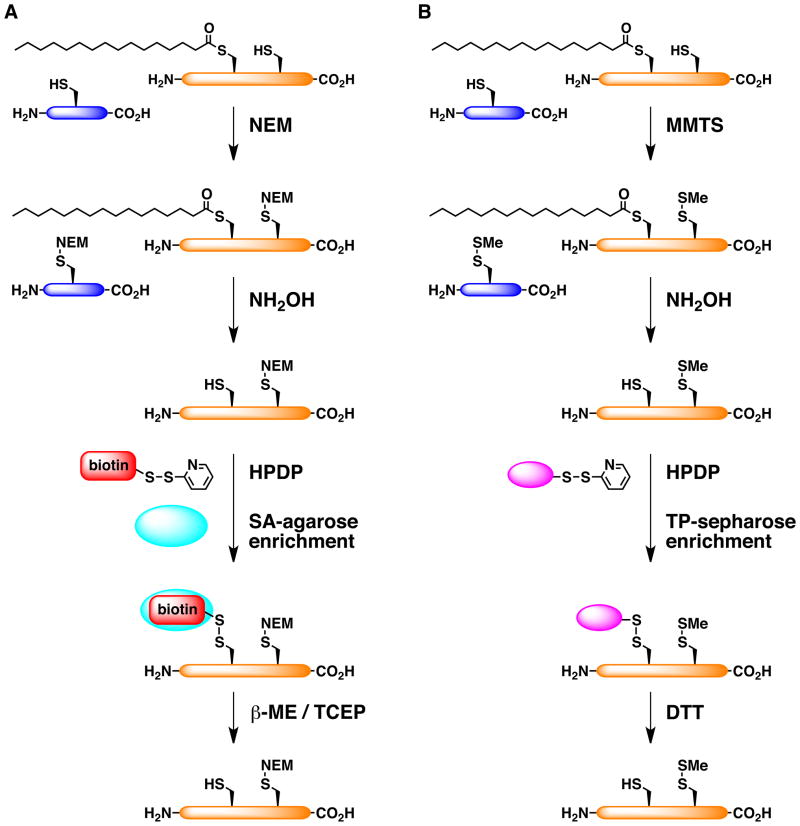
The dynamic nature of S-palmitoylation and limited primary sequence similarity for the site of modification has made this form of protein fatty acylation particularly difficult to analyze (Fig. 1F). Predictive algorithms such as CSS-Palm (http://csspalm.biocuckoo.org/) provide confidence scores for potential S-palmitoylation sites of a given protein that can be validated biochemically by radioactive palmitate labeling. The limited sensitivity of radioactive labeling has demanded more efficient methods for analyzing S-palmitoylation biochemically. One unique chemical feature of S-palmitoylation compared to amide-linked fatty acids on N-myristoylated proteins is the differential sensitivity of thioesters to strong nucleophiles such as hydroxylamine (NH2OH).79 Researchers have developed S-acyl-biotin exchange (ABE) protocols to exploit the hydroxylamine-sensitivity of S-acylated proteins to selectively label these proteins with biotin for non-radioactive detection and enrichment with streptavidin reagents (Fig. 9A).80 An important feature of ABE proteomics is the reversible elution of captured proteins using thiol-reactive and cleavable reagents such as N-[6-(Biotinamido)hexyl]-3′-(2′-pyridyldithio)propionamide (Biotin-HPDP) (Fig. 9A).80b The application of ABE to budding yeast enabled large-scale profiling of known and many new S-palmitoylated proteins (~75 substrates) using multidimensional protein identification technology (MudPIT)-based proteomics.16 The comparative proteomic analysis of wild-type and mutant budding yeast strains with ABE revealed distinct and overlapping protein substrates for DHHC-PATs that provides an important foundation understanding the specificity of this enzyme family.16 Notably, the large-scale analysis of S-acylated proteins by ABE identified ~200 hundred S-acylated proteins in primary neurons, including S-palmitoylation of the brain isoform of the GTPase CDC42, as well as differential changes in S-palmitoylation of proteins upon neuronal stimulation.81 Application of ABE proteomics at the protein and peptide level in various mammalian cell lines has also revealed many new candidate S-palmitoylated proteins as well as their sites of modification.70,82 Initial ABE enrichment of S-acylated peptides in HeLa cells recovered ~57 sites of modification.82a The fractionation of lipid-raft associated proteins from a prostate cancer cell line (DU145), enrichment of S-acylated proteins and peptides, as well as label-free MS quantitation revealed ~398 S-acylated proteins and 168 S-acylation sites (Fig. 9).70 ABE analysis of RAW264.7 macrophages has also yielded many candidate S-acylated proteins and revealed that S-palmitoylation facilitates mitochondrial targeting and pro-apoptotic activity of phospholipid scramblase 3 (Plscr3).83 Alternatively, the analysis of HEK293 cells using S-acyl resin-assisted capture (acyl-RAC) and stable-isotope proteomics methods identified ~93 S-acylation sites (Fig. 9B).82b It should also be noted that S-acylation proteomics are complementary to other large-scale studies of cysteine modifications such as S-nitrosylation84 and sulfenic acid modification85. Interestingly, S-nitrosylation and S-palmitoylation have been reported to reciprocally regulate the synaptic targeting of PSD-95 in neurons.86 These studies highlight the remarkable advances in protein S-palmitoylation with improved methods for biochemical analysis.
FIGURE 9.
Enrichment protocols for S-acylated proteins. A) Acyl-biotin exchange (ABE) with HPDP. B) Acyl-resin assisted capture (Acyl-RAC) with thiopropyl (TP)-sepharose. β-mercaptoethanol (β-ME). Dithiothreitol (DTT). Methylmethanethiosulfonate (MMTS). Tris(2-carboxyethyl)phosphine (TCEP).
9. Bioorthogonal Detection of Lipid-Modified Proteins
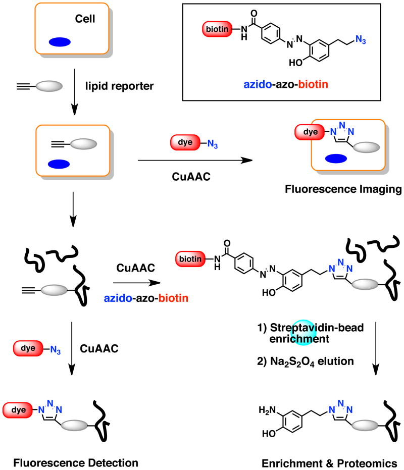
The development of bioorthogonal chemical ligation reactions has afforded new technology to monitor the biosynthesis of nucleic acids, protein and posttranslational modifications,87 including lipid-modified proteins76(Fig. 10). The Staudinger ligation88 and CuI-catalyzed [3 + 2] azide-alkyne cycloaddition (CuAAC) or “click chemistry”89 have provided bioorthogonal ligation reactions that are specific for alkyl-azides and alkynes that exhibit minimal reactivity with endogenous functionality on biomolecules. Since azides and alkynes are relatively small, non-polar and stable functional groups, they can be readily installed onto metabolites such as lipids with minimal structural perturbation for metabolic incorporation and protein modification in cells. Bioorthogonal chemical ligation reactions have therefore enabled two-step labeling approaches using small azide/alkyne-functionalized lipid reporters and complementary detection tags to monitor protein lipidation as well as lipid trafficking (Fig. 10).76
FIGURE 10.
Schematic for bioorthogonal detection and enrichment of lipidated proteins using alkynyl chemical reporters and CuAAC.
9.1 Bioorthogonal Reporters for Protein S-Prenylation
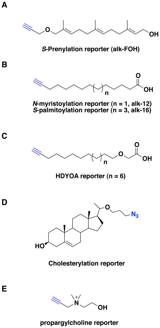
The promiscuity of protein S-prenyltransferases for NBD- and other functionalized-substrates suggested that azide- or alkyne-functionalized isoprenoids could be used for bioorthogonal detection of protein S-prenylation.76a,b,90 Indeed, metabolic labeling of mammalian cells with azido-farnesol (az-FOH) and its pyrophosphate derivative (az-FPP) demonstrated that S-prenylated proteins could be visualized following Staudinger ligation with biotinylated phosphine-reagents.91 Affinity enrichment and proteomic analysis of az-FPP labeled proteins resulted in the identification of known and 18 putatively S-farnesylated proteins from COS-1 cells.91 Several other azide and alkyne-isoprenoids and their pyrophosphate analogs have also been shown to function as S-prenylation reporters in vitro and in cells.92 Proteomic analysis of azido-geranylgeraniol (az-GGOH) labeled polypeptides after CuAAC revealed 10 previously described S-geranylgeranylated proteins of the Rab and Ras families from MCF-7 cells.92e In contrast to NBD- or biotinylated isoprenoid analogs, azide/alkyne-isoprenoid reporters are utilized more efficiently by wild-type S-prenyltransferases and can label proteins at endogenous levels. However, proteomic studies with BGPP and engineered RabGGTase has afforded more efficient recovery and profiling of S-prenylated substrates78 compared to bioorthogonal proteomics with isoprenoid reporters so far.91,92e. Prenylome profiling with both methods is currently limited by the need to deplete endogenous isoprenoids with statins for efficient labeling of S-prenylated proteins. Alkynyl-isoprenoids that afford more sensitive detection of S-prenylated proteins compared to their azide counterparts (Fig. 11A)93, other isoprenoid reporters 94 and improved affinity enrichment methods 95 may enable large-scale analysis of prenylated proteins without significant perturbation of endogenous isoprenoid levels in cells.
FIGURE 11.
Bioorthogonal chemical reporters for analysis of lipid-modified proteins. A) Alkynyl-isoprenoid reporter. B). Alkynyl-fatty acid reporters. C) HDYOA reporter. D) Azido cholesteroylation reporter. E) Propargylcholine reporter.
9.2 Bioorthogonal Reporters for Protein Fatty Acylation
Azide and alkyne-functionalized fatty acids can also function as lipid reporters for visualizing and identifying diverse classes of fatty-acylated proteins.76 For example, azido-fatty acid labeling of mammalian cells enables the bioorthogonal detection of N-myristoylated or S-palmitoylated proteins depending on the fatty acid chain length.96 Alkynyl-fatty acids also proved to be efficient lipid reporters for monitoring fatty acylation in mammalian cells (Fig. 11B).76b,97 The direct comparison of lipid reporters, bioorthogonal ligation methods and detection modes demonstrated that alkynyl-fatty acid reporters in conjunction with CuAAC and in-gel fluorescence detection afford the most sensitive protocol for visualizing lipidated proteins.76b In addition to cytosolic forms of fatty acylation, these fatty acid reporters can be used to visualize lipidation of secreted morphogens such as Wnt- and Hedgehog.98
The improved sensitivity of bioorthogonal fatty acid reporters has provided unique opportunities to evaluate alterations in protein fatty acylation in different cell types, cellular states as well as their mechanisms of regulation. For example, in-gel fluorescence profiling of mammalian cell lines with alkynyl-fatty acids revealed diverse N-myristoylated or S-palmitoylated proteins between various cell types.76b The cellular fractionation along with fatty acid reporter labeling has also revealed discrete profiles of fatty-acylated proteins in the mitochondria96c,99 as well as posttranslationally N-myristoylated proteins during apoptosis100. Fluorescence microscopy analysis of PC3 tumor cells undergoing cytokinesis revealed an enrichment of alkynyl-palmitate reporter labeling at the cleavage furrow, suggesting that S-palmitoylated proteins may be recruited to specific membranes during cell division.97b To explore dynamic changes in protein S-palmitoylation in different cellular states, a dual pulse-chase labeling protocol with alkynyl-fatty acids and azidohomoalanine or azido-myristic acid followed by sequential CuAAC reaction with orthogonal fluorophores was developed to simultaneously monitor depalmitoylation and protein turnover of specific protein substrates.101 This tandem imaging protocol revealed accelerated depalmitoylation of Lck upon T-cell activation, which suggests dynamic membrane targeting of this Src-family kinase may be crucial for cell signaling.101 Interestingly, pulse-chase analysis of the β1-adrenergic receptor with alkynyl-palmitate reporter revealed differential depalmitoylation rates of two S-palmitoylation sites,102 suggesting unique factors may regulate dynamic S-palmitoylation at specific Cys residues on the same protein.
Many new fatty-acylated proteins have also been identified with fatty acid reporters and bioorthogonal proteomics. Bioorthogonal proteomic analysis of membrane fractions from Jurkat T cells labeled with an alkynyl-palmitic acid reporter identified 125 high-confidence candidate fatty-acylated proteins and revealed novel S-palmitoylation of serine hydrolases.97c The large-scale proteomic analysis of total cell lysates from Jurkat T cells revealed 178 high-confidence candidate fatty-acylated proteins as well as S-acylation of histone H3 variants.103 Interestingly, O-palmitoylation of histone H4 at Ser47 by acyl-CoA: lysophosphatidylcholine acyltransferase has also been reported.104 Palmitoylome profiling of a dendritic cell line (DC2.4) identified 157 high-confidence candidate fatty-acylated proteins and uncovered a family of S-palmitoylated interferon-induced transmembrane proteins (IFITMs).105 Notably, S-palmitoylation of IFITM3 appears to be crucial for cellular resistance to influenza virus infection.105 Metabolic labeling of bacteria with alkynyl-fatty acids also allows profiling of canonical lipoproteins and identification of unpredicted fatty-acylated substrates.106 It is important to note that in addition to fatty-acylated proteins, these alkynyl-fatty acid proteomic studies also recovered many enzymes involved in fatty acid metabolism,97c,103,105,106 suggesting fatty acid reporters may be broken down and label non-lipidated proteins at lower levels. Indeed, short chain alkynyl-fatty acids (ω-butynyl and pentynyl acids) and their corresponding acyl-CoA derivatives are efficient bioorthogonal chemical reporters for monitoring lysine protein acetylation. 95 To circumvent the potential degradation of alkynyl-fatty acids via β-oxidation pathway, 15-hexadecynyloxyacetic acid (HDYOA) (Fig. 11C), a fatty acid chemical reporter with an oxy-ether linkage has also been synthesized and demonstrated to label known S-palmitoylated proteins and fewer metabolic enzymes.107 For proteomic analysis of alkyne-modified proteins,70,103,105–107 the use of clickable and cleavable biotinylated tags such as azido-azo-biotin has been particularly helpful for elution of captured polypeptides from streptavidin beads for proteomics and western blot validation (Fig. 10).70,95 Bioorthogonal fatty acid reporter97c,103,105,107 and ABE proteomic studies16,70,81–82 have revealed many new candidate S-palmitoylated proteins and suggest that ~1–2% of the protein encoding open-reading frames in eukaryotes are covalently modified with fatty acids.
The comparative analyses of chemical/enzymatic methods and bioorthogonal lipid reporter studies have revealed important advantages and limitations of these methods. For example, less sterically demanding azide/alkyne-isoprenoid reporters were utilized by all three S-prenyltransferases in cells. Alternatively, the unique reactivity of BGPP with mutant RabGGTase may provide new opportunities for profiling specific S-prenyltransferase protein substrates. For protein fatty acylation studies, ABE provides an excellent method for analyzing S-acylated proteins in primary tissues that is challenging with lipid chemical reporters, which are rapidly distributed to tissues such as the liver and metabolized in vivo (Hang lab unpublished results). ABE on peptides has also revealed many sites of S-acylation in mammalian proteomes. 82a 82b ABE does have some limitations, as the multiple protein labeling and precipitation steps take several days to execute that also complicate quantitative analyses. It should be noted that the hydroxylamine-sensitivity exploited by ABE is not specific for S-palmitoylation but selective for S-acylation, which also includes S-acylated proteins such as E2 and E3 ligase proteins involved in the ubiquitin conjugation cascade. Alternatively, bioorthogonal lipid reporters allow rapid fluorescence detection of lipidated proteins and pulse-labeling to analyze dynamics of protein lipidation rates in cells. Large-scale proteomic analysis can also be executed with bioorthogonal lipid reporters, but researchers should be cautious of off target protein labeling due to lipid metabolism. Despite the limitations of individual methods, these biochemical approaches have collectively provided powerful and complementary methods for monitoring protein lipidation in vitro and in vivo.
9.3 Other Bioorthogonal Lipid Reporters
Lipid chemical reporters can also be used to visualize other classes of protein lipidation and lipid trafficking. The synthesis of an azide-modified cholesterol reporter has enabled metabolic labeling and fluorescence detection of Shh cholesterylation after CuAAC (Fig. 11D).98b In addition, several azide/alkyne lipid reporters have been developed for biochemical analysis and imaging lipids in membranes.108 Azide-analogs of diacylglycerol (DAG) have been synthesized and can be incorporated into vesicles for biochemical studies with membrane-binding proteins.109 Alternatively, metabolic labeling with propargylcholine allows bioorthogonal fluorescence imaging of phosphatidylcholine lipids in mammalian cells and in mice (Fig. 11E).108a Surface exposed and internalized choline reporters in membranes can be distinguished using imaging reagents with differential cell permeability.108a Three alkynyl-phosphatidic acid reporters modified with an S-acetylthioethyl group (SATE) have also been synthesized for efficient cellular uptake and visualization of membranes.108b Fluorescence imaging of the terminal alkynyl-bearing phosphatidic acid reporters yielded general labeling of membranes in RAW264.7 macrophages by CuAAC after fixation.108b In addition, a cyclooctyne-functionalized phosphatidic acid analog allowed live cell imaging of membranes using a fluorogenic azide-functionalized coumarin dye via strain-promoted azide-alkyne cycloaddition (SPAAC).108b
9.4 Biotechnology Applications of Lipid Reporters
Bioorthogonal lipid chemical reporters have also been adapted for biotechnology applications. For example, the addition of CaaX-motifs on the C-terminus of recombinant proteins allows metabolic installation of azides or alkynes for specific conjugation of fluorophores for imaging applications or affinity tags for immobilization of proteins on defined surfaces for microarray applications.92b–d,110 Alternatively, recombinant proteins bearing an N-terminal NMT recognition sequence can be coexpressed with NMT in E. coli and labeled with azido/alkynyl-fatty acids for site-specific protein labeling as well.111 Site-specific attachment of lipid reporters can also be used for protein trafficking studies in cells, as demonstrated with lipoic acid ligase labeling of tagged proteins with azido-caprylic acid followed by SPAAC with fluorophores.112
10. Protein Lipidation of Bacterial Effectors
As improved methods for protein lipidation studies are now available, the roles of lipid-modified proteins in biology are becoming more prevalent. One emerging area is the impact of host lipidation on the function of bacterial protein effectors that are injected into host cells during infection. A variety of genetic and biochemical studies have revealed that many bacterial pathogens utilized specialized secretion systems to inject a few to over a hundred bacterial protein effectors into host cells during infection.113 These bacterial protein effectors encode diverse biological activities that remodel host cytoskeleton, membrane trafficking and signaling pathways to subvert host defenses.113 Once injected into host cells these bacterial proteins can co-opt posttranslational mechanisms such as protein lipidation to regulate their function.
Bacterial protein effectors can be regulated by host fatty acylation and S-prenylation. An early example of host lipidation of bacterial effectors was revealed by biochemical analysis of AvrRpm1 and AvrB, type III secretion system (T3SS) effector from Pseudomonas syringae.114 The examination of AvrRpm1 and AvrB amino acid sequences revealed N-myristoylation sites that when mutated to Ala abrogated fatty acylation, plasma membrane localization and attenuated the virulence properties of these bacterial effectors.114b In addition to these bacterial effectors, other P. syringae effectors such as AvrPphB, ORF4, NopT, and RipT can undergo proteolytic processing to reveal cryptic N-myristoylation sites (Table 1).114a Site-directed mutagenesis of adjacent Cys residues for some these effectors suggest they may also be S-palmitoylated.114a Notably, fatty acylation of AvrPphB, ORF4 and NopT is crucial for their membrane targeting and P. syringae avirulence in plants. In Salmonella typhimurium, T3SS effector SifA that is involved in remodeling host membranes and intracellular bacterial replication is S-palmitoylated and S-prenylated in host cells (Table 1).115 Although mutation of individual Cys residues associated with host lipid-modifications does not significantly alter membrane partitioning of SifA or bacterial virulence, deletion of all six amino acids at the C-terminus that encodes all the lipidation sites significantly impairs SifA activity and Salmonella infection in vivo.115 Using more sensitive alkynyl-isoprenoid reporters of protein S-prenylation, it appears that mutation of individual Cys residues of SifA does not fully abrogate protein lipidation, which may explain why single Cys mutants did not fully recapitulate the activity of the C-terminal deletion construct.93a S-Palmitoylation has also been discovered on two other Salmonella bacterial effectors SspH2 and SseI, which share a conserved N-terminal domain encoding the site of lipid modification (Table 1).116 Mutation of the conserved Cys residues abrogated plasma membrane targeting of these bacterial effectors and perturbed the inhibitory activity of SseI on cell migration. Co-overexpression of SspH2 with murine DHHC-PATs and alk-16 revealed a distinct subset of acyltransferases (3, 5, 7, 10 and 14) capable of increasing S-palmitoylation of these bacterial effectors. Interestingly, pulse-chase analysis with alk-16 suggests that S-palmitoylation of these bacterial effectors is a relatively stable lipid modification.
Table 1.
Summary of lipidated bacterial protein effectors
| Bacterial effector | Proposed function | Type of lipidation | Site of modification | Reference |
|---|---|---|---|---|
| P. syringae | ||||
| AvrRpm1 | RIN4 antagonist | Dual fatty-acylation | MGCVSSTSRS | 114b |
| AvrB | RIN4 antagonist | Dual fatty-acylation | MGCVSSKSTT | 114b |
| AvrPphB | Cysteine protease | Dual fatty-acylation | KGCASSSGVS | 114q |
| ORF4 | Cysteine protease | Dual fatty-acylation | RGCSSSKALS | 114a |
| NopT | Cysteine protease | Dual fatty-acylation | MGCCASKPQA | 114a |
| RipT | Cysteine protease | N-Myristoylation | RGDRYSSEPG | 114a |
| S. typhimurium | ||||
| SifA | G-protein antagonist | S-Geranylgeranylation | QQSGCLCCFL | 115, 93a |
| S-Palmitoylation | ||||
| SspH2 | S-Palmitoylation | MPFHIGSGCL | 116 | |
| SseI | S-Palmitoylation | MPFHIGSGCL | 116 | |
| S-Geranylgeranylation | QQSGCLCCFL | 115, 93a | ||
| L. pneumophila | ||||
| Lpp1863 | unknown | Not applicable | IISSNNCSLL | 93c |
| Lpp2144 | F-box ligase | S-Farnesylation | KIAQSKCLVC | 93c |
| Lpg2375 | unknown | S-Prenylation | PKFTEQCSIL | 93c |
| Lpg1976 | unknown | S-Geranylgeranylation | ISKFSPCNLL | 93c |
| Lpg0254 | unknown | S-Prenylation | PEKESVCVLM | 93c |
| Lpg2541 | unknown | S-Farnesylation | DTKKHRCTIM | 93c |
| Lpg2477 | Phosphatase | S-Farnesylation | SDPDPKCTIM | 93c |
| Lpg2806 | unknown | S-Farnesylation | EHKNNACVIS | 93c |
| Lpg2525 | F-box ligase | S-Prenylation | EKTNNFCSIL | 93c |
| Lpg1312 | unknown | Not completed | EFEPPCCTII | 93c |
The bioinformatic analysis of potential Legionella pneumophila effectors revealed that several substrates of type 4 secretion system (T4SS) contain CaaX-motifs (Table 1).93c Biochemical fractionation, alkynyl-isoprenoid labeling and cellular localization studies revealed that these L. pneumophila T4SS effectors could be S-farnesylated or S-geranylgeranylated in host cells.93c Furthermore, S-prenylation of these T4SS effectors was crucial for their targeting to intracellular membranes and maintenance of Legionella-containing vacuoles inside host cells.93c,117 Interestingly, farnesyltransferase and geranylgeranyltransferase inhibitors perturbed the membrane targeting of these T4SS effectors, which is likely to perturb their functions in mammalian cells. These observations raise the possibility of repurposing previously developed S-prenyltransferase inhibitors to target host enzymes for attenuating bacterial virulence.
Bacterial pathogens also encode protein effectors that can target lipid-modified proteins in host cells. For example, the Yersinia T3SS effector YopT encodes a cysteine protease that cleaves RhoA, Rac and Cdc42 directly N-terminal to their S-prenylation sites to inhibit the functions of these small GTPases in phagocytosis.118 Likewise, the T3SS effector AvrPpt2 from P. syringae is cysteine protease that targets the lipid-modified domain of RIN4, an Arabidopsis protein that may be involved in pathogen sensing.119 These studies highlight the crucial roles for host lipidation on bacterial effector function as well as lipid-modified host proteins that are directly targeted by bacterial pathogens.
11. Concluding remarks
The impact of chemical biology on the field of protein lipidation in the last decade has been substantial. In the era of “–omes”, the application of bioorthoganol chemistry, acylbiotin exchange, and chemoenzymatic methods to protein lipidation has expanded the catalogue of proteins modified with lipids. This has been particularly important for S-palmitoylation, which lacks well-defined consensus sequences for bioinformatic predictions. The identification of many new integral membrane proteins as substrates for S-palmitoylation81 underscores the importance of expanding our understanding of the functional signficance of lipidating a protein already embedded in the membrane.
The reversibility of protein S-palmitoylation is a feature that distinguishes it from other lipid modifications. Progress in elucidating how basal and stimulated turnover of palmitate on proteins is regulated and how it contributes to function has been accelerated due to advances in live cell imaging of genetically encoded and semisynthetic fluorescent proteins.25b,29,57 Bioorthogonal fatty acid reporters show promise for cell imaging applications and assessment of the kinetics of palmitate turnover.101 An important goal for the future will be development of site-specific incorporation of bioorthogonal lipid reporters into individual proteins. The ability to monitor protein trafficking and lipidation in concert would facilitate functional studies. Direct spectroscopic imaging of lipid chemical reporters in cells may be feasible by adapting techniques in infrared and Raman spectroscopy that have enabled the visualization of azide-labeled proteins in membranes.120 Imaging lipidated proteins in living animals may be achieveable through bioorthogonal chemistry, as recently demonstrated with glycan reporters.121
Protein engineering and synthetic isoprenoids made enzyme-specific profiling of S-prenyltransferases possible, permitting detection of protein substrates and evaluation of inhibitors.77 Development of enzyme-specific chemical reporters for DHHC-PATs would be an enormous asset in elucidating the substrate specificity of this family of enzymes, a number of which have been linked to disease states.66
Over two decades of research have gone into development of small molecule inhibitors of S-prenyltransferases for cancer therapy. The wealth of compounds, preclinical studies, and clinical trials from pharmaceutical programs for farnesyl transferase inhibitors, along with outstanding enzymology and structural biology, is being exploited in “piggy-back” efforts to leverage these resources for treatments of infectious disease122 and the genetic disorder progeria123. NMT inhibitor programs also draw from a large body of functional and structural information.43a There is much less information available for the enzymes that mediate S-palmitoylation. APT1 and PPT1 structures are known, and in the case of APT1, used for inhibitor design.21 Interference of Ras trafficking and function with the inhibitor Palmostatin B is an encouraging sign that the enzymes that regulate S-palmitoylation may have value as therapeutic targets.21 Structures of DHHC-PATs will require overcoming the challenges associated with crystallizing integral membrane proteins and high throughput screening of the enzymes may be a more feasible approach in the short term. The abundance of receptors and signaling proteins that are modified with palmitate encourages discovery and design of molecules that can be used to interrogate pathways regulated by S-palmitoylation to elucidate its biological consequences in health and disease.
Acknowledgments
H.C.H acknowledges Irma T. Hirschl/Monique Weill-Caulier Trust, Ellison Medical Foundation and NIH/NIGMS (1R01GM087544) for support. M.E.L. acknowledges support from Cornell University and NIH/NIGMS (5R01GM051466).
Biographies

Howard C. Hang is an Assistant Professor and Head of the Laboratory of Chemical Biology and Microbial Pathogenesis. He obtained his B.S. degree in chemistry from the University of California, Santa Cruz 1998 with Professor Joseph P. Konopelski. In 2003, he completed his Ph.D. in chemistry at University of California, Berkeley with Professor Carolyn Bertozzi. During his graduate studies he was awarded an American Chemical Society, Organic Division Graduate Fellowship. He then worked with Professor Hidde Ploegh at Harvard Medical School and the Whitehead Institute of Biomedical Research at Massachusetts Institute of Technology from 2004 through 2006 as Damon Runyon Cancer Research Foundation Postdoctoral Fellow. He joined the faculty at The Rockefeller University in 2007.

Maurine E. Linder is a Professor of Pharmacology and Chair of the Department of Molecular Medicine in the College of Veterinary Medicine at Cornell University. She received her Ph.D. in 1987 following graduate training in molecular and cell biology at the University of Texas at Dallas with Dr. John Burr. Her postdoctoral training with Dr. Alfred Gilman was in the Department of Pharmacology at the University of Texas Southwestern Medical School. In 1993 she joined the faculty of the Department of Cell Biology and Physiology at Washington University School of Medicine in St. Louis where she moved through the ranks, becoming a full Professor in 2006. She moved to Cornell University in 2009. Dr. Linder was an Established Investigator of the American Heart Association from 2001–2004 and was elected as an AAAS Fellow in 2009.
Contributor Information
Howard C. Hang, Email: hhang@mail.rockefeller.edu, Laboratory of Chemical Biology and Microbial Pathogenesis, The Rockefeller University, 1230 York Avenue, New York, NY 10065 (USA)
Maurine E. Linder, Department of Molecular Medicine, College of Veterinary Medicine, Cornell University, Ithaca, NY 14853 (USA).
References
- 1.(a) Paulick MG, Bertozzi CR. Biochemistry. 2008;47:6991. doi: 10.1021/bi8006324. [DOI] [PMC free article] [PubMed] [Google Scholar]; (b) Mann RK, Beachy PA. Annu Rev Biochem. 2004;73:891. doi: 10.1146/annurev.biochem.73.011303.073933. [DOI] [PubMed] [Google Scholar]
- 2.Kleuss C, Krause E. EMBO J. 2003;22:826. doi: 10.1093/emboj/cdg095. [DOI] [PMC free article] [PubMed] [Google Scholar]
- 3.Ichimura Y, Kirisako T, Takao T, Satomi Y, Shimonishi Y, Ishihara N, Mizushima N, Tanida I, Kominami E, Ohsumi M, Noda T, Ohsumi Y. Nature. 2000;408:488. doi: 10.1038/35044114. [DOI] [PubMed] [Google Scholar]
- 4.Zhang FL, Casey PJ. Annu Rev Biochem. 1996;65:241. doi: 10.1146/annurev.bi.65.070196.001325. [DOI] [PubMed] [Google Scholar]
- 5.Lane KT, Beese LS. J Lipid Res. 2006;47:681. doi: 10.1194/jlr.R600002-JLR200. [DOI] [PubMed] [Google Scholar]
- 6.Leung KF, Baron R, Seabra MC. J Lipid Res. 2006;47:467. doi: 10.1194/jlr.R500017-JLR200. [DOI] [PubMed] [Google Scholar]
- 7.(a) Blum R, Cox AD, Kloog Y. Recent Pat Anticancer Drug Discov. 2008;3:31. doi: 10.2174/157489208783478702. [DOI] [PubMed] [Google Scholar]; (b) Konstantinopoulos PA, Karamouzis MV, Papavassiliou AG. Nat Rev Drug Discov. 2007;6:541. doi: 10.1038/nrd2221. [DOI] [PubMed] [Google Scholar]
- 8.(a) Farazi T, Waksman G, Gordon J. J Biol Chem. 2001;276:39501. doi: 10.1074/jbc.R100042200. [DOI] [PubMed] [Google Scholar]; (b) Resh MD. Nat Chem Biol. 2006;2:584. doi: 10.1038/nchembio834. [DOI] [PubMed] [Google Scholar]
- 9.(a) Zha J, Weiler S, Oh K, Wei M, Korsmeyer S. Science. 2000;290:1761. doi: 10.1126/science.290.5497.1761. [DOI] [PubMed] [Google Scholar]; (b) Martin DD, Beauchamp E, Berthiaume LG. Biochimie. 2011;93:18. doi: 10.1016/j.biochi.2010.10.018. [DOI] [PubMed] [Google Scholar]
- 10.Duronio RJ, Towler DA, Heuckeroth RO, Gordon JI. Science. 1989;243:796. doi: 10.1126/science.2644694. [DOI] [PubMed] [Google Scholar]
- 11.Ntwasa M, Aapies S, Schiffmann DA, Gay NJ. Exp Cell Res. 2001;262:134. doi: 10.1006/excr.2000.5086. [DOI] [PubMed] [Google Scholar]
- 12.Yang SH, Shrivastav A, Kosinski C, Sharma RK, Chen MH, Berthiaume LG, Peters LL, Chuang PT, Young SG, Bergo MO. J Biol Chem. 2005;280:18990. doi: 10.1074/jbc.M412917200. [DOI] [PubMed] [Google Scholar]
- 13.(a) Smotrys JEME. Annu Rev Biochem. 2004;73:559. doi: 10.1146/annurev.biochem.73.011303.073954. [DOI] [PubMed] [Google Scholar]; (b) Resh MD. Sci STKE. 2006;2006:re14. doi: 10.1126/stke.3592006re14. [DOI] [PubMed] [Google Scholar]
- 14.Linder ME, Deschenes RJ. Nat Rev Mol Cell Biol. 2007;8:74. doi: 10.1038/nrm2084. [DOI] [PubMed] [Google Scholar]
- 15.(a) Lobo S, Greentree W, Linder M, Deschenes R. J Biol Chem. 2002;277:41268. doi: 10.1074/jbc.M206573200. [DOI] [PubMed] [Google Scholar]; (b) Roth A, Feng Y, Chen L, Davis N. J Cell Biol. 2002;159:23. doi: 10.1083/jcb.200206120. [DOI] [PMC free article] [PubMed] [Google Scholar]
- 16.Roth AF, Wan J, Bailey AO, Sun B, Kuchar JA, Green WN, Phinney BS, Yates JR, 3rd, Davis NG. Cell. 2006;125:1003. doi: 10.1016/j.cell.2006.03.042. [DOI] [PMC free article] [PubMed] [Google Scholar]
- 17.Mitchell DA, Vasudevan A, Linder ME, Deschenes RJ. J Lipid Res. 2006;47:11188. doi: 10.1194/jlr.R600007-JLR200. [DOI] [PubMed] [Google Scholar]
- 18.Vesa J, Hellsten E, Verkruyse LA, Camp LA, Rapola J, Santavuori P, Hofmann SL, Peltonen L. Nature. 1995;376:584. doi: 10.1038/376584a0. [DOI] [PubMed] [Google Scholar]
- 19.Duncan JA, Gilman AG. J Biol Chem. 1998;273:15830. doi: 10.1074/jbc.273.25.15830. [DOI] [PubMed] [Google Scholar]
- 20.Duncan J, Gilman A. J Biol Chem. 2002;277:31740. doi: 10.1074/jbc.M202505200. [DOI] [PubMed] [Google Scholar]
- 21.Dekker FJ, Rocks O, Vartak N, Menninger S, Hedberg C, Balamurugan R, Wetzel S, Renner S, Gerauer M, Scholermann B, Rusch M, Kramer JW, Rauh D, Coates GW, Brunsveld L, Bastiaens PI, Waldmann H. Nat Chem Biol. 2010;6:449. doi: 10.1038/nchembio.362. [DOI] [PubMed] [Google Scholar]
- 22.(a) Linder ME, Middleton P, Hepler JR, Taussig R, Gilman AG, Mumby SM. Proc Natl Acad Sci USA. 1993;90:3675. doi: 10.1073/pnas.90.8.3675. [DOI] [PMC free article] [PubMed] [Google Scholar]; (b) Parenti M, Vigano MA, Newman CM, Milligan G, Magee AI. Biochem J. 1993;291(Pt 2):349. doi: 10.1042/bj2910349. [DOI] [PMC free article] [PubMed] [Google Scholar]
- 23.(a) Paige LA, Nadler MJ, Harrison ML, Cassady JM, Geahlen RL. J Biol Chem. 1993;268:8669. [PubMed] [Google Scholar]; (b) Shenoy-Scaria AM, Dietzen DJ, Kwong J, Link DC, Lublin DM. J Cell Biol. 1994;126:353. doi: 10.1083/jcb.126.2.353. [DOI] [PMC free article] [PubMed] [Google Scholar]; (c) Alland L, Peseckis SM, Atherton RE, Berthiaume L, Resh MD. J Biol Chem. 1994;269:16701. [PubMed] [Google Scholar]
- 24.Appolloni A, Prior I, Lindsay M, Parton R, Hancock J. Mol Cell Biol. 2000;20:2475. doi: 10.1128/mcb.20.7.2475-2487.2000. [DOI] [PMC free article] [PubMed] [Google Scholar]
- 25.(a) Schroeder H, Leventis R, Shahinian S, Walton PA, Silvius JR. J Cell Biol. 1996;134:647. doi: 10.1083/jcb.134.3.647. [DOI] [PMC free article] [PubMed] [Google Scholar]; (b) Rocks O, Peyker A, Kahms M, Verveer PJ, Koerner C, Lumbierres M, Kuhlmann J, Waldmann H, Wittinghofer A, Bastiaens PI. Science. 2005;307:1746. doi: 10.1126/science.1105654. [DOI] [PubMed] [Google Scholar]
- 26.(a) Reents R, Wagner M, Kuhlmann J, Waldmann H. Angewandte Chemie. 2004;43:2711. doi: 10.1002/anie.200353265. [DOI] [PubMed] [Google Scholar]; (b) Brunsveld L, Kuhlmann J, Alexandrov K, Wittinghofer A, Goody RS, Waldmann H. Angew Chem Int Ed Engl. 2006;45:6622. doi: 10.1002/anie.200600855. [DOI] [PubMed] [Google Scholar]
- 27.Shahinian S, Silvius JR. Biochemistry. 1995;34:3813. doi: 10.1021/bi00011a039. [DOI] [PubMed] [Google Scholar]
- 28.Bader B, Kuhn K, Owen DJ, Waldmann H, Wittinghofer A, Kuhlmann J. Nature. 2000;403:223. doi: 10.1038/35003249. [DOI] [PubMed] [Google Scholar]
- 29.Rocks O, Gerauer M, Vartak N, Koch S, Huang ZP, Pechlivanis M, Kuhlmann J, Brunsveld L, Chandra A, Ellinger B, Waldmann H, Bastiaens PI. Cell. 2010;141:458. doi: 10.1016/j.cell.2010.04.007. [DOI] [PubMed] [Google Scholar]
- 30.Chen YX, Koch S, Uhlenbrock K, Weise K, Das D, Gremer L, Brunsveld L, Wittinghofer A, Winter R, Triola G, Waldmann H. Angew Chem Int Ed Engl. 2010;49:6090. doi: 10.1002/anie.201001884. [DOI] [PubMed] [Google Scholar]
- 31.(a) Muir TW. Annu Rev Biochem. 2003;72:249. doi: 10.1146/annurev.biochem.72.121801.161900. [DOI] [PubMed] [Google Scholar]; (b) Flavell RR, Muir TW. Acc Chem Res. 2009;42:107. doi: 10.1021/ar800129c. [DOI] [PubMed] [Google Scholar]
- 32.(a) Pylypenko O, Rak A, Durek T, Kushnir S, Dursina BE, Thomae NH, Constantinescu AT, Brunsveld L, Watzke A, Waldmann H, Goody RS, Alexandrov K. EMBO J. 2006;25:13. doi: 10.1038/sj.emboj.7600921. [DOI] [PMC free article] [PubMed] [Google Scholar]; (b) Rak A, Pylypenko O, Durek T, Watzke A, Kushnir S, Brunsveld L, Waldmann H, Goody RS, Alexandrov K. Science. 2003;302:646. doi: 10.1126/science.1087761. [DOI] [PubMed] [Google Scholar]; (c) Rak A, Pylypenko O, Niculae A, Pyatkov K, Goody RS, Alexandrov K. Cell. 2004;117:749. doi: 10.1016/j.cell.2004.05.017. [DOI] [PubMed] [Google Scholar]
- 33.Wu YW, Oesterlin LK, Tan KT, Waldmann H, Alexandrov K, Goody RS. Nat Chem Biol. 2010;6:534. doi: 10.1038/nchembio.386. [DOI] [PubMed] [Google Scholar]
- 34.Durek T, Alexandrov K, Goody RS, Hildebrand A, Heinemann I, Waldmann H. J Am Chem Soc. 2004;126:16368. doi: 10.1021/ja046164n. [DOI] [PubMed] [Google Scholar]
- 35.Wu YW, Tan KT, Waldmann H, Goody RS, Alexandrov K. Proc Natl Acad Sci USA. 2007;104:12294. doi: 10.1073/pnas.0701817104. [DOI] [PMC free article] [PubMed] [Google Scholar]
- 36.Stenmark H. Nat Rev Mol Cell Biol. 2009;10:513. doi: 10.1038/nrm2728. [DOI] [PubMed] [Google Scholar]
- 37.Lee MT, Mishra A, Lambright DG. Traffic. 2009;10:1377. doi: 10.1111/j.1600-0854.2009.00942.x. [DOI] [PMC free article] [PubMed] [Google Scholar]
- 38.Sivars U, Aivazian D, Pfeffer SR. Nature. 2003;425:856. doi: 10.1038/nature02057. [DOI] [PubMed] [Google Scholar]
- 39.(a) Calero M, Collins RN. Biochem Biophys Res Commun. 2002;290:676. doi: 10.1006/bbrc.2001.6242. [DOI] [PubMed] [Google Scholar]; (b) Calero M, Chen CZ, Zhu W, Winand N, Havas KA, Gilbert PM, Burd CG, Collins RN. Mol Biol Cell. 2003;14:1852. doi: 10.1091/mbc.E02-11-0707. [DOI] [PMC free article] [PubMed] [Google Scholar]
- 40.(a) Machner MP, Isberg RR. Science. 2007;318:974. doi: 10.1126/science.1149121. [DOI] [PubMed] [Google Scholar]; (b) Ingmundson A, Delprato A, Lambright DG, Roy CR. Nature. 2007;450:365. doi: 10.1038/nature06336. [DOI] [PubMed] [Google Scholar]
- 41.Schoebel S, Oesterlin LK, Blankenfeldt W, Goody RS, Itzen A. Mol Cell. 2009;36:1060. doi: 10.1016/j.molcel.2009.11.014. [DOI] [PubMed] [Google Scholar]
- 42.Johnson JL, Erickson JW, Cerione RA. J Biol Chem. 2009;284:23860. doi: 10.1074/jbc.M109.031815. [DOI] [PMC free article] [PubMed] [Google Scholar]
- 43.(a) Frearson JA, Brand S, McElroy SP, Cleghorn LA, Smid O, Stojanovski L, Price HP, Guther ML, Torrie LS, Robinson DA, Hallyburton I, Mpamhanga CP, Brannigan JA, Wilkinson AJ, Hodgkinson M, Hui R, Qiu W, Raimi OG, van Aalten DM, Brenk R, Gilbert IH, Read KD, Fairlamb AH, Ferguson MA, Smith DF, Wyatt PG. Nature. 2010;464:728. doi: 10.1038/nature08893. [DOI] [PMC free article] [PubMed] [Google Scholar]; (b) Georgopapadakou NH. Expert Opin Investig Drugs. 2002;11:1117. doi: 10.1517/13543784.11.8.1117. [DOI] [PubMed] [Google Scholar]
- 44.Philips MR, Cox AD. J Clin Invest. 2007;117:1223. doi: 10.1172/JCI32108. [DOI] [PMC free article] [PubMed] [Google Scholar]
- 45.Resh MD. Methods. 2006;40:191. doi: 10.1016/j.ymeth.2006.04.013. [DOI] [PMC free article] [PubMed] [Google Scholar]
- 46.Coleman RA, Rao P, Fogelsong RJ, Bardes ES. Biochim Biophys Acta. 1992;1125:203. doi: 10.1016/0005-2760(92)90046-x. [DOI] [PubMed] [Google Scholar]
- 47.Patterson SI, Skene JH. J Cell Biol. 1994;124:521. doi: 10.1083/jcb.124.4.521. [DOI] [PMC free article] [PubMed] [Google Scholar]
- 48.Jennings BC, Nadolski MJ, Ling Y, Baker MB, Harrison ML, Deschenes RJ, Linder ME. J Lipid Res. 2008;50:233. doi: 10.1194/jlr.M800270-JLR200. [DOI] [PMC free article] [PubMed] [Google Scholar]
- 49.Ducker CE, Griffel LK, Smith RA, Keller SN, Zhuang Y, Xia Z, Diller JD, Smith CD. Mol Cancer Ther. 2006;5:1647. doi: 10.1158/1535-7163.MCT-06-0114. [DOI] [PMC free article] [PubMed] [Google Scholar]
- 50.(a) Sugimoto H, Hayashi H, Yamashita S. J Biol Chem. 1996;271:7705. doi: 10.1074/jbc.271.13.7705. [DOI] [PubMed] [Google Scholar]; (b) Wang A, Deems RA, Dennis EA. J Biol Chem. 1997;272:12723. doi: 10.1074/jbc.272.19.12723. [DOI] [PubMed] [Google Scholar]
- 51.Devedjiev Y, Dauter Z, Kuznetsov S, Jones T, Derewenda Z. Structure. 2000;8:1137. doi: 10.1016/s0969-2126(00)00529-3. [DOI] [PubMed] [Google Scholar]
- 52.Simon GM, Cravatt BF. J Biol Chem. 2010;285:11051. doi: 10.1074/jbc.R109.097600. [DOI] [PMC free article] [PubMed] [Google Scholar]
- 53.Tomatis VM, Trenchi A, Gomez GA, Daniotti JL. PLoS One. 2010;5:e15045. doi: 10.1371/journal.pone.0015045. [DOI] [PMC free article] [PubMed] [Google Scholar]
- 54.(a) Biel M, Deck P, Giannis A, Waldmann H. Chemistry. 2006;12:4121. doi: 10.1002/chem.200501128. [DOI] [PubMed] [Google Scholar]; (b) Deck P, Pendzialek D, Biel M, Wagner M, Popkirova B, Ludolph B, Kragol G, Kuhlmann J, Giannis A, Waldmann H. Angew Chem Int Ed Engl. 2005;44:4975. doi: 10.1002/anie.200462625. [DOI] [PubMed] [Google Scholar]
- 55.Hadvary P, Sidler W, Meister W, Vetter W, Wolfer H. J Biol Chem. 1991;266:2021. [PubMed] [Google Scholar]
- 56.Wright LP, Philips MR. J Lipid Res. 2006;47:883. doi: 10.1194/jlr.R600004-JLR200. [DOI] [PubMed] [Google Scholar]
- 57.Goodwin JS, Drake KR, Rogers C, Wright L, Lippincott-Schwartz J, Philips MR, Kenworthy AK. J Cell Biol. 2005;170:261. doi: 10.1083/jcb.200502063. [DOI] [PMC free article] [PubMed] [Google Scholar]
- 58.Choy E, Chiu VK, Silletti J, Feoktistov M, Morimoto T, Michaelson D, Ivanov IE, Philips MR. Cell. 1999;98:69. doi: 10.1016/S0092-8674(00)80607-8. [DOI] [PubMed] [Google Scholar]
- 59.Tsutsumi R, Fukata Y, Noritake J, Iwanaga T, Perez F, Fukata M. Mol Cell Biol. 2009;29:435. doi: 10.1128/MCB.01144-08. [DOI] [PMC free article] [PubMed] [Google Scholar]
- 60.Chisari M, Saini DK, Kalyanaraman V, Gautam N. J Biol Chem. 2007;282:24092. doi: 10.1074/jbc.M704246200. [DOI] [PMC free article] [PubMed] [Google Scholar]
- 61.Kanaani J, Patterson G, Schaufele F, Lippincott-Schwartz J, Baekkeskov S. J Cell Sci. 2008;121:437. doi: 10.1242/jcs.011916. [DOI] [PubMed] [Google Scholar]
- 62.Ohno Y, Kihara A, Sano T, Igarashi Y. Biochim Biophys Acta. 2006;1761:474. doi: 10.1016/j.bbalip.2006.03.010. [DOI] [PubMed] [Google Scholar]
- 63.Keller CA, Yuan X, Panzanelli P, Martin ML, Alldred M, Sassoe-Pognetto M, Luscher B. J Neurosci. 2004;24:5881. doi: 10.1523/JNEUROSCI.1037-04.2004. [DOI] [PMC free article] [PubMed] [Google Scholar]
- 64.Ohta E, Misumi Y, Sohda M, Fujiwara T, Yano A, Ikehara Y. J Biol Chem. 2003;278:51957. doi: 10.1074/jbc.M310014200. [DOI] [PubMed] [Google Scholar]
- 65.Swarthout JT, Lobo S, Farh L, Croke MR, Greentree WK, Deschenes RJ, Linder ME. J Biol Chem. 2005;280:31141. doi: 10.1074/jbc.M504113200. [DOI] [PubMed] [Google Scholar]
- 66.Greaves J, Chamberlain LH. Trends Biochem Sci. 2011 doi: 10.1016/j.tibs.2011.01.003. [DOI] [PubMed] [Google Scholar]
- 67.(a) Noritake J, Fukata Y, Iwanaga T, Hosomi N, Tsutsumi R, Matsuda N, Tani H, Iwanari H, Mochizuki Y, Kodama T, Matsuura Y, Bredt DS, Hamakubo T, Fukata M. J Cell Biol. 2009;186:147. doi: 10.1083/jcb.200903101. [DOI] [PMC free article] [PubMed] [Google Scholar]; (b) Jia L, Linder ME, Blumer KJ. J Biol Chem. 2011;286:13695. doi: 10.1074/jbc.M110.193763. [DOI] [PMC free article] [PubMed] [Google Scholar]
- 68.Greaves J, Carmichael JA, Chamberlain LH. Mol Biol Cell. 2011 doi: 10.1091/mbc.E10-11-0924. [DOI] [PMC free article] [PubMed] [Google Scholar]
- 69.Ahearn IM, Tsai FD, Court H, Zhou M, Jennings BC, Ahmed M, Fehrenbacher N, Linder ME, Philips MR. Mol Cell. 2011;41:173. doi: 10.1016/j.molcel.2011.01.001. [DOI] [PMC free article] [PubMed] [Google Scholar]
- 70.Yang W, Di Vizio D, Kirchner M, Steen H, Freeman MR. Mol Cell Proteomics. 2010;9:54. doi: 10.1074/mcp.M800448-MCP200. [DOI] [PMC free article] [PubMed] [Google Scholar]
- 71.Mitchell DA, Mitchell G, Ling Y, Budde C, Deschenes RJ. J Biol Chem. 2010;285:38104. doi: 10.1074/jbc.M110.169102. [DOI] [PMC free article] [PubMed] [Google Scholar]
- 72.Nadolski MJ, Linder ME. J Biol Chem. 2009;284:17720. doi: 10.1074/jbc.M109.005447. [DOI] [PMC free article] [PubMed] [Google Scholar]
- 73.Huang K, Sanders S, Singaraja R, Orban P, Cijsouw T, Arstikaitis P, Yanai A, Hayden MR, El-Husseini A. FASEB J. 2009;23:2605. doi: 10.1096/fj.08-127399. [DOI] [PMC free article] [PubMed] [Google Scholar]
- 74.Misaki R, Morimatsu M, Uemura T, Waguri S, Miyoshi E, Taniguchi N, Matsuda M, Taguchi T. J Cell Biol. 2010;191:23. doi: 10.1083/jcb.200911143. [DOI] [PMC free article] [PubMed] [Google Scholar]
- 75.(a) Grecco HE, Schmick M, Bastiaens PI. Cell. 2011;144:897. doi: 10.1016/j.cell.2011.01.029. [DOI] [PubMed] [Google Scholar]; (b) Salaun C, Greaves J, Chamberlain LH. J Cell Biol. 2010;191:1229. doi: 10.1083/jcb.201008160. [DOI] [PMC free article] [PubMed] [Google Scholar]
- 76.(a) Hannoush RN, Sun J. Nat Chem Biol. 2010;6:498. doi: 10.1038/nchembio.388. [DOI] [PubMed] [Google Scholar]; (b) Charron G, Wilson J, Hang HC. Curr Opin Chem Biol. 2009;13:382. doi: 10.1016/j.cbpa.2009.07.010. [DOI] [PubMed] [Google Scholar]; (c) Hang HC, Wilson JP, Charron G. Acc Chem Res. 2011 doi: 10.1021/ar200063v. [DOI] [PMC free article] [PubMed] [Google Scholar]
- 77.Dursina B, Reents R, Delon C, Wu Y, Kulharia M, Thutewohl M, Veligodsky A, Kalinin A, Evstifeev V, Ciobanu D, Szedlacsek SE, Waldmann H, Goody RS, Alexandrov K. J Am Chem Soc. 2006;128:2822. doi: 10.1021/ja052196e. [DOI] [PubMed] [Google Scholar]
- 78.Nguyen UT, Guo Z, Delon C, Wu Y, Deraeve C, Franzel B, Bon RS, Blankenfeldt W, Goody RS, Waldmann H, Wolters D, Alexandrov K. Nat Chem Biol. 2009;5:227. doi: 10.1038/nchembio.149. [DOI] [PubMed] [Google Scholar]
- 79.Schmidt MF, Schlesinger MJ. Cell. 1979;17:813. doi: 10.1016/0092-8674(79)90321-0. [DOI] [PubMed] [Google Scholar]
- 80.(a) Drisdel RC, Green WN. Biotechniques. 2004;36:276. doi: 10.2144/04362RR02. [DOI] [PubMed] [Google Scholar]; (b) Wan J, Roth AF, Bailey AO, Davis NG. Nat Protoc. 2007;2:1573. doi: 10.1038/nprot.2007.225. [DOI] [PubMed] [Google Scholar]
- 81.Kang R, Wan J, Arstikaitis P, Takahashi H, Huang K, Bailey AO, Thompson JX, Roth AF, Drisdel RC, Mastro R, Green WN, Yates JR, 3rd, Davis NG, El-Husseini A. Nature. 2008;456:904. doi: 10.1038/nature07605. [DOI] [PMC free article] [PubMed] [Google Scholar]
- 82.(a) Zhang J, Planey SL, Ceballos C, Stevens SM, Jr, Keay SK, Zacharias DA. Mol Cell Proteomics. 2008;7:1378. doi: 10.1074/mcp.M800069-MCP200. [DOI] [PMC free article] [PubMed] [Google Scholar]; (b) Forrester MT, Hess DT, Thompson JW, Hultman R, Moseley MA, Stamler JS, Casey PJ. J Lipid Res. 2011;52:393. doi: 10.1194/jlr.D011106. [DOI] [PMC free article] [PubMed] [Google Scholar]
- 83.Merrick BA, Dhungana S, Williams JG, Aloor JJ, Peddada S, Tomer KB, Fessler MB. Mol Cell Proteomics. 2011 doi: 10.1074/mcp.M110.006007. [DOI] [PMC free article] [PubMed] [Google Scholar]
- 84.Forrester MT, Thompson JW, Foster MW, Nogueira L, Moseley MA, Stamler JS. Nat Biotechnol. 2009;27:557. doi: 10.1038/nbt.1545. [DOI] [PMC free article] [PubMed] [Google Scholar]
- 85.Leonard SE, Reddie KG, Carroll KS. ACS Chem Biol. 2009;4:783. doi: 10.1021/cb900105q. [DOI] [PubMed] [Google Scholar]
- 86.Ho GP, Selvakumar B, Mukai J, Hester LD, Wang Y, Gogos JA, Snyder SH. Neuron. 2011;71:131. doi: 10.1016/j.neuron.2011.05.033. [DOI] [PMC free article] [PubMed] [Google Scholar]
- 87.Sletten EM, Bertozzi CR. Angew Chem Int Ed Engl. 2009;48:6974. doi: 10.1002/anie.200900942. [DOI] [PMC free article] [PubMed] [Google Scholar]
- 88.Saxon E, Bertozzi CR. Science. 2000;287:2007. doi: 10.1126/science.287.5460.2007. [DOI] [PubMed] [Google Scholar]
- 89.(a) Tornoe CW, Christensen C, Meldal M. J Org Chem. 2002;67:3057. doi: 10.1021/jo011148j. [DOI] [PubMed] [Google Scholar]; (b) Rostovtsev VV, Green LG, Fokin VV, Sharpless KB. Angew Chem Int Ed Engl. 2002;41:2596. doi: 10.1002/1521-3773(20020715)41:14<2596::AID-ANIE2596>3.0.CO;2-4. [DOI] [PubMed] [Google Scholar]
- 90.Nguyen UT, Goody RS, Alexandrov K. Chembiochem. 2010;11:1194. doi: 10.1002/cbic.200900727. [DOI] [PubMed] [Google Scholar]
- 91.Kho Y, Kim SC, Jiang C, Barma D, Kwon SW, Cheng JK, Jaunbergs J, Weinbaum C, Tamanoi F, Falck J, Zhao YM. Proc Natl Acad Sci USA. 2004;101:12479. doi: 10.1073/pnas.0403413101. [DOI] [PMC free article] [PubMed] [Google Scholar]
- 92.(a) Rose MW, Rose ND, Boggs J, Lenevich S, Xu J, Barany G, Distefano MD. J Pept Res. 2005;65:529. doi: 10.1111/j.1399-3011.2005.00261.x. [DOI] [PubMed] [Google Scholar]; (b) Rose MW, Xu JH, Kale TA, O’Doherty G, Barany G, Distefano MD. Biopolymers. 2005;80:164. doi: 10.1002/bip.20239. [DOI] [PubMed] [Google Scholar]; (c) Duckworth BP, Zhang Z, Hosokawa A, Distefano MD. Chembiochem. 2007;8:98. doi: 10.1002/cbic.200600340. [DOI] [PubMed] [Google Scholar]; (d) Gauchet C, Labadie GR, Poulter CD. J Am Chem Soc. 2006;128:9274. doi: 10.1021/ja061131o. [DOI] [PMC free article] [PubMed] [Google Scholar]; (e) Chan LN, Hart C, Guo L, Nyberg T, Davies BSJ, Fong LG, Young SG, Agnew BJ, Tamanoi F. Electrophoresis. 2009;30:3598. doi: 10.1002/elps.200900259. [DOI] [PMC free article] [PubMed] [Google Scholar]; (f) Labadie GR, Viswanathan R, Poulter CD. J Org Chem. 2007;72:9291. doi: 10.1021/jo7017747. [DOI] [PMC free article] [PubMed] [Google Scholar]; (g) Berry AFH, Heal WP, Tarafder AK, Tolmachova T, Baron RA, Seabra MC, Tate EW. Chembiochem. 2010;11:771. doi: 10.1002/cbic.201000087. [DOI] [PubMed] [Google Scholar]
- 93.(a) Charron G, Tsou LK, Maguire W, Yount JS, Hang HC. Mol Biosyst. 2011;7:67. doi: 10.1039/c0mb00183j. [DOI] [PMC free article] [PubMed] [Google Scholar]; (b) DeGraw AJ, Palsuledesai C, Ochocki JD, Dozier JK, Lenevich S, Rashidian M, Distefano MD. Chem Biol Drug Des. 2010;76:460. doi: 10.1111/j.1747-0285.2010.01037.x. [DOI] [PMC free article] [PubMed] [Google Scholar]; (c) Ivanov SS, Charron G, Hang HC, Roy CR. J Biol Chem. 2010 doi: 10.1074/jbc.M110.170746. [DOI] [PMC free article] [PubMed] [Google Scholar]
- 94.Placzek AT, Gibbs RA. Org Lett. 2011;13:3576. doi: 10.1021/ol201069x. [DOI] [PMC free article] [PubMed] [Google Scholar]
- 95.Yang YY, Ascano JM, Hang HC. J Am Chem Soc. 2010;132:3640. doi: 10.1021/ja908871t. [DOI] [PMC free article] [PubMed] [Google Scholar]
- 96.(a) Hang HC, Geutjes EJ, Grotenbreg G, Pollington AM, Bijlmakers MJ, Ploegh HL. J Am Chem Soc. 2007;129:2744. doi: 10.1021/ja0685001. [DOI] [PubMed] [Google Scholar]; (b) Charron G, Zhang MM, Yount JS, Wilson J, Raghavan AS, Shamir E, Hang HC. J Am Chem Soc. 2009;131:4967. doi: 10.1021/ja810122f. [DOI] [PubMed] [Google Scholar]; (c) Kostiuk MA, Corvi MM, Keller BO, Plummer G, Prescher JA, Hangauer MJ, Bertozzi CR, Rajaiah G, Falck JR, Berthiaume LG. FASEB J. 2008;22:721. doi: 10.1096/fj.07-9199com. [DOI] [PMC free article] [PubMed] [Google Scholar]
- 97.(a) Yap MC, Kostiuk MA, Martin DD, Perinpanayagam MA, Hak PG, Siddam A, Majjigapu JR, Rajaiah G, Keller BO, Prescher JA, Wu P, Bertozzi CR, Falck JR, Berthiaume LG. J Lipid Res. 2010;51:1566. doi: 10.1194/jlr.D002790. [DOI] [PMC free article] [PubMed] [Google Scholar]; (b) Hannoush RN, Arenas-Ramirez N. ACS Chem Biol. 2009;4:581. doi: 10.1021/cb900085z. [DOI] [PubMed] [Google Scholar]; (c) Martin BR, Cravatt BF. Nat Methods. 2009;6:135. doi: 10.1038/nmeth.1293. [DOI] [PMC free article] [PubMed] [Google Scholar]
- 98.(a) Ching W, Hang HC, Nusse R. J Biol Chem. 2008;283:17092. doi: 10.1074/jbc.M802059200. [DOI] [PMC free article] [PubMed] [Google Scholar]; (b) Heal WP, Jovanovic B, Bessin S, Wright MH, Magee AI, Tate EW. Chem Commun (Camb) 2011;47:4081. doi: 10.1039/c0cc04710d. [DOI] [PubMed] [Google Scholar]; (c) Gao X, Arenas-Ramirez N, Scales SJ, Hannoush RN. FEBS Lett. 2011;585:2501. doi: 10.1016/j.febslet.2011.06.033. [DOI] [PubMed] [Google Scholar]
- 99.Kostiuk MA, Keller BO, Berthiaume LG. Meth Enzymol. 2009;457:149. doi: 10.1016/S0076-6879(09)05009-5. [DOI] [PubMed] [Google Scholar]
- 100.Martin DDO, Vilas GL, Prescher JA, Rajaiah G, Falck JR, Bertozzi CR, Berthiaume LG. FASEB J. 2008;22:797. doi: 10.1096/fj.07-9198com. [DOI] [PMC free article] [PubMed] [Google Scholar]
- 101.Zhang MM, Tsou LK, Charron G, Raghavan AS, Hang HC. Proc Natl Acad Sci USA. 2010;107:8627. doi: 10.1073/pnas.0912306107. [DOI] [PMC free article] [PubMed] [Google Scholar]
- 102.Zuckerman DM, Hicks SW, Charron G, Hang HC, Machamer CE. J Biol Chem. 2011;286:19014. doi: 10.1074/jbc.M110.189977. [DOI] [PMC free article] [PubMed] [Google Scholar]
- 103.Wilson JP, Raghavan AS, Yang YY, Charron G, Hang HC. Mol Cell Proteomics. 2011;10:M110.001198. doi: 10.1074/mcp.M110.001198. [DOI] [PMC free article] [PubMed] [Google Scholar]
- 104.Zou C, Ellis BM, Smith RM, Chen BB, Zhao Y, Mallampalli RK. J Biol Chem. 2011;286:28019. doi: 10.1074/jbc.M111.253385. [DOI] [PMC free article] [PubMed] [Google Scholar]
- 105.Yount JS, Moltedo B, Yang YY, Charron G, Moran TM, Lopez CB, Hang HC. Nat Chem Biol. 2010;6:610. doi: 10.1038/nchembio.405. [DOI] [PMC free article] [PubMed] [Google Scholar]
- 106.Rangan KJ, Yang YY, Charron G, Hang HC. J Am Chem Soc. 2010;132:10628. doi: 10.1021/ja101387b. [DOI] [PMC free article] [PubMed] [Google Scholar]
- 107.Yount JS, Charron G, Hang HC. Bioorg Med Chem. 2011 doi: 10.1016/j.bmc.2011.03.062. in press. [DOI] [PubMed] [Google Scholar]
- 108.(a) Jao CY, Roth M, Welti R, Salic A. Proc Natl Acad Sci USA. 2009;106:15332. doi: 10.1073/pnas.0907864106. [DOI] [PMC free article] [PubMed] [Google Scholar]; (b) Neef AB, Schultz C. Angew Chem Int Ed Engl. 2009;48:1498. doi: 10.1002/anie.200805507. [DOI] [PubMed] [Google Scholar]
- 109.(a) Smith MD, Gong DH, Sudhahar CG, Reno JC, Stahelin RV, Best MD. Bioconjugate Chem. 2008;19:1855. doi: 10.1021/bc8001002. [DOI] [PubMed] [Google Scholar]; (b) Smith MD, Sudhahar CG, Gong D, Stahelin RV, Best MD. Mol Biosyst. 2009;5:962. doi: 10.1039/b901420a. [DOI] [PMC free article] [PubMed] [Google Scholar]
- 110.(a) Nguyen UT, Cramer J, Gomis J, Reents R, Gutierrez-Rodriguez M, Goody RS, Alexandrov K, Waldmann H. Chembiochem. 2007;8:408. doi: 10.1002/cbic.200600440. [DOI] [PubMed] [Google Scholar]; (b) Wollack JW, Silverman JM, Petzold CJ, Mougous JD, Distefano MD. Chembiochem. 2009;10:2934. doi: 10.1002/cbic.200900566. [DOI] [PMC free article] [PubMed] [Google Scholar]
- 111.(a) Heal WP, Wickramasinghe SR, Bowyer PW, Holder AA, Smith DF, Leatherbarrow RJ, Tate EW. Chem Commun. 2008:480. doi: 10.1039/b716115h. [DOI] [PubMed] [Google Scholar]; (b) Heal WP, Wickramasinghe SR, Leatherbarrow RJ, Tate EW. Org Biomol Chem. 2008;6:2308. doi: 10.1039/b803258k. [DOI] [PubMed] [Google Scholar]
- 112.Fernandez-Suarez M, Baruah H, Martinez-Hernandez L, Xie KT, Baskin JM, Bertozzi CR, Ting AY. Nat Biotechnol. 2007;25:1483. doi: 10.1038/nbt1355. [DOI] [PMC free article] [PubMed] [Google Scholar]
- 113.Galan JE. Cell Host Microbe. 2009;5:571. doi: 10.1016/j.chom.2009.04.008. [DOI] [PMC free article] [PubMed] [Google Scholar]
- 114.(a) Dowen RH, Engel JL, Shao F, Ecker JR, Dixon JE. J Biol Chem. 2009;284:15867. doi: 10.1074/jbc.M900519200. [DOI] [PMC free article] [PubMed] [Google Scholar]; (b) Nimchuk Z, Marois E, Kjemtrup S, Leister RT, Katagiri F, Dangl JL. Cell. 2000;101:353. doi: 10.1016/s0092-8674(00)80846-6. [DOI] [PubMed] [Google Scholar]
- 115.Reinicke AT, Hutchinson JL, Magee AI, Mastroeni P, Trowsdale J, Kelly AP. J Biol Chem. 2005;280:14620. doi: 10.1074/jbc.M500076200. [DOI] [PubMed] [Google Scholar]
- 116.Hicks SW, Charron G, Hang HC, Galan JE. Cell Host Microbe. 2011;10:9. doi: 10.1016/j.chom.2011.06.003. [DOI] [PMC free article] [PubMed] [Google Scholar]
- 117.Price CT, Al-Quadan T, Santic M, Jones SC, Abu Kwaik Y. J Exp Med. 2010;207:1713. doi: 10.1084/jem.20100771. [DOI] [PMC free article] [PubMed] [Google Scholar]
- 118.Shao F, Vacratsis PO, Bao Z, Bowers KE, Fierke CA, Dixon JE. Proc Natl Acad Sci USA. 2003;100:904. doi: 10.1073/pnas.252770599. [DOI] [PMC free article] [PubMed] [Google Scholar]
- 119.Kim HS, Desveaux D, Singer AU, Patel P, Sondek J, Dangl JL. Proc Natl Acad Sci USA. 2005;102:6496. doi: 10.1073/pnas.0500792102. [DOI] [PMC free article] [PubMed] [Google Scholar]
- 120.(a) Ye S, Huber T, Vogel R, Sakmar TP. Nat Chem Biol. 2009;5:397. doi: 10.1038/nchembio.167. [DOI] [PMC free article] [PubMed] [Google Scholar]; (b) Ye S, Zaitseva E, Caltabiano G, Schertler GF, Sakmar TP, Deupi X, Vogel R. Nature. 2010;464:1386. doi: 10.1038/nature08948. [DOI] [PubMed] [Google Scholar]; (c) Taskent-Sezgin H, Marek P, Thomas R, Goldberg D, Chung J, Carrico I, Raleigh DP. Biochemistry. 2010;49:6290. doi: 10.1021/bi100932p. [DOI] [PMC free article] [PubMed] [Google Scholar]
- 121.Laughlin ST, Bertozzi CR. Proc Natl Acad Sci USA. 2009;106:12. doi: 10.1073/pnas.0811481106. [DOI] [PMC free article] [PubMed] [Google Scholar]
- 122.(a) Eastman RT, Buckner FS, Yokoyama K, Gelb MH, Van Voorhis WC. J Lipid Res. 2006;47:233. doi: 10.1194/jlr.R500016-JLR200. [DOI] [PubMed] [Google Scholar]; (b) Hast MA, Fletcher S, Cummings CG, Pusateri EE, Blaskovich MA, Rivas K, Gelb MH, Van Voorhis WC, Sebti SM, Hamilton AD, Beese LS. Chem Biol. 2009;16:181. doi: 10.1016/j.chembiol.2009.01.014. [DOI] [PMC free article] [PubMed] [Google Scholar]
- 123.Yang SH, Meta M, Qiao X, Frost D, Bauch J, Coffinier C, Majumdar S, Bergo MO, Young SG, Fong LG. J Clin Invest. 2006;116:2115. doi: 10.1172/JCI28968. [DOI] [PMC free article] [PubMed] [Google Scholar]


