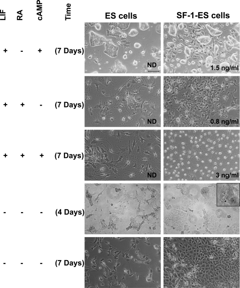
Fig. 4.
Effects of different treatment protocols on the differentiation of SF-1-ES cells. ES cells and SF-1-ES cells were differentiated in modified culture media as indicated at the left side of the panel. Progesterone production was measured in case of the cells differentiated in presence of LIF and absence of an external source of cholesterol. The amount of progesterone produced is indicated in the bottom right of the corresponding figures. Progesterone production in the absence of LIF is described in Fig. 5. ND, Not detected.

