Abstract
Background Knee prosthesis instability (KPI) is a frequent cause of failure of total knee arthroplasty. Moreover, the degree of constraint required to achieve immediate and long-term stability in total knee arthroplasty (TKA) is frequently debated. Questions This review aims to define the problem, analyze risk factors, and review strategies for prevention and treatment of KPI. Methods A PubMed (MEDLINE) search of the years 2000 to 2010 was performed using two key words: TKA and instability. One hundred and sixty-five initial articles were identified. The most important (17) articles as judged by the author were selected for this review. The main criteria for selection were that the articles addressed and provided solutions to the diagnosis and treatment of KPI. Results Patient-related risk factors predisposing to post-operative instability include deformity requiring a large surgical correction and aggressive ligament release, general or regional neuromuscular pathology, and hip or foot deformities. KPI can be prevented in most cases with appropriate selection of implants and good surgical technique. When ligament instability is anticipated post-operatively, the need for implants with a greater degree of constraint should be anticipated. In patients without significant varus or valgus malalignment and without significant flexion contracture, the posterior cruciate ligament (PCL) can be retained. However, the PCL should be sacrificed when deformity exists particularly in patients with rheumatoid arthritis, previous patellectomy, previous high tibial osteotomy or distal femoral osteotomy, and posttraumatic osteoarthritis with disruption of the PCL. In most cases, KPI requires revision surgery. Successful outcomes can only be obtained if the cause of KPI is identified and addressed. Conclusions Instability following TKA is a common cause of the need for revision. Typically, knees with deformity, rheumatoid arthritis, previous patellectomy or high tibial osteotomy, and posttraumatic arthritis carry higher risks of post-operative instability and are indications for more constrained TKA designs. Instability following TKA usually requires revision surgery which must address the cause of the instability for success.
Keywords: knee, arthroplasty, instability, risk factors, prevention, treatment
Introduction
Knee prosthesis instability is cited as the third most frequent cause of failure of total knee arthroplasty. It has been reported that 10–22% of revision surgeries after TKA are due to instability [15, 16]. Unfortunately, there is confusing information in the literature concerning definitions, risk factors and prevention, and treatment and outcomes [3, 5]. This review has three purposes: first is to define the common causes of KPI, second is to analyze risk factors for KPI which would allow prevention of knee arthroplasty instability, and third is to review treatment options for KPI and their results.
Methods
PubMed articles (MEDLINE) in English related to TKA and instability were searched using the key words TKA and instability from the years 2000 to 2010. One hundred and sixty-five initial articles were identified. The quality of the articles chosen was based on the judgment of the author. Articles were included if they specifically addressed the diagnosis of KPI, strategies which effectively prevent KPI and principles of treatment including revision total knee arthroplasty.
Results
Definition of KPI
KPI is defined as the abnormal and excessive displacement of the articular elements that leads to clinical failure of the arthroplasty and is one of the most common causes of aseptic failure following total knee replacement [15]. Instability may be early or late, and may involve global instability or instability in flexion or extension.
Early instability is that which occurs relatively early (weeks to months) after TKA. The etiology of these early symptoms is multiple. Early instability is typically caused by malalignment of the components, failure of restoration of the mechanical axis of the limb, improper balancing of the flexion–extension space, rupture of the posterior cruciate ligament (PCL) or medial collateral ligament (MCL), and patellar tendon rupture or patella fracture.
There are also multiple causes of late instability following TKA. The most common is usually related to polyethylene (PE) wear either alone or in combination with ligamentous instability. PE wear is often a function of malalignment, and it is not unusual to see an asymmetric wear pattern either on the medial or the posteromedial aspect of the implant. Medial wear of the PE can result in a relative MCL contracture and subsequent varus deformity and instability.
Instability in extension may be symmetric or asymmetric. Isolated symmetric extension instability may be due to excessive bone removal from the distal part of the femur. If flexion instability is also present, this implies excessive proximal tibial resection which affects the space between the femur and tibia equally. When this is recognized during the operation, the potential instability is corrected by using a thicker tibial insert.
Managing isolated excessive bone removal from the distal part of the femur can be challenging. A thicker tibial insert will not solve this problem and leads to elevation of the joint line and excessively tightens the flexion space resulting in the potential for achievement of poor post-op flexion and patellar maltracking. Marked elevation of the joint line limits knee flexion, affects patellar function, and contributes to midflexion instability. In this situation, the solution requires the addition of distal femoral augments.
Asymmetric extension instability is much more common and is typically related to a preoperative angular deformity of the knee and is caused by persistent or iatrogenic ligamentous asymmetry. The most common mistake leading to asymmetric instability is inadequate medial or lateral release. Additionally, iatrogenic malalignment of the femoral or tibial components in the coronal plane during surgery as well as post-operative polyethylene wear or change of component position due to loosening or subsidence can lead to medial or lateral asymmetric instability [15, 16].
Instability in flexion results from a flexion gap that is larger than the extension gap. Historically, this problem has been underdiagnosed in patients with cruciate-retaining knee implants where injury or release of the PCL can selectively aggravate an already loose flexion gap. Late insufficiency of the PCL can develop and cause instability symptoms in previously well-functioning cruciate-retaining knees. The manifestations of flexion instability range from a mere sense of instability to frank dislocation. Additional manifestations of flexion instability are recurrent synovitis and hemarthroses as well as anterior knee pain when reciprocating stairs or standing up from a seated position. Mild flexion instability is often underdiagnosed. When the PCL is injured during the time of surgery, instability usually presents late (months after) because the rest of the knee structures have a temporary protective effect.
Cruciate-retaining (CR) ligament designs require integrity of the PCL for the adequate translation of the femoral and tibial surfaces during flexion–extension and anteroposterior stability in flexion. When PCL insufficiency is present, CR prostheses should be avoided. PCL substituting designs (posterior stabilization designs or posteriorly stabilized or PS) increase anteroposterior stability in flexion, but do not guarantee stability in flexion. In fact, some authors suggest that the potential for an imbalance between flexion and extension may be greater in PCL substituting designs. In general, the flexion gap exhibits increased laxity compared to extension in these knees. An excessive posterior inclination of the tibial component also can contribute to flexion gap laxity. Other causes of flexion–extension imbalance include varus or valgus malalignment or malrotation of the femoral component [1].
Global instability is a pattern of instability that is clearly detectable in multiple planes and is a combination of loose flexion and extension gaps. There are several causes of global instability including PE wear that results in laxity of the surrounding soft tissue envelope, implant migration, motor dysfunction, and extensor mechanism disruption. The treatment options for global instability generally requires revisions to constrained or linked implants as treatment of gross instability with insert exchange and bracing tend to produce unsatisfactory results [1, 5].
Risk Factors and Prevention
Some patients are prone to instability. Those who have greater preoperative deformities, especially if compounded by extra-articular deformity or dynamic aberrations of gait, require large surgical corrections and aggressive ligament releases and may be difficult to stabilize [16].
Several factors can produce instability after total knee replacement (Table 1). Specific patient-related risk factors are a large surgical correction including an aggressive ligament release, general or regional neuromuscular pathology (quadriceps weakness inducing recurvatum or weak hip abductors that impart a medial thrust to the knee), hip or foot deformities typified by posterior tibial tendon rupture and pes planus. These deformities induce valgus moments at the knee. Clinical obesity is also a risk factor because it complicates surgical exposure, jeopardizes the collateral ligaments (8% incidence of avulsion of the medial collateral ligament in obese patients) and makes it difficult to appreciate component position [16].
Table 1.
Main causes of knee prosthesis instability (KPI)
| Ligament imbalance |
| Component disalignment |
| Component failure |
| Implant design |
| Mediolateral instability |
| Bone loss from over resection of the distal femur |
| Bone loss from femoral or tibial component loosening |
| Soft tissue laxity of the medial and lateral collateral ligaments |
| Connective tissue disorders (rheumatoid arthritis or Ehlers–Danlos syndrome) |
| Inaccurate femoral or tibial bone resection |
| Collateral ligament imbalance (under release, over release, or traumatic disruption) |
Instability of the knee can be prevented in most cases with an adequate selection of implants and a good surgical technique. Preoperative physical examination allows for evaluation of the state of the LCL, MCL, and PCL in order to select the adequate implant for each patient.
PS implants should be utilized in those patients with PCL insufficiency and in those with increased risk of posterior instability (rheumatoid arthritis, previous patellectomy, or the need to resect the PCL to correct a ligamentous imbalance, flexion contracture, or previous tibial osteotomy). If the choice is made to preserve the PCL, it is important to take special care in maintaining its integrity when the tibial cut is made. In case of doubt, it is preferable to convert the arthroplasty to a PS design. Careful attention to the balance of soft tissues and the correct implantation of the components in every plane, including the rotation of the femoral component, is essential to achieve symmetric spaces on flexion and extension. In some patients with marked instability (knee with valgus and complete insufficiency of the PCL, poliomielitis, or Charcot arthropathy), a primary constrained or linked hinge implants may be indicated.
Treatment Options and Results
Most of the patients with KPI require surgical treatment and the use of preoperative planning is very important. An implant with the required constraint can be determined pre-operatively [8]. As a general rule, it is recommended that the minimum amount of constraint necessary to achieve stability should be used [7, 13]. With many choices of component designs and levels of constraint, it can be a very difficult process to select the optimum implant for a given patient. Successful outcomes can be obtained in many of these cases, but without identifying the cause of instability, the surgeon risks repeating the mistakes that led to the instability after the initial TKA. KPI can be prevented in most cases with an adequate selection of implants and a good surgical technique (Figs. 1, 2, and 3).
Fig. 1.
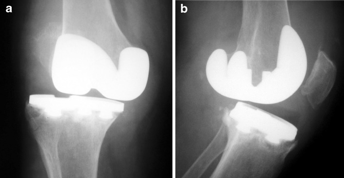
a, b Radiographs of an unstable total knee arthroplasty due to ligament insufficiency. a Anteroposterior view. b Lateral view
Fig. 2.

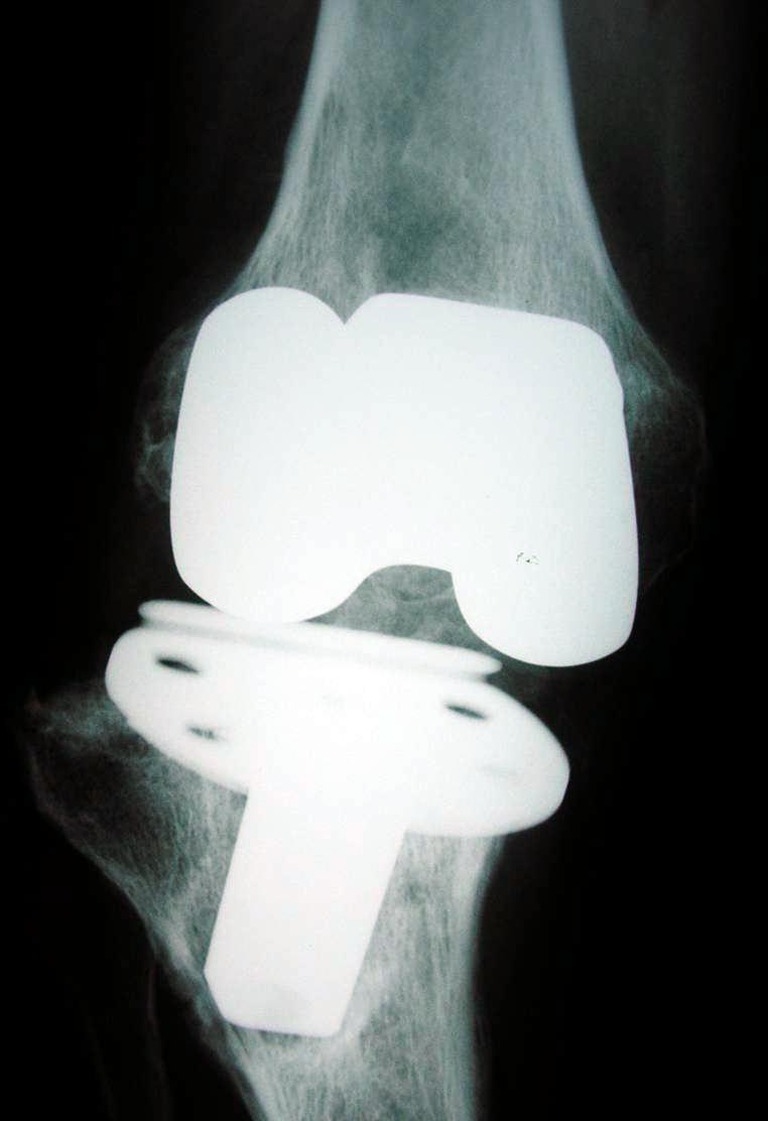
Anteroposterior radiograph of an unstable knee prosthesis due to loosening of the tibial component
Fig. 3.
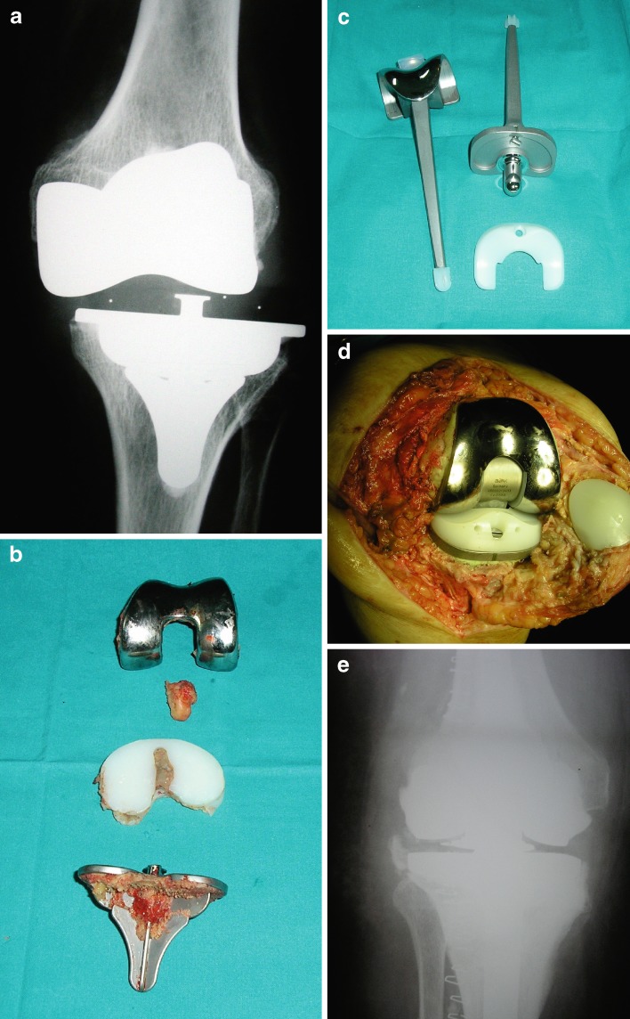
a–e Unstable knee prosthesis which required revision arthroplasty by means of a rotating hinged prosthesis. a Preoperative radiograph. b Intraoperative view of the removed components. c View of the components of the rotational hinge prosthesis to be implanted. d Intraoperative view of the rotating hinged prosthesis already implanted. e Anteroposterior post-operative view of the new prosthesis (satisfactory result)
Conservative treatment can be useful in a small percentage of patients with knee instability. Closed reduction and brace immobilization are used in patients with acute prosthesis dislocation. Bracing and rehabilitation programs are effective to strengthen the quadriceps and the hamstring and reduce the symptoms of some patients with mild and moderate instability. However, in many cases it is necessary to turn to surgical treatment, especially if other problems such as malalignment of the components, polyethylene wear, or loosening are noted [15].
Planning for a stable revision knee arthroplasty must include not only how to “stabilize” the knee but how to eliminate the source of instability: malalignment and gap imbalance. Unchecked, these forces will ultimately destroy any constrained device, hinged or non-hinged by breakage or loosening. Revision surgery for instability requires the ability to (1) correct the mechanical axis of the limb, (2) equalize the flexion and extension gaps, and (3) assess ligament integrity. The surgeon must correctly diagnose the cause of instability and have available proper implants at the time of surgery. [16].
CR implants designs represent the least amount of component constraint. Successful use of CR implants requires the presence of good quality bone with minimal defects, intact soft tissues, and a PCL that remains functional and balanced. In most revision situations, cruciate-retaining implants are not indicated.
The next level in constraint is PCL substituting designs. Many surgeons favor this option because the technical and judgment issues of balancing the PCL are eliminated. There is no gain in varus–valgus stability, and realistically speaking, minimal rotational stability. Thus, for a PS implant to succeed, a functional soft tissue envelope is needed to provide varus–valgus stability. However, the need for good flexion–extension balancing is also important because a residually loose flexion space can result in posterior tibio-femoral dislocation.
The next level of constraint is nonlinked hinge implant such varus–valgus constrained (VVC) or constrained condylar knee (CCK). Such components provide a significant degree of rotational control and more significantly a great deal of constraint to varus–valgus angulation. The trade-off is the theoretical disadvantage of increased stress transmission to the component–bone interfaces. Because these implants limit varus–valgus angulation between the femoral and tibial components, it would seem intuitive that they could be used in cases of severe medial or lateral instability. One must not forget that flexion instability is still a limitation for these implants [14].
With the absence soft tissue support or in the presence of gross flexion extension instability, linked hinge components are indicated [2]. Unfortunately, disappointing results have historically been associated with these implants predominantly because of implant loosening, significant patellar pain and high infection rates. However, newer rotating hinge designs have produced more encouraging clinical and radiographic results [2, 14, 17] (Fig. 3).
There are instances where constrained primary total knee arthroplasty is often required. Knees with a severe valgus or varus deformity which require extensive release to achieve soft tissue balance are often best treated with constrained designs. Some studies support the use of primary constrained total knee implants in patients with severe deformity or in patients requiring complex reconstructions, particularly if they are elderly and have lower physical demands. Easley et al. reviewed primary CCK prostheses in older patients with severe genu valgum and reported excellent clinical results with no failure at an 8-year follow-up [4]. Intraoperative disruption of the MCL during primary TKA also requires a prosthesis with additional varus–valgus constraint, although this has been addressed by primary ligament repair and use of a less constrained prosthesis in select cases [12]. Finally, there are some other situations in primary TKA in which more constraint is indicated, for example in patients with poor neuromuscular control, such as poliomyelitis or neuropathic arthropathy (in which the patients surrounding soft tissues will not confer sufficient stability), or patients who have had a prior high tibial osteotomy or patellectomy [7, 9, 11].
Discussion
KPI is the third most frequent cause of failure of total knee arthroplasty. Moreover, the degree of constraint required to achieve immediate and long-term stability in TKA is frequently debated. This review has tried to answer three questions: first is to define the common causes of KPI, second is to analyze risk factors for KPI which would allow prevention of knee arthroplasty instability, and third is to review the treatment options for KPI and their results.
KPI is defined as the abnormal and excessive displacement of the articular elements that leads to clinical failure of the arthroplasty and is one of the most common causes of aseptic failure following TKA. Instability may be early or late, but also may be in extension, in flexion, or global. Several specific patient-related risk factors have been identified and include gross deformity, general or regional neuromuscular pathology, hip or foot deformities, and obesity. Instability of the knee can be prevented in most cases with an adequate selection of implants and a good surgical technique. It is imperative to pre-operatively plan for anticipated instability to insure correct implant selection. The degree of constraint of the articulation in TKA should be dictated by the degree of disease and associated deformity. The prevention of instability after TKA is paramount. In this regard, careful femoral component size selection and placement can help balance flexion and extension gaps.
Most cases of KPI require surgical treatment. Successful outcomes can be obtained in many of these cases, but without identifying the cause of instability, the surgeon risks repeating the mistakes that led to the instability after the initial total knee arthroplasty. Primary indications for a hinge include medial or lateral collateral loss, massive bone loss including the femoral condyles and the origins or insertions of the collateral ligaments, and severe flexion gap imbalance. Additionally, patients with neuromuscular deficits such as polio or flail knee, who require the hyperextension, stop benefit from hinged primary TKA. Surgeons should have the option of modifying the degree of constraint at the time of surgical intervention. Currently, many TKA implant systems offer such flexibility. Nowadays there are several levels of implant constraint apart from the classical designs (CR, PS, CCK, rotating hinges): highly conforming cruciate-retaining designs, post-less cruciate-substituting implants, medial-pivot designs [6, 10], and PS plus components.
Unfortunately, the literature neither clarifies which design is most appropriate for KPI nor defines the rates of component loosening associated with use of more constrained implants. Future studies should define the rates of recurrent instability after revision using implants with various levels of constraint. As a general rule, it is recommended that the minimum amount of constraint necessary to achieve stability should be used. With many choices of component designs and levels of constraint, it can be a very difficult process to select the optimum implant for a given patient. Surgeons should have the option of modifying the degree of constraint at the time of surgical intervention. Currently, many TKA implant systems offer such flexibility.
Footnotes
The author certifies that he has no commercial associations (e.g., consultancies, stock ownership, equity interest, patent/licensing arrangements, etc.) that might pose a conflict of interest in connection with the submitted article.
References
- 1.Babis GC, Trousdale RT, Morrey BF. The effectiveness of isolated tibial insert exchange in revision total knee arthroplasty. J Bone Joint Surg Am. 2002;84:64–68. doi: 10.2106/00004623-200201000-00010. [DOI] [PubMed] [Google Scholar]
- 2.Barrack RL. Evolution of the rotating hinge for complex total knee arthroplasty. Clin Orthop Relat Res. 2001;392:292–299. doi: 10.1097/00003086-200111000-00038. [DOI] [PubMed] [Google Scholar]
- 3.Callaghan JJ, O’Rourke MR, Liu SS. The role of implant constraint in revision total knee arthroplasty: Not too little, not too much. J Arthroplasty. 2005;20:41–43. doi: 10.1016/j.arth.2005.03.008. [DOI] [PubMed] [Google Scholar]
- 4.Easley ME, Insall JN, Scuderi GR, Bullek DD. Primary constrained condylar knee arthroplasty for the arthritic valgus knee. Clin Orthop Relat Res. 2000;380:58–64. doi: 10.1097/00003086-200011000-00008. [DOI] [PubMed] [Google Scholar]
- 5.Engh GA, Koralewicz LM, Pereles TR. Clinical results of modular polyethylene insert exchange with retention of total knee arthroplasty components. J Bone Joint Surg Am. 2000;82:516–523. doi: 10.2106/00004623-200004000-00007. [DOI] [PubMed] [Google Scholar]
- 6.Fan CY, Hsieh JT, Hsieh MS, Shih YC, Lee CH. Primitive results after medial-pivot knee arthroplasties: a minimum 5-year follow-up study. J Arthroplasty. 2010;25:492–496. doi: 10.1016/j.arth.2009.05.008. [DOI] [PubMed] [Google Scholar]
- 7.Giori NJ, Lewallen DG. Total knee arthroplasty in limbs affected by poliomyelitis. J Bone Joint Surg Am. 2002;84:1157–1161. doi: 10.2106/00004623-200207000-00010. [DOI] [PubMed] [Google Scholar]
- 8.Gustke KA. Preoperative planning for revision total knee arthroplasty: Avoiding chaos. J Arthroplasty. 2005;20:37–40. doi: 10.1016/j.arth.2005.03.026. [DOI] [PubMed] [Google Scholar]
- 9.Kim YH, Kim JS, Oh SW. Total knee arthroplasty in neuropathic arthropathy. J Bone Joint Surg Br. 2002;84:216–219. doi: 10.1302/0301-620X.84B2.12312. [DOI] [PubMed] [Google Scholar]
- 10.Kim YH, Yoon SH, Kim JS. Early outcome of TKA with a medial pivot fixed-bearing prosthesis is worse than with a PFC mobile-bearing prosthesis. Clin Orthop Relat Res. 2009;467:493–503. doi: 10.1007/s11999-008-0221-8. [DOI] [PMC free article] [PubMed] [Google Scholar]
- 11.Lachiewicz PF, Soileau ES. Ten year survival and clinical results of constrained components in primary total knee arthroplasty. J Arthroplasty. 2006;21:803–808. doi: 10.1016/j.arth.2005.09.008. [DOI] [PubMed] [Google Scholar]
- 12.Leopold SS, McStay C, Klafeta K, Jacobs JJ, Berger RA, Rosenberg AG. Primary repair of intraoperative disruption of the medial collateral ligament during total knee arthroplasty. J Bone Joint Surg Am. 2001;83:86–91. doi: 10.2106/00004623-200101000-00012. [DOI] [PubMed] [Google Scholar]
- 13.Lombardi AV, Jr, Berend KR. Posterior cruciate ligament-retaining, posterior stabilized, and varus/valgus posterior stabilized constrained articulations in total knee arthroplasty. Instr Course Lect. 2006;55:419–427. [PubMed] [Google Scholar]
- 14.McAuley JP, Engh GA. Constraint in total knee arthroplasty: When and what? J Arthroplasty. 2003;18:51–54. doi: 10.1054/arth.2003.50103. [DOI] [PubMed] [Google Scholar]
- 15.Parrate S, Pagnano MW. Instability after total knee arthroplasty. J Bone Joint Surg Am. 2008;90:184–194. [PubMed] [Google Scholar]
- 16.Vince KG, Abdeen A, Sugimori T. The unstable total knee arthroplasty: Causes and cures. J Arthroplasty. 2006;21:44–49. doi: 10.1016/j.arth.2006.02.101. [DOI] [PubMed] [Google Scholar]
- 17.Westrich GH, Mollano AV, Sculco TP, Buly RL, Laskin RS, Windsor R. Rotating hinge total knee arthroplasty in severely affected knees. Clin Orthop Relat Res. 2000;379:195–208. doi: 10.1097/00003086-200010000-00023. [DOI] [PubMed] [Google Scholar]


