Abstract
Background
Although the reliability of determining acromial morphology has been examined, to date, there has not been an analysis of interobserver and intraobserver reliability on determining the presence and measuring the size of an acromial enthesophyte.
Questions/Purposes
The hypothesis of this study was that there will be poor intraobserver and interobserver reliability in the (1) determination of the presence of an acromial enthesophyte, (2) determination of the size of an acromial enthesophyte, and (3) determination of acromial morphology.
Patients and Methods
Fifteen fellowship-trained orthopedic shoulder surgeons reviewed the radiographs of 15 patients at two different intervals. Measurement of acromial enthesophytes was performed using two techniques: (1) enthesophyte length and (2) enthesophyte–humeral distance. Acromial morphology was also determined. Interobserver and intraobserver agreement was determined using intraclass correlation and kappa statistical methods.
Results
The interobserver reliability was fair to moderate and the intraobserver reliability moderate for determining the presence of an acromial enthesophyte. The measurement of the enthesophyte length showed poor interobserver and intraobserver reliability. The measurement of the enthesophyte–humeral distance showed poor interobserver reliability and moderate intraobserver reliability. The interobserver and intraobserver reliability in determining acromial morphology was found to be moderate and good, respectively.
Conclusions
There is fair to moderate reliability among fellowship-trained shoulder surgeons in determining the presence of an acromial enthesophyte. However, there is poor reliability among observers in measuring the size of the enthesophyte. This study suggests that the enthesophyte–humeral distance may be more reliable than the enthesophyte length when measuring the size of the enthesophyte.
Keywords: enthesophyte, acromion, reliability, morphology
Introduction
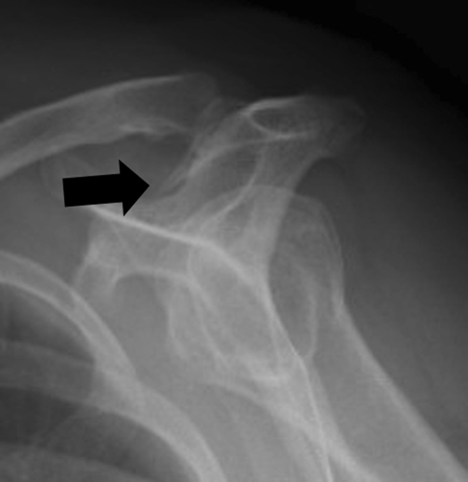
Acromial enthesophytes are thought to be an ossification of the insertion of the coracoacromial ligament where it attaches to the anterior acromial surface [5, 7, 12, 27] (Fig. 1). The terminology acromial osteophyte or acromial spur is commonly used by patients, physicians, and shoulder specialists when describing acromial anatomy after radiographic or arthroscopic assessment. An acromial enthesophyte is a more correct anatomical term since the bone under the acromion originates from the ligament or tendon attachment rather than the lateral margin of the joint [6, 22].
Fig. 1.
A supraspinatus outlet view demonstrating an acromial enthesophyte (black arrow)
The distinction between acromial enthesophytes and the native acromial morphology (type I = flat, type II = curved, type III = hooked) as described by Bigliani et al. [3] has not always been clearly drawn in the literature. Descriptions of the same morphology have varied between authors, and this may lead to confusion when interpreting radiological data and considering surgical treatment of such conditions [6]. Type III acromions have often been referred to as large spurs [6, 41, 43].
To date, there is no published study found in the Medline database that examines the effect of an acromial enthesophyte on the outcome of patients with full-thickness rotator cuff tears treated with non-operative techniques. One study performed in Iran suggested that the presence of an acromial enthesophyte was not a predictor of outcome in patients with impingement syndrome that underwent non-operative management [40]. The remaining studies found examined acromial morphology with respect to the non-operative management of impingement syndrome [28, 42].
Prior to undertaking studies that examine the relationship of acromial enthesophytes and the outcomes of treatment of rotator cuff disease, it is essential to determine the reliability of identifying and measuring the size of acromial enthesophytes. To the best of the authors’ knowledge, there is no published study that evaluates the reliability of determining the presence or measuring the length of an acromial enthesophyte. The purpose of this study was to examine the ability of fellowship-trained orthopedic shoulder specialists in differentiating between anatomic acromial morphology and acquired acromial enthesophytes. Interobserver and intraobserver reliability measurements will be performed to assess the consistency among and between observers in differentiating between acromial morphology and determining the presence of an acquired acromial enthesophyte. In addition, this study will determine the consistency among and between observers in the measurement of the size of the acromial enthesophyte. Based on reviews of previous studies [4, 19, 21, 36, 44], we hypothesized that there will be poor intraobserver and interobserver reliability in the (1) determination of the presence of an acromial enthesophyte, (2) determination of the size of an acromial enthesophyte, and (3) determination of acromial morphology.
Materials and Methods
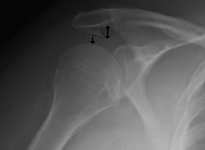
After institutional review board approval (Avera Institutional Review Board #2007.060E), a plain radiographic database was examined by the principal investigator in order to identify radiographic series that were thought to be representative of the three acromial morphologies initially described by Bigliani [3]. Fifteen patients were included in the study. The presenting complaints of these patients requiring radiographic examination of the shoulder were unknown. Exclusion criteria were having an incomplete radiographic series (anterior–posterior (AP), true AP, axillary view, and supraspinatus outlet view). Fifteen fellowship-trained shoulder surgeons were first instructed to judge the acromial morphology based on the classification of Bigliani et al. [3]. After determination of the acromial morphology, the observers were asked to record whether an acromial enthesophyte was present. Measurement of the length of the enthesophyte was performed as described by Kitay et al. [21]. In this method, the distance between the inferior aspect of the acromion to the distal tip of the enthesophyte is measured (enthesophyte length). In addition, the distance between the inferior tip of the enthesophyte and the humeral head was measured on the AP view (enthesophyte–humeral distance; Fig. 2). A minimum of 4 months later, the observers were asked to rate the same series of radiographs again. All observers were blinded to their original answers and the order of radiographs examined was randomized.
Fig. 2.
An AP view revealing an acromial enthesophyte (outlined in black). The enthesophyte length (double arrowhead) and the enthesophyte–humeral distance (single arrowhead) are demonstrated
After each surgeon reviewed the 15 radiographic series twice, the data were entered into a spreadsheet for statistical analysis. Ordinal interobserver and intraobserver data agreement was determined using the kappa statistical methods [13]. Kappa statistics measure agreement beyond the agreement due to chance alone. The kappa statistic was then interpreted by the method of Landis and Koch: 0–0.2 = poor; 0.21–0.40 = fair; 0.41–0.60 = moderate; 0.61–0.80 = good; 0.80–1.0 = excellent [23]. Continuous interobserver and intraobserver agreement (enthesophyte length and enthesophyte–humeral distance) was determined using intraclass correlation (ICC) and interpreted using the method of Portney and Watkins [38].
Analyses were performed using free open source R statistical software (http://www.r-project.org). Since each surgeon reviewed each radiographic series twice, interobserver reliability was calculated twice. Therefore, interobserver reliability is presented as the mean and standard deviation of the two calculations.
Results
Interobserver reliability in determining the presence of an acromial enthesophyte was found to range from fair to moderate (kappa value = 0.36 ± 0.15). Interobserver reliability for determining enthesophyte length was poor (ICC = 0.08 ± 0.05). The interobserver reliability for determining the enthesophyte–humeral distance was poor (ICC = 0.24 ± 0.03). The interobserver reliability for determining acromial morphology was moderate (kappa value = 0.53 ± 0.04).
The intraobserver reliability in determining the presence of an acromial enthesophyte was moderate (kappa value = 0.49). Intraobserver reliability for determining enthesophyte length was also poor (ICC = 0.04). The intraobserver reliability for determining the enthesophyte–humeral distance was moderate (ICC = 0.50). The intraobserver reliability for determining acromial morphology was good (kappa value = 0.66).
Of 15 radiographic series examined, at least one observer recorded the presence of an acromial enthesophyte on 12 of the radiographic series. The mean lengths of the enthesophytes measured ranged from 4.8 to 14.4 mm. The mean distance measured between the enthesophyte and the humeral head ranged from 1.1 to 10.9 mm.
Discussion
This study demonstrated that orthopedic surgeons with fellowship training in shoulder surgery have fair to moderate interobserver reliability in determining the presence of an enthesophyte. However, the measurement of these enthesophytes showed only poor interobserver reliability. The interobserver reliability in determining acromial morphology was found to be moderate.
As in most reliability studies, the intraobserver reliability was generally better than the interobserver reliability [20]. The intraobserver reliability for determining the presence of an enthesophyte was moderate. Similar to the interobserver findings, intraobserver reliability using the enthesophyte length technique was poor. However, the intraobserver reliability of measuring the enthesophyte–humeral distance was moderate. Intraobserver reliability in determining acromial morphology was determined to be good.
It is not clear why the radiographic methods of measuring the size of the acromial enthesophyte were unreliable. It is possible that it is difficult to reliably determine the inferior cortex of the acromion on two-dimensional radiographs. It is also possible that the reliability of this method would be improved using digital radiographs with a computerized measuring device. Computed tomography may be a more reliable method of measuring acromial enthesophytes, but this does not appear to be practical from a clinical standpoint.
This study was potentially limited by the inclusion of non-consecutive patients. The methodology of this study would be improved by having the investigators review a series of consecutive radiographs that were not previewed by the principal investigator. The difficulty with this proposed study design is that the true incidence of acromial enthesophytes is not well known. It may take >100 consecutive patients to have enough radiographic images to evaluate the presence of an acromial enthesophyte and all three acromial morphologies. In an attempt to limit the inclusion of potential bias, all of the investigators were blinded to the classifications determined by all other investigators including the principal investigator that selected the radiographic series.
The presence of an acromial enthesophyte was first noted in 1922 when Graves described a plaque of bone on the undersurface of the acromion. Graves suggested that it was an ossification of the coracoacromial ligament [6, 16]. Further studies have noted the presence of the acromial enthesophyte [1, 4, 6, 30]. The presence of enthesophytes ranges from 7% to 43.7% [3, 9, 25, 29, 31, 34, 39]. While most studies suggest that acromial spur incidence and size increase with age [14, 31, 33, 34], two studies indicate no correlation of age and the prevalence of acromial enthesophytes [32, 39]. Shoulders with type III acromial morphology have been associated with a statistically increased presence of an entheophyte [14, 29, 34, 35].
Bigliani et al. stressed the importance of distinguishing between spurs, which were probably acquired, and the variations in the native architectural type of the acromion [6]. Others have supported this distinction between native acromial morphology and the presence of an acquired acromial enthesophyte [14, 31, 34]. However, some believe that the hooked acromion (type III) is primarily an acquired characteristic and is a result of a degenerative change that occurs with aging [8, 15, 24, 43]. Edelson [8] supports this theory through his finding that no hooked acromions were found in cadaveric specimens under the age of 30 and that as age increased hooks became more common and larger. No longitudinal studies on acromial morphology have been recorded, and so the impact of the aging process on acromial morphology remains theoretical [31].
Although there are no other studies found in the literature that assess reliability in determining the presence or size of an acromial enthesophyte, there are several studies that have examined the reliability of determining acromial morphology [4, 17, 19, 26, 36]. The interobserver reliability of this study was as good as or better than that of the previously published studies. An author of one of these studies felt that one of the difficulties with determining acromial morphology is the presence of acromial spurs which may confound the classification of the acromial morphology [4]. When a spur is present, the true acromial edge may appear indistinct, which may influence its classification. The current study is the first published reliability study that attempts to differentiate between native acromial morphology and the presence of an acromial enthesophyte.
Several studies have noted a correlation with the presence of a type III acromion and the increased prevalence and severity of rotator cuff disease [2, 10, 11, 15, 18, 34]. Similar findings have been found correlating the presence of acromial enthesophytes and rotator cuff disease [2, 11, 33]. However, other studies debate the correlation of rotator cuff disease with acromial morphology or acromial enthesophytes [18, 37]. When examining the success of non-operative treatment of impingement syndrome, acromial morphology has been shown to influence the outcome scores and the need for surgical intervention [28, 40, 42]. The presence of an acromial enthesophyte was not found to be statistically related to worse outcomes of the non-operative management of impingement syndrome [40].
It is possible that the enthesophyte size and the distance between the enthesophyte and the humeral head is correlated with rotator cuff pathology; however, it has not been documented thus far. The authors believe that future studies should be performed to examine the role of the enthesophyte in the development of symptoms and the ability of treating rotator cuff tears associated with acromial enthesophytes. This current study may be helpful in developing these future studies.
Conclusions
When developing and interpreting future longitudinal studies that examine the presence of acromial enthesophytes on outcomes of rotator cuff disease, it is important to account for the reliability of determining and measuring the enthesophyte. This study demonstrates that there is fair to moderate reliability among observers in determining the presence of an acromial enthesophyte. However, there is poor reliability among observers in measuring the size of the enthesophyte. We recommend incorporating the enthesophyte–humeral distance in future studies that examine the role of the enthesophyte on the outcomes of rotator cuff disease. This study suggests that it may be more reliable than the enthesophyte length when measuring the enthesophyte. Additional methods of measuring the enthesophyte may need to be developed in the future to improve the reliability of measuring the acromial enthesophyte.
Acknowledgements
This research was supported by Grant Number 5 K23 AR052392-04 from the National Institute of Arthritis and Musculoskeletal and Skin Diseases. This network has received research funding from Arthrex and NFL Charities.
Footnotes
Each author certifies that he has no commercial associations (e.g., consultancies, stock ownership, equity interest, patent/licensing arrangements, etc.) that might pose a conflict of interest in connection with the submitted article.
Multi-centered Orthopaedic Outcomes Network (MOON) for the Shoulder
The corresponding author certifies that his institution has approved the human protocol for this investigation and that all investigations were conducted in conformity with ethical principles of research (Avera Institutional Review Board #2007.060E).
Level of Evidence: Level III Diagnostic Study
References
- 1.Aoki M, Ishii S, Usui M. The morphology of the acromion and its relationship to rotator cuff impingement. Orthop Trans. 1986;10:228. [Google Scholar]
- 2.Banas MP, Miller RJ, Totterman S. Relationship between the lateral acromion angle and rotator cuff disease. J Shoulder Elbow Surg. 1995;4(6):454–461. doi: 10.1016/S1058-2746(05)80038-2. [DOI] [PubMed] [Google Scholar]
- 3.Bigliani LU, Morrison DS, April EW. The morphology of the acromion and its relationship to rotator cuff tears. Orthop Trans. 1986;10:216. [Google Scholar]
- 4.Bright AS, Torpey B, Magid D, Codd T, McFarland EG. Reliability of radiographic evaluation for acromial morphology. Skeletal Radiol. 1997;26(12):718–721. doi: 10.1007/s002560050317. [DOI] [PubMed] [Google Scholar]
- 5.Chambler AF, Bull AM, Reilly P, Amis AA, Emery RJ. Coracoacromial ligament tension in vivo. J Shoulder Elbow Surg. 2003;12(4):365–367. doi: 10.1016/S1058-2746(03)00031-4. [DOI] [PubMed] [Google Scholar]
- 6.Chambler AF, Emery RJ. Acromial morphology: the enigma of terminology. Knee Surg Sports Traumatol Arthrosc. 1997;5(4):268–272. doi: 10.1007/s001670050062. [DOI] [PubMed] [Google Scholar]
- 7.Chambler AF, Pitsillides AA, Emery RJ. Acromial spur formation in patients with rotator cuff tears. J Shoulder Elbow Surg. 2003;12(4):314–321. doi: 10.1016/S1058-2746(03)00030-2. [DOI] [PubMed] [Google Scholar]
- 8.Edelson JG. The ‘hooked’ acromion revisited. J Bone Joint Surg Br. 1995;77(2):284–287. doi: 10.1302/0301-620X.77B2.7706348. [DOI] [PubMed] [Google Scholar]
- 9.Edelson JG, Taitz C. Anatomy of the coraco-acromial arch. Relation to degeneration of the acromion. J Bone Joint Surg Br. 1992;74(4):589–594. doi: 10.1302/0301-620X.74B4.1624522. [DOI] [PubMed] [Google Scholar]
- 10.Epstein RE, Schweitzer ME, Frieman BG, Fenlin JM, Jr, Mitchell DG. Hooked acromion: prevalence on MR images of painful shoulders. Radiology. 1993;187(2):479–481. doi: 10.1148/radiology.187.2.8475294. [DOI] [PubMed] [Google Scholar]
- 11.Farley TE, Neumann CH, Steinbach LS, Petersen SA. The coracoacromial arch: MR evaluation and correlation with rotator cuff pathology. Skeletal Radiol. 1994;23(8):641–645. doi: 10.1007/BF02580386. [DOI] [PubMed] [Google Scholar]
- 12.Fealy S, April EW, Khazzam M, Armengol-Barallat J, Bigliani LU. The coracoacromial ligament: morphology and study of acromial enthesopathy. J Shoulder Elbow Surg. 2005;14(5):542–548. doi: 10.1016/j.jse.2005.02.006. [DOI] [PubMed] [Google Scholar]
- 13.Fleiss J. Statistical methods for rates and proportions. 2. New York: Wiley; 1981. [DOI] [PubMed] [Google Scholar]
- 14.Getz JD, Recht MP, Piraino DW, Schils JP, Latimer BM, Jellema LM, et al. Acromial morphology: relation to sex, age, symmetry, and subacromial enthesophytes. Radiology. 1996;199(3):737–742. doi: 10.1148/radiology.199.3.8637998. [DOI] [PubMed] [Google Scholar]
- 15.Gill TJ, McIrvin E, Kocher MS, Homa K, Mair SD, Hawkins RJ. The relative importance of acromial morphology and age with respect to rotator cuff pathology. J Shoulder Elbow Surg. 2002;11(4):327–330. doi: 10.1067/mse.2002.124425. [DOI] [PubMed] [Google Scholar]
- 16.Graves WW. Observation on ages changes in the scapula. Am j Phys Anthrop. 1922;5:21–34. doi: 10.1002/ajpa.1330050125. [DOI] [Google Scholar]
- 17.Haygood TM, Langlotz CP, Kneeland JB, Iannotti JP, Williams GR, Jr, Dalinka MK. Categorization of acromial shape: interobserver variability with MR imaging and conventional radiography. AJR Am J Roentgenol. 1994;162(6):1377–1382. doi: 10.2214/ajr.162.6.8192003. [DOI] [PubMed] [Google Scholar]
- 18.Hirano M, Ide J, Takagi K. Acromial shapes and extension of rotator cuff tears: magnetic resonance imaging evaluation. J Shoulder Elbow Surg. 2002;11(6):576–578. doi: 10.1067/mse.2002.127097. [DOI] [PubMed] [Google Scholar]
- 19.Jacobson SR, Speer KP, Moor JT, Janda DH, Saddemi SR, MacDonald PB, et al. Reliability of radiographic assessment of acromial morphology. J Shoulder Elbow Surg. 1995;4(6):449–453. doi: 10.1016/S1058-2746(05)80037-0. [DOI] [PubMed] [Google Scholar]
- 20.Karanicolas PJ, Bhandari M, Kreder H, Moroni A, Richardson M, Walter SD, et al. Evaluating agreement: conducting a reliability study. J Bone Joint Surg Am. 2009;91(Suppl 3):99–106. doi: 10.2106/JBJS.H.01624. [DOI] [PubMed] [Google Scholar]
- 21.Kitay GS, Iannotti JP, Williams GR, Haygood T, Kneeland BJ, Berlin J. Roentgenographic assessment of acromial morphologic condition in rotator cuff impingement syndrome. J Shoulder Elbow Surg. 1995;4(6):441–448. doi: 10.1016/S1058-2746(05)80036-9. [DOI] [PubMed] [Google Scholar]
- 22.Kramer M. Undersurface acromial osteophyte or deltoid tendon attachment to the acromion? AJR Am J Roentgenol. 2008;190(6):W376–W377. doi: 10.2214/AJR.07.3523. [DOI] [PubMed] [Google Scholar]
- 23.Landis JR, Koch GG. The measurement of observer agreement for categorical data. Biometrics. 1977;33(1):159–174. doi: 10.2307/2529310. [DOI] [PubMed] [Google Scholar]
- 24.MacGillivray JD, Fealy S, Potter HG, O’Brien SJ. Multiplanar analysis of acromion morphology. Am J Sports Med. 1998;26(6):836–840. doi: 10.1177/03635465980260061701. [DOI] [PubMed] [Google Scholar]
- 25.Mahakkanukrauh P, Surin P. Prevalence of osteophytes associated with the acromion and acromioclavicular joint. Clin Anat. 2003;16(6):506–510. doi: 10.1002/ca.10182. [DOI] [PubMed] [Google Scholar]
- 26.Mayerhoefer ME, Breitenseher MJ, Roposch A, Treitl C, Wurnig C. Comparison of MRI and conventional radiography for assessment of acromial shape. AJR Am J Roentgenol. 2005;184(2):671–675. doi: 10.2214/ajr.184.2.01840671. [DOI] [PubMed] [Google Scholar]
- 27.Milz S, Jakob J, Buttner A, Tischer T, Putz R, Benjamin M. The structure of the coracoacromial ligament: fibrocartilage differentiation does not necessarily mean pathology. Scand J Med Sci Sports. 2008;18:16–22. doi: 10.1111/j.1600-0838.2007.00644.x. [DOI] [PubMed] [Google Scholar]
- 28.Morrison DS, Frogameni AD, Woodworth P. Non-operative treatment of subacromial impingement syndrome. J Bone Joint Surg Am. 1997;79(5):732–737. doi: 10.2106/00004623-199705000-00013. [DOI] [PubMed] [Google Scholar]
- 29.Natsis K, Tsikaras P, Totlis T, Gigis I, Skandalakis P, Appell HJ, et al. Correlation between the four types of acromion and the existence of enthesophytes: a study on 423 dried scapulas and review of the literature. Clin Anat. 2007;20(3):267–272. doi: 10.1002/ca.20320. [DOI] [PubMed] [Google Scholar]
- 30.Neer CS., 2nd Anterior acromioplasty for the chronic impingement syndrome in the shoulder: a preliminary report. J Bone Joint Surg Am. 1972;54(1):41–50. doi: 10.2106/00004623-197254010-00003. [DOI] [PubMed] [Google Scholar]
- 31.Nicholson GP, Goodman DA, Flatow EL, Bigliani LU. The acromion: morphologic condition and age-related changes. A study of 420 scapulas. J Shoulder Elbow Surg. 1996;5(1):1–11. doi: 10.1016/S1058-2746(96)80024-3. [DOI] [PubMed] [Google Scholar]
- 32.Ogata S, Uhthoff HK. Acromial enthesopathy and rotator cuff tear. A radiologic and histologic postmortem investigation of the coracoacromial arch. Clin Orthop Relat Res. 1990;254:39–48. [PubMed] [Google Scholar]
- 33.Ogawa K, Yoshida A, Inokuchi W, Naniwa T. Acromial spur: relationship to aging and morphologic changes in the rotator cuff. J Shoulder Elbow Surg. 2005;14(6):591–598. doi: 10.1016/j.jse.2005.03.007. [DOI] [PubMed] [Google Scholar]
- 34.Panni AS, Milano G, Lucania L, Fabbriciani C, Logroscino CA. Histological analysis of the coracoacromial arch: correlation between age-related changes and rotator cuff tears. Arthroscopy. 1996;12(5):531–540. doi: 10.1016/S0749-8063(96)90190-5. [DOI] [PubMed] [Google Scholar]
- 35.Paraskevas G, Tzaveas A, Papaziogas B, Kitsoulis P, Natsis K, Spanidou S. Morphological parameters of the acromion. Folia Morphol (Warsz). 2008;67(4):255–260. [PubMed] [Google Scholar]
- 36.Park TS, Park DW, Kim SI, Kweon TH. Roentgenographic assessment of acromial morphology using supraspinatus outlet radiographs. Arthroscopy. 2001;17(5):496–501. doi: 10.1053/jars.2001.23579. [DOI] [PubMed] [Google Scholar]
- 37.Pearsall AWt, Bonsell S, Heitman RJ, Helms CA, Osbahr D, Speer KP. Radiographic findings associated with symptomatic rotator cuff tears. J Shoulder Elbow Surg. 2003;12(2):122–127. doi: 10.1067/mse.2003.19. [DOI] [PubMed] [Google Scholar]
- 38.Portney L, Watkins M. Foundations of Clinical Research: Applications to Practice. New Jersey: Prentice Hall Health; 2000. [Google Scholar]
- 39.Sangiampong A, Chompoopong S, Sangvichien S, Thongtong P, Wongjittraporn S. The acromial morphology of Thais in relation to gender and age: study in scapular dried bone. J Med Assoc Thai. 2007;90(3):502–507. [PubMed] [Google Scholar]
- 40.Taheriazam A, Sadatsafavi M, Moayyeri A. Outcome predictors in nonoperative management of newly diagnosed subacromial impingement syndrome: a longitudinal study. MedGenMed. 2005;7(1):63. [PMC free article] [PubMed] [Google Scholar]
- 41.Toivonen DA, Tuite MJ, Orwin JF. Acromial structure and tears of the rotator cuff. J Shoulder Elbow Surg. 1995;4(5):376–383. doi: 10.1016/S1058-2746(95)80022-0. [DOI] [PubMed] [Google Scholar]
- 42.Wang JC, Horner G, Brown ED, Shapiro MS. The relationship between acromial morphology and conservative treatment of patients with impingement syndrome. Orthopedics. 2000;23(6):557–559. doi: 10.3928/0147-7447-20000601-12. [DOI] [PubMed] [Google Scholar]
- 43.Wang JC, Shapiro MS. Changes in acromial morphology with age. J Shoulder Elbow Surg. 1997;6(1):55–59. doi: 10.1016/S1058-2746(97)90071-9. [DOI] [PubMed] [Google Scholar]
- 44.Zuckerman JD, Kummer FJ, Cuomo F, Greller M. Interobserver reliability of acromial morphology classification: an anatomic study. J Shoulder Elbow Surg. 1997;6(3):286–287. doi: 10.1016/S1058-2746(97)90017-3. [DOI] [PubMed] [Google Scholar]


