Abstract
Chemokines are a family of chemotactic cytokines that function in host defense by orchestrating cellular movement during infection. In addition to this function, many chemokines have also been found to mediate the direct killing of a range of pathogenic microorganisms through an as-yet-undefined mechanism. As an understanding of the molecular mechanism and microbial targets of chemokine-mediated antimicrobial activity is likely to lead to the identification of unique, broad-spectrum therapeutic targets for effectively treating infection, we sought to investigate the mechanism by which the chemokine CXCL10 mediates bactericidal activity against the Gram-positive bacterium Bacillus anthracis, the causative agent of anthrax. Here, we report that disruption of the gene ftsX, which encodes the transmembrane domain of a putative ATP-binding cassette transporter, affords resistance to CXCL10-mediated antimicrobial effects against vegetative B. anthracis bacilli. Furthermore, we demonstrate that in the absence of FtsX, CXCL10 is unable to localize to its presumed site of action at the bacterial cell membrane, suggesting that chemokines interact with specific, identifiable bacterial components to mediate direct microbial killing. These findings provide unique insight into the mechanism of CXCL10-mediated bactericidal activity and establish, to our knowledge, the first description of a bacterial component critically involved in the ability of host chemokines to target and kill a bacterial pathogen. These observations also support the notion of chemokine-mediated antimicrobial activity as an important foundation for the development of innovative therapeutic strategies for treating infections caused by pathogenic, potentially multidrug-resistant microorganisms.
Keywords: CXC chemokine, CXCR3, innate immune system, transposon mutant library, ABC transporter
Chemokines are a family of small, structurally homologous host proteins originally recognized for their ability to facilitate the directed movement of leukocytes to sites of inflammation (1). Although the ability of chemokines to orchestrate cellular trafficking is central to numerous biological processes in both health and disease, chemokines do not function solely as chemotactic factors. Indeed, chemokines have been found to mediate many significant biological activities including, among others, participation in hematopoiesis, angiogenesis/angiostasis, cancer biology, and host defense against microbial infection (2–4). The latter, in particular, has been found to be especially important as chemokines exert pleiotropic effects that influence nearly every aspect of the host–immune response (5, 6).
Chemokines elicit biological activity primarily through receptor-dependent interactions in which chemokine binding to specific G protein-coupled receptors expressed by responsive cells triggers intracellular signaling and cell-specific functional activation (7). Although these ligand–receptor interactions are well established as being important in host defense (5), numerous chemokines have also been found to mediate direct antimicrobial activity against a broad range of bacterial and fungal pathogens in vitro (8). Among some of the best described examples of antimicrobial chemokines are CXCL9, CXCL10, and CXCL11, members of an IFN-inducible group of tripeptide motif Glu-Leu-Arg-negative (ELR−) CXC chemokines. Each of these chemokines is produced by epithelial and phagocytic cells in response to proinflammatory stimuli, especially IFN-γ, and bind the shared cellular receptor CXCR3 expressed primarily by activated T cells and natural killer cells (9). As suggested above, CXCL9, CXCL10, and CXCL11 each possess the ability to mediate the direct killing of pathogenic microorganisms, including Staphylococcus aureus, Listeria monocytogenes, and Escherichia coli (8, 10). Recent studies by our laboratory have also shown each of these CXC chemokines to exert antimicrobial effects against Bacillus anthracis, the Gram-positive, spore-forming bacterium that causes the disease anthrax. More particularly, CXCL9, CXCL10, and CXCL11 were found to target both the spore and bacillus forms of B. anthracis Sterne strain, causing disruptions in spore germination and marked reductions in spore and bacilli viability (11); additionally, human CXCL10 has also been found to directly kill encapsulated Ames strain bacilli (12). In vivo, these chemokines likely reach antimicrobial concentrations at local sites of infection as previously discussed (11). In support of this notion, neutralization of CXCL9, CXCL10, and CXCL11, but not their shared cellular receptor CXCR3, significantly increased host susceptibility to inhalational anthrax in a murine model of infection (12), indicating that these host chemokines mediate biologically relevant contributions to host defense against pulmonary B. anthracis infection.
Although the mechanistic details of chemokine-mediated microbial killing remain to be defined, structure-function studies have shown bactericidal activity to be associated primarily with the C-terminal α-helical region of individual chemokines (13). In agreement with these findings, the C-terminal region of many chemokines shares structural similarities with classical α-helical antimicrobial peptides, and molecular properties, including cationicity and amphipathicity, common among antimicrobial peptides that function in host defense (14). These observations suggest that chemokines mediate microbicidal activity through a mechanism in which electrostatic interactions between positively charged regions at the chemokine's surface and negatively charged moieties present on the microbial cell provide for chemokine localization to the cytoplasmic membrane and, subsequently, membrane permeabilization and cell lysis (8). Absent from this relatively nonspecific model, however, are potential roles for microbial components in supporting chemokine localization and the disruption of membrane barrier function. Indeed, although rarely addressed in mechanistic studies, the identities and functional roles of microbial components important in susceptibility to chemokine-mediated killing may represent broad-spectrum antimicrobial targets and lead to new therapeutic avenues for treating a range of infections caused by pathogenic, potentially multidrug-resistant microorganisms.
In this study, we hypothesized that specific bacterial protein targets are involved in facilitating chemokine-mediated antimicrobial activity. Therefore, using an innovative genetic approach, we sought to identify genome-encoded bacterial targets important in CXCL10-mediated killing of B. anthracis by screening a transposon mutant library for vegetative B. anthracis bacilli resistant to CXCL10-mediated bactericidal activity. From this screen, we identified three separate genomic loci that, when disrupted, significantly increase B. anthracis resistance to CXCL10: ftsX [ATP-binding cassette (ABC) transporter permease], BAS0651 (conserved hypothetical protein), and lytE (cell wall autolysin). Additionally, we report on the functional and structural properties that may allow FtsX to interact directly with CXCL10 and promote chemokine localization to its presumed site of antimicrobial action at the bacterial membrane. These data provide unique insight into the mechanism by which CXCL10 mediates bactericidal activity against B. anthracis and support the notion that specific bacterial components are important in the targeting and/or killing of microorganisms by host chemokines.
Results
Transposon Mutant Screen for B. anthracis Bacilli Resistant to CXCL10-Mediated Bactericidal Activity.
To identify bacterial targets of chemokine-mediated antimicrobial activity, we developed an approach to isolate transposon mutants resistant to the bactericidal effects of human CXCL10. In this system, vegetative bacilli were prepared from a B. anthracis Sterne strain 7702 transposon mutant library generated by random insertion of the mariner transposable element Himar1 into genetic loci of B. anthracis. This pooled library of transposon mutants (≈100,000 mutants per screen) was treated with CXCL10 at 48 μg/mL (5.6 μM), a chemokine concentration shown to consistently and completely kill Sterne inocula (11). In this way, CXCL10 would eliminate organisms lacking the targeted phenotype, CXCL10 resistance, and allow the isolation of surviving bacilli harboring Himar1-disrupted genes whose products are important in the ability of CXCL10 to effectively kill vegetative B. anthracis.
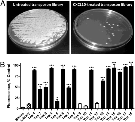
Two independent primary screens were performed (Fig. 1A), resulting in the recovery of 18 individual isolates, designated Tnx1–Tnx18. To confirm resistance to CXCL10, each recovered Tnx mutant was tested individually in an Alamar blue-based assay of bacterial growth and viability, a measure shown to correlate well with viable cfu levels (11). Of the 18 primary screen isolates, 15 were confirmed to be significantly resistant to CXCL10 compared with B. anthracis Sterne strain, and 10 of these isolates demonstrated near complete resistance to CXCL10-mediated bactericidal activity at the chemokine concentration examined (Fig. 1B). To further verify that CXCL10 resistance was related to transposon insertion, phage transduction was performed. Phage-mediated transduction of the transposon erythromycin resistance marker from Tnx18 into B. anthracis Sterne strain was accomplished, and the resulting transductant was found to be similarly resistant to CXCL10 (Fig. S1), indicating that resistance is associated with transposon insertion at specific genetic loci.
Fig. 1.
Isolation of B. anthracis transposon mutants resistant to CXCL10-mediated bactericidal activity. (A) Primary screen: Vegetative bacilli were prepared from a pooled B. anthracis transposon mutant library and treated with vehicle alone (untreated) (Left) or 48 μg/mL human CXCL10 (Right). Viable transposon mutants that survived CXCL10 treatment are shown and represent the mutants isolated from one of two independent screens. (B) Secondary screen: Isolated transposon mutants were examined individually for CXCL10 resistance by using Alamar blue analysis. Vegetative bacilli were treated with 12 μg/mL CXCL10 and Alamar blue reduction was determined 6 h after treatment. Data are expressed as a percentage of the isolate-specific untreated control and represent mean values ± SEs of the means (SEM); n = 3 independent experiments. ***P < 0.001, *P < 0.05 compared with CXCL10-treated Sterne bacilli (checkered bar); open bars represent isolates that were not significantly resistant to CXCL10.
Identification of Himar1-Disrupted Genes Among Tnx Isolates.
The transposon insertion site for each Tnx mutant was determined through specific amplification of DNA flanking Himar1 insertion sites. A single transposon insertion site was identified in each mutant and these amplification products were sequenced; BLASTn analysis was then used to determine the precise site of insertion within the genomes of the Tnx1–Tnx18 isolates (Table S1). Six individual insertion sites were identified and, with a single exception (Tnx10), each was found to be located within a putative coding region of the B. anthracis genome. Among the isolates demonstrated to be resistant to CXCL10, three unique and independent genomic loci were identified: BAS5033 (ftsX), BAS0651 (conserved, hypothetical), and BAS5043 (lytE); none of these three genes have been characterized in B. anthracis.
The gene ftsX is well conserved across bacterial species (15) and encodes FtsX, an integral membrane protein thought to form the substrate translocation channel of a putative ABC transporter (predicted topology; Fig. S2). FtsX has been examined primarily in E. coli, where it has been shown to localize at the inner membrane and interact with FtsE, a cytoplasmic ATPase believed to power transport across the cellular membrane (16). Sequence comparisons indicate that the FtsEX transporter complex functions as an importer, yet its ability to mediate transport and the identity of possible substrates remain undefined. Although divergent activities have been attributed to FtsEX, this transporter complex has been found to support bacterial cell division in several microorganisms including E. coli and Neisseria gonorrheae (15, 17). Similarly, studies in B. subtilis have shown FtsEX to be involved in asymmetric cellular division, facilitating the initiation of sporulation (18).
Considerably less is known about the genes BAS0651 and lytE. BAS0651 encodes a conserved hypothetical protein; BLASTp analysis of the predicted amino acid sequence identified two conserved domains: a “3D” domain containing three conserved aspartate residues and shown to be part of the catalytic domain of a lytic murein transglycosylase (19), and a structural maintenance of chromosomes (SMC) domain involved in organizing and segregating chromosomes for partition (20). In common with BAS0651, B. anthracis lytE, the third gene identified as being involved in susceptibility to CXCL10, is also predicted to contain a SMC domain and the gene's product, LytE, has been shown to contribute to cell wall lytic activity in B. subtilis (21). These similarities may indicate a related functional role for BAS0651 and LytE in facilitating susceptibility to CXCL10-mediated bactericidal activity.
CXC Chemokine Susceptibility Among B. anthracis ΔftsX Bacilli and Spores.
Of the Tnx isolates recovered from the primary screen, those harboring a transposon within the gene ftsX were found to display the greatest resistance to direct killing by CXCL10. Additionally, ftsX-disrupted organisms represented two distinct Himar1 insertion sites (Table S1), suggesting that the gene's high representation among resistant isolates was not simply a result of clonal expansion. Because of the evident importance of FtsX in antimicrobial susceptibility, we continued to characterize the role of B. anthracis FtsX in chemokine-mediated bactericidal activity. To preclude possible difficulties arising from the characterization of a transposon mutant, we generated an in-frame ftsX deletion mutant (ΔftsX) in B. anthracis Sterne strain by using markerless allelic exchange.
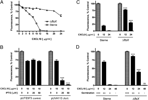
CXCL10 resistance by ΔftsX bacilli was examined over a range of chemokine concentrations using Alamar blue analysis. Compared with B. anthracis Sterne strain, ΔftsX vegetative cells exhibited significantly increased resistance to CXCL10-mediated killing with considerable numbers of ΔftsX organisms surviving at CXCL10 concentrations capable of killing Sterne inocula (Fig. 2A). Although highly resistant to CXCL10, ΔftsX bacilli were found to display an FtsX-independent killing effect at higher chemokine concentrations, suggesting that additional bacterial components (potentially those identified in the primary CXCL10 screen) or nonspecific electrostatic interactions also contribute to the ability of CXCL10 to target and kill vegetative bacilli. The specific importance of FtsX in the ability of CXCL10 to kill B. anthracis bacilli was further demonstrated by using transcomplementation. In this system, the native B. anthracis Sterne strain ftsX gene was inserted into the plasmid pUTE973 (22) to generate the plasmid pUVA113 in which ftsX is under the control of an IPTG-inducible promoter. Transformation and subsequent expression of the empty vector (pUTE973) by ftsX-null bacilli was not observed to affect the resistant phenotype. In contrast, IPTG-induced expression of the ftsX-containing vector (pUVA113) by ftsX-null bacilli restored the ability of CXCL10 to mediate significant killing of B. anthracis (Fig. 2B). These data support our hypothesis that specific bacterial components facilitate chemokine-mediated antimicrobial activity and identify FtsX as being important in the ability of CXCL10 to exert bactericidal effects.
Fig. 2.
Direct chemokine-mediated antimicrobial activity against B. anthracis ΔftsX bacilli and spores. (A) ΔftsX bacilli are resistant to human CXCL10; B. anthracis Sterne and ΔftsX bacilli were treated with increasing concentrations of CXCL10 for 6 h. Alamar blue reduction is expressed as a percentage of the strain-specific untreated control and data points represent mean values ± SEM; n = 4 independent experiments, curve comparison P < 0.001. (B) Complementation of ftsX restores CXCL10 susceptibility; ftsX-null bacilli were transformed with the IPTG-inducible plasmids pUTE973 (empty vector control) or pUVA113 (ftsX complementation vector) and treated with 10 μg/mL CXCL10 in the absence or presence of increasing IPTG concentrations. Data are expressed as a percentage of the appropriate untreated control (checkered bars) and represent mean values ± SEM; n = 3 independent experiments, ***P < 0.001 compared with similarly treated empty vector controls. (C) ΔftsX bacilli are resistant to murine CXCL9; Sterne and ΔftsX bacilli were treated with murine CXCL9 for 6 h. Alamar blue reduction is expressed as a percentage of the untreated control and represents mean values ± SEM; n = 3 independent experiments, ***P < 0.001 compared with murine CXCL9-treated Sterne bacilli. (D) ΔftsX spores are not resistant to CXCL10; Sterne and ΔftsX spores were treated with human CXCL10, and are shown together with the corresponding levels of relative spore germination ranging from >90% germination (+++) to no detectable germination (-) as determined visually. Alamar blue reduction, as an index of the resumption of metabolic activity, is expressed as percent control ± SEM; n = 3 independent experiments, ***P < 0.001, ns, not significant compared with CXCL10-treated Sterne spores.
To determine whether FtsX is important in B. anthracis susceptibility to other IFN-inducible ELR− CXC chemokines, we examined the bactericidal activity of murine CXCL9 against Sterne and ΔftsX bacilli; murine CXCL9 has been shown to kill both unencapsulated and encapsulated B. anthracis bacilli (12). Treatment of Sterne bacilli with murine CXCL9 resulted in marked, concentration-dependent reductions in vegetative growth and bacterial cell viability; ΔftsX organisms were found to be significantly more resistant to murine CXCL9 at each chemokine concentration examined (Fig. 2C). In contrast, Sterne and ΔftsX bacilli were found to be equally susceptible to the cationic antimicrobial peptide protamine (Fig. S3), discounting a generalized resistance to bactericidal peptides due to potential changes in membrane structure and/or function in the absence of FtsX. These results suggest that the IFN-inducible ELR− CXC chemokines mediate antimicrobial activity through a common mechanism, which may be specific to this class of host mediator.
We also examined a potential role for FtsX in B. anthracis spore susceptibility to chemokine-mediated disruptions in spore germination and the maintenance of viability, as reported (11). Spores were prepared from Sterne and ΔftsX bacteria and treated with several concentrations of CXCL10. When treated with 48 μg/mL CXCL10, shown to effectively block spore germination (11), spores from both the Sterne and ΔftsX strains failed to germinate and resume vegetative growth (Fig. 2D), indicating that FtsX is not required for the ability of CXCL10 to target the spore form of B. anthracis. As the CXCL10 concentration was reduced, both strains demonstrated increasing levels of spore germination, leading to the appearance of germinated, vegetative bacilli. Under these conditions, in which vegetative bacilli became an increasing portion of the bacterial population examined, ΔftsX organisms were found to be significantly more resistant to CXCL10 than Sterne bacteria. This observation likely reflects the ability of ΔftsX vegetative bacilli to escape CXCL10-mediated killing and suggests that FtsX functions distinctly in bacilli susceptibility to CXCL10.
Influence of Growth Phase on Susceptibility to Chemokine-Mediated Antimicrobial Activity.
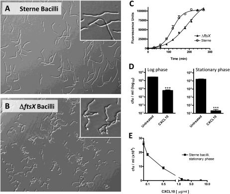
Visualization of ΔftsX vegetative bacilli by light microscopy revealed mutant cells to exhibit a distinct morphology in which individual bacilli appeared “kinked” with frequent curves and sharp angles among chains of bacilli (Fig. 3 A and B); expression of the ftsX complementation vector pUVA113 by these organisms corrected this phenotype and restored Sterne morphology (Fig. S4). Unusual morphologies have been described for several bacterial species in the absence of ftsX and/or ftsE and have been proposed to reflect aberrant cellular division (16, 17). To determine whether FtsX contributes to cellular division in B. anthracis, we compared the growth rates of Sterne and ΔftsX bacilli. Bacterial growth rates were examined in conditions analogous to those used during chemokine treatment (Fig. 3C), as well as in brain-heart infusion bacteriologic medium (Fig. S5). Although no gross defects in bacterial viability were observed, ΔftsX bacilli were found to grow more slowly than Sterne bacteria under each condition tested. Because reduced bacterial growth rates are associated with the ability of bacteria to evade the killing activity of many antibiotics (23), we examined the consequences of decreased growth rate on CXCL10-mediated antimicrobial activity. Of note, to our knowledge, the requirement and potential influence of bacterial growth on susceptibility to antimicrobial chemokines has not been investigated.
Fig. 3.
Influence of bacterial growth phase on CXCL10 susceptibility. (A and B) ΔftsX bacilli exhibit a distinct morphology; Sterne and ΔftsX bacilli were visualized by light microscopy in the absence of chemokine treatment. Representative fields from five independent experiments are shown at 200× magnification; Insets show expanded views of bacilli. (C) ΔftsX bacilli grow more slowly than Sterne bacilli; bacterial growth rates were determined in tissue culture medium and analyzed by using Alamar blue analysis. Data are expressed as relative fluorescence units and represent mean values ± SEM; n = 3 independent experiments, curve comparison P < 0.001. (D) Nongrowing bacilli are highly susceptible to CXCL10; log phase and stationary phase Sterne strain bacilli were treated with 8 μg/mL CXCL10 for 30 min before viability was assessed by cfu determination. Data are expressed as cfu/mL (log10 scale) and represent mean values ± SEM; n = 3 independent experiments, ***P < 0.001 compared with untreated controls. (E) CXCL10 susceptibility among nongrowing Sterne bacilli; stationary phase bacilli were treated with CXCL10 for 30 min before cfu determination. Viability is expressed as cfu/mL (x104) and represents mean values ± SEM; n = 3 independent experiments.
In this set of experiments, B. anthracis Sterne strain bacilli were prepared from log phase (rapidly growing) and stationary phase (nongrowing) cultures and treated with 8 μg/mL CXCL10 for 30 min before bacterial viability was assessed by cfu determination. As expected from prior studies (11), including those presented above, log phase Sterne bacteria were susceptible to CXCL10 at the chemokine concentration examined (Fig. 3D). Interestingly, CXCL10 treatment of stationary phase Sterne bacilli demonstrated nongrowing cells to be markedly more susceptible to direct killing by CXCL10 than actively growing organisms. Concentration-response studies demonstrated the half maximal effective concentration (EC50) for CXCL10 to be 0.33 ± 0.05 μg/mL against stationary phase bacilli (Fig. 3E), a >10-fold reduction in chemokine concentration compared with the EC50 reported for log phase B. anthracis bacilli, ≈4.00 μg/mL (11). That CXCL10 is more effective in killing nongrowing B. anthracis bacilli suggests that reduced bacterial growth rates among ΔftsX vegetative cells do not account for increased resistance to CXCL10; indeed, ΔftsX bacilli were resistant to CXCL10-mediated bactericidal activity regardless of their growth phase (Fig. S6). These data indicate that FtsX significantly contributes to the ability of CXCL10 to kill both rapidly growing and nongrowing B. anthracis bacilli and also support the notion that CXCL10 mediates a unique antimicrobial activity distinct from many traditional antibiotics.
CXCL10 Requires FtsX for Chemokine Localization to the Bacterial Cell Membrane.
Because the bacterial cell membrane is the presumed site of chemokine-mediated antimicrobial activity, we sought to examine whether CXCL10 localizes to the membrane of B. anthracis and, if so, whether an absence of FtsX affects the ability of CXCL10 to similarly target ΔftsX organisms. To determine chemokine localization, silver-enhanced immunogold labeling of CXCL10 was performed. In contrast to untreated Sterne and ΔftsX bacteria, which showed negligible binding of labeling reagents (Fig. S7), visualization of CXCL10-treated Sterne bacilli by transmission EM revealed widespread CXCL10 localization to the bacterial cell membrane as judged by considerable deposition of gold particles at this site (Fig. 4A); cytological changes associated with chemokine-mediated bactericidal activity, including disruptions in cellular integrity, were also apparent in Sterne bacilli after CXCL10 treatment. Importantly, in the presence of CXCL10, ΔftsX bacilli demonstrated a near complete absence of CXCL10 binding with no detectable chemokine localization to the cell membrane (Fig. 4B); expression of the ftsX complementation vector pUVA113 by ftsX-null bacilli restored CXCL10 localization to the bacterial membrane (Fig. 4C). These observations indicate that CXCL10 localization to the bacterial membrane is necessary for efficient bacterial killing and that B. anthracis FtsX mediates an important role in this localization.
Fig. 4.
B. anthracis FtsX is required for efficient CXCL10 localization to the bacterial membrane. Chemokine localization was determined by using silver-enhanced immunogold labeling of CXCL10 and visualized by transmission EM. (A) CXCL10-treated Sterne bacilli demonstrated considerable CXCL10 localization to the bacterial membrane; gold particle deposition is indicated by black arrows. (B) CXCL10-treated ΔftsX bacilli did not demonstrate detectable CXCL10 localization to the bacterial surface including the cell membrane. (C) Expression of the ftsX complementation vector pUVA113 by ftsX-null bacilli restored CXCL10 localization to the B. anthracis membrane. Representative fields from two independent experiments are shown at 30,000× magnification. (Scale bars: 0.5 μm.)
The striking lack of chemokine binding to ΔftsX bacilli suggested that CXCL10 might target B. anthracis by directly interacting with FtsX. To examine the potential for such an interaction, we used in silico protein analysis to compare B. anthracis FtsX to human CXCR3, the native cellular receptor responsible for binding CXCL10. Primary amino acid sequence alignment revealed that FtsX and CXCR3 share a 27 residue extracellular region (45% similarity as determined by BLASTp analysis) that contains numerous (≥10) charged amino acid residues. Notably, this region of homology (FtsX amino acids 54–80; CXCR3 amino acids 9–35) comprises much of CXCR3's N-terminal chemokine binding domain that is responsible for stable binding of CXCL10 by this receptor (24). Taken together, these data suggest that FtsX represents a unique bacterial target of chemokine-mediated bactericidal activity, which may interact directly with CXCL10 to facilitate chemokine localization and subsequent cell death.
Discussion
In this study, we have established that specific bacterial components are important in facilitating efficient chemokine-mediated antimicrobial activity against B. anthracis. Using an innovative genetic approach, we identified three nonessential B. anthracis genes as being significantly involved in the ability of CXCL10 to mediate bactericidal activity: BAS5033 (ftsX), BAS0651 (conserved, hypothetical), and BAS5043 (lytE). Prominent among these genetic loci was ftsX, a gene that encodes the integral membrane permease of the putative ABC transporter complex FtsEX (16). Functional disruption of ftsX, via transposon insertion or gene deletion, afforded vegetative B. anthracis bacilli with a marked resistance to chemokine-mediated killing, a characteristic lost after transcomplementation and expression of the native ftsX gene. In addition to highlighting the utility of transposon mutagenesis in identifying bacterial targets of antimicrobial activity, these data also demonstrate a role for B. anthracis FtsX as a unique target of CXCL10-mediated bactericidal activity.
Based on previous reports that FtsX participates in bacterial cell division and differentiation (15, 18), we investigated the effects of bacterial growth rate on CXCL10 susceptibility. Although a decreased rate of growth was not found to account for CXCL10 resistance among ΔftsX organisms, studies using B. anthracis Sterne strain showed CXCL10 to be distinctly effective in killing nongrowing bacterial cells. This bactericidal activity exceeded that observed for CXCL10 against rapidly growing bacilli and was also found to require FtsX. The ability of CXCL10 to target and kill stationary phase bacterial cells stands in contrast to many traditional antibiotics, including penicillin, that fail to mediate bactericidal activity against slowly growing and nongrowing bacteria (23). Indeed, most antibiotics target biosynthetic processes that become substantially down-regulated in the absence of growth, leading to an incomplete eradication of clinical infection that may result in persistent or recurrent disease (25). In addition, surviving bacterial populations are more likely to develop inheritable, genetic antibiotic resistance and thereby further exacerbate the emergence and spread of multidrug-resistant pathogens. That bacterial cell membrane integrity is essential for the maintenance of cell viability regardless of bacterial growth rate (25) supports the notion that chemokine-mediated disruptions in membrane barrier function, and the role of FtsX therein, may serve as a substantive template for the development of therapeutic strategies capable of successfully targeting slowly growing and/or nongrowing bacterial populations during infection.
Immunoelectron microscopy demonstrated that CXCL10 localizes to the cell membrane of B. anthracis bacilli and that chemokine binding is associated with the loss of cellular integrity. CXCL10 was not found to bind ΔftsX bacilli, indicating that FtsX significantly contributes to chemokine localization at the bacterial surface. In support of this conclusion, B. anthracis FtsX was found to share an extracellular region with CXCR3, the native CXCL10 receptor. Importantly, this N-terminal region of CXCR3 has been reported to interact with amino acid residues N-terminal to the first conserved cysteine residue of CXCL10 (24). Although it is not known whether CXCL10 interacts with FtsX, these data suggest that CXCL10 may target B. anthracis FtsX through the chemokine's N-terminal receptor binding domain(s); this type of interaction would localize CXCL10 to the bacterial surface while leaving the chemokine's C terminus free to interact with the bacterial cell membrane and facilitate cell death. Alternatively, interaction between FtsX and CXCL10 may influence the biological function of FtsX in such a way as to directly mediate bactericidal activity. Along these lines, the FtsEX transporter complex has been shown to be a major component involved in initiating B. subtilis sporulation (18). FtsEX likely has a similar function in B. anthracis, raising the possibility that CXCL10 targets FtsX and, thereby, facilitates the inappropriate activation of sporulation. This type of mechanism would be expected to trigger a cascade of improperly controlled events, resulting in a considerable disruption of cellular processes; interestingly, DNA condensation occurs early in sporulation (26) and may explain the identification of BAS0651 and LytE (each predicted to contain SMC domains and function in genomic partitioning) as bacterial components involved in CXCL10-mediated bactericidal activity.
In addition to providing unique insight into the mechanism of CXCL10-mediated antimicrobial activity, the data presented here also support the emerging concept that chemokines function directly in host–pathogen interaction by specifically targeting bacterial components. Although further studies are required to determine whether FtsX represents a broad-spectrum target of bactericidal activity among diverse pathogens, it is interesting to note that other bacteria have been reported to interact directly with host chemokines, including members of the IFN-inducible ELR− CXC chemokine family. For example, CXCL9 was recently reported to bind several surface proteins expressed by Chlamydophila pneumoniae, an activity that may promote antimicrobial activity against this organism (27). Also, S. aureus has been found to interact with numerous host chemokines; interestingly, these binding events are recognized by the bacteria and trigger the release of the virulence factor protein A (28). Indeed, it appears that host chemokines may be significantly involved in a previously unappreciated area of dynamic and, possibly, complex host–pathogen interaction that substantially influences bacterial pathogenesis and the outcome of infection.
Materials and Methods
Bacterial Strains and Growth Conditions.
B. anthracis Sterne strain 7702 and its derivatives were used throughout; E. coli strains Alpha-select (Bioline) and GM119 were used in molecular cloning. Growth conditions are detailed in SI Materials and Methods.
Transposon Library Generation, Insertion Site Determination, and Phage Transduction.
The mariner-based transposon library was generated in B. anthracis by using plasmid pJZ037, as described for use in L. monocytogenes (29). Insertion sites were determined by PCR, and phage-transduction was accomplished by using the bacteriophage CP-51ts45. Details for each procedure are provided in SI Materials and Methods.
Antimicrobial Assays and Microscopic Visualization.
B. anthracis bacilli or spores were treated with recombinant human CXCL10 or murine CXCL9 (Peprotech) in DMEM supplemented with 10% FBS. Alamar blue (Invitrogen) analysis, cfu determination, and microscopic visualization were performed as described in SI Materials and Methods.
Markerless Gene Deletion and Complementation.
Gene deletion was performed by the method of Janes and Stibitz (30); ftsX complementation was accomplished by inserting the native B. anthracis ftsX gene into plasmid pUTE973 (22). Procedures for ftsX deletion and complementation are described in SI Materials and Methods.
Examination of Bacterial Growth Rates.
Bacterial growth rates were determined by using standard methodology as described in SI Materials and Methods.
Silver-Enhanced Immunogold Labeling and Transmission EM.
CXCL10 immunogold labeling with silver enhancement was performed by using a preembedding protocol and subsequently prepared for transmission EM as described (11).
Statistical Analysis.
Statistical analysis and graphing were performed by using GraphPad Prism 4.0 software as described in SI Materials and Methods; a P value <0.05 was considered to be significant.
Supplementary Material
Acknowledgments
We thank Theresa M. Koehler for kindly providing plasmid pUTE973, David S. Weiss and Marie D. Burdick for helpful discussions, and Stephanie M. Zilora for assistance with preliminary studies. This work was supported by the Virginia Commonwealth Health Research Board (M.A.H.), National Institutes of Health/National Institute of Allergy and Infectious Diseases Grant R21 AI072469 (M.A.H.), and National Institutes of Health Grant T32 AI055432-08 (M.A.C.).
Footnotes
The authors declare no conflict of interest.
This article is a PNAS Direct Submission.
This article contains supporting information online at www.pnas.org/lookup/suppl/doi:10.1073/pnas.1108495108/-/DCSupplemental.
References
- 1.Luster AD. Chemokines—chemotactic cytokines that mediate inflammation. N Engl J Med. 1998;338:436–445. doi: 10.1056/NEJM199802123380706. [DOI] [PubMed] [Google Scholar]
- 2.Chensue SW. Molecular machinations: Chemokine signals in host-pathogen interactions. Clin Microbiol Rev. 2001;14:821–835. doi: 10.1128/CMR.14.4.821-835.2001. [DOI] [PMC free article] [PubMed] [Google Scholar]
- 3.Mehrad B, Keane MP, Strieter RM. Chemokines as mediators of angiogenesis. Thromb Haemost. 2007;97:755–762. [PMC free article] [PubMed] [Google Scholar]
- 4.Raman D, Baugher PJ, Thu YM, Richmond A. Role of chemokines in tumor growth. Cancer Lett. 2007;256:137–165. doi: 10.1016/j.canlet.2007.05.013. [DOI] [PMC free article] [PubMed] [Google Scholar]
- 5.Esche C, Stellato C, Beck LA. Chemokines: Key players in innate and adaptive immunity. J Invest Dermatol. 2005;125:615–628. doi: 10.1111/j.0022-202X.2005.23841.x. [DOI] [PubMed] [Google Scholar]
- 6.Campbell DJ, Kim CH, Butcher EC. Chemokines in the systemic organization of immunity. Immunol Rev. 2003;195:58–71. doi: 10.1034/j.1600-065x.2003.00067.x. [DOI] [PubMed] [Google Scholar]
- 7.Allen SJ, Crown SE, Handel TM. Chemokine: Receptor structure, interactions, and antagonism. Annu Rev Immunol. 2007;25:787–820. doi: 10.1146/annurev.immunol.24.021605.090529. [DOI] [PubMed] [Google Scholar]
- 8.Yang D, et al. Many chemokines including CCL20/MIP-3alpha display antimicrobial activity. J Leukoc Biol. 2003;74:448–455. doi: 10.1189/jlb.0103024. [DOI] [PubMed] [Google Scholar]
- 9.Groom JR, Luster AD. CXCR3 ligands: Redundant, collaborative and antagonistic functions. Immunol Cell Biol. 2011;89:207–215. doi: 10.1038/icb.2010.158. [DOI] [PMC free article] [PubMed] [Google Scholar]
- 10.Cole AM, et al. Cutting edge: IFN-inducible ELR- CXC chemokines display defensin-like antimicrobial activity. J Immunol. 2001;167:623–627. doi: 10.4049/jimmunol.167.2.623. [DOI] [PubMed] [Google Scholar]
- 11.Crawford MA, et al. Antimicrobial effects of interferon-inducible CXC chemokines against Bacillus anthracis spores and bacilli. Infect Immun. 2009;77:1664–1678. doi: 10.1128/IAI.01208-08. [DOI] [PMC free article] [PubMed] [Google Scholar]
- 12.Crawford MA, et al. Interferon-inducible CXC chemokines directly contribute to host defense against inhalational anthrax in a murine model of infection. PLoS Pathog. 2010;6:e1001199. doi: 10.1371/journal.ppat.1001199. [DOI] [PMC free article] [PubMed] [Google Scholar]
- 13.Liu B, Wilson E. The antimicrobial activity of CCL28 is dependent on C-terminal positively-charged amino acids. Eur J Immunol. 2010;40:186–196. doi: 10.1002/eji.200939819. [DOI] [PMC free article] [PubMed] [Google Scholar]
- 14.Yount NY, Bayer AS, Xiong YQ, Yeaman MR. Advances in antimicrobial peptide immunobiology. Biopolymers. 2006;84:435–458. doi: 10.1002/bip.20543. [DOI] [PubMed] [Google Scholar]
- 15.Schmidt KL, et al. A predicted ABC transporter, FtsEX, is needed for cell division in Escherichia coli. J Bacteriol. 2004;186:785–793. doi: 10.1128/JB.186.3.785-793.2004. [DOI] [PMC free article] [PubMed] [Google Scholar]
- 16.de Leeuw E, et al. Molecular characterization of Escherichia coli FtsE and FtsX. Mol Microbiol. 1999;31:983–993. doi: 10.1046/j.1365-2958.1999.01245.x. [DOI] [PubMed] [Google Scholar]
- 17.Bernatchez S, et al. Genomic, transcriptional and phenotypic analysis of ftsE and ftsX of Neisseria gonorrhoeae. DNA Res. 2000;7:75–81. doi: 10.1093/dnares/7.2.75. [DOI] [PubMed] [Google Scholar]
- 18.Garti-Levi S, Hazan R, Kain J, Fujita M, Ben-Yehuda S. The FtsEX ABC transporter directs cellular differentiation in Bacillus subtilis. Mol Microbiol. 2008;69:1018–1028. doi: 10.1111/j.1365-2958.2008.06340.x. [DOI] [PubMed] [Google Scholar]
- 19.van Straaten KE, Dijkstra BW, Vollmer W, Thunnissen AM. Crystal structure of MltA from Escherichia coli reveals a unique lytic transglycosylase fold. J Mol Biol. 2005;352:1068–1080. doi: 10.1016/j.jmb.2005.07.067. [DOI] [PubMed] [Google Scholar]
- 20.Milutinovich M, Koshland DE. Molecular biology. SMC complexes—wrapped up in controversy. Science. 2003;300:1101–1102. doi: 10.1126/science.1084478. [DOI] [PubMed] [Google Scholar]
- 21.Margot P, Wahlen M, Gholamhoseinian A, Piggot P, Karamata D. The lytE gene of Bacillus subtilis 168 encodes a cell wall hydrolase. J Bacteriol. 1998;180:749–752. doi: 10.1128/jb.180.3.749-752.1998. [DOI] [PMC free article] [PubMed] [Google Scholar]
- 22.Pflughoeft KJ, Sumby P, Koehler TM. Bacillus anthracis sin locus and regulation of secreted proteases. J Bacteriol. 2011;193:631–639. doi: 10.1128/JB.01083-10. [DOI] [PMC free article] [PubMed] [Google Scholar]
- 23.Eng RH, Padberg FT, Smith SM, Tan EN, Cherubin CE. Bactericidal effects of antibiotics on slowly growing and nongrowing bacteria. Antimicrob Agents Chemother. 1991;35:1824–1828. doi: 10.1128/aac.35.9.1824. [DOI] [PMC free article] [PubMed] [Google Scholar]
- 24.Booth V, Keizer DW, Kamphuis MB, Clark-Lewis I, Sykes BD. The CXCR3 binding chemokine IP-10/CXCL10: Structure and receptor interactions. Biochemistry. 2002;41:10418–10425. doi: 10.1021/bi026020q. [DOI] [PubMed] [Google Scholar]
- 25.Hurdle JG, O'Neill AJ, Chopra I, Lee RE. Targeting bacterial membrane function: An underexploited mechanism for treating persistent infections. Nat Rev Microbiol. 2011;9:62–75. doi: 10.1038/nrmicro2474. [DOI] [PMC free article] [PubMed] [Google Scholar]
- 26.Setlow B, et al. Condensation of the forespore nucleoid early in sporulation of Bacillus species. J Bacteriol. 1991;173:6270–6278. doi: 10.1128/jb.173.19.6270-6278.1991. [DOI] [PMC free article] [PubMed] [Google Scholar]
- 27.Balogh EP, Faludi I, Virók DP, Endrész V, Burián K. Chlamydophila pneumoniae induces production of the defensin-like MIG/CXCL9, which has in vitro antichlamydial activity. Int J Med Microbiol. 2011;301:252–259. doi: 10.1016/j.ijmm.2010.08.020. [DOI] [PubMed] [Google Scholar]
- 28.Yung SC, Parenti D, Murphy PM. Host chemokines bind to Staphylococcus aureus and stimulate protein A release. J Biol Chem. 2011;286:5069–5077. doi: 10.1074/jbc.M110.195180. [DOI] [PMC free article] [PubMed] [Google Scholar]
- 29.Zemansky J, et al. Development of a mariner-based transposon and identification of Listeria monocytogenes determinants, including the peptidyl-prolyl isomerase PrsA2, that contribute to its hemolytic phenotype. J Bacteriol. 2009;191:3950–3964. doi: 10.1128/JB.00016-09. [DOI] [PMC free article] [PubMed] [Google Scholar]
- 30.Janes BK, Stibitz S. Routine markerless gene replacement in Bacillus anthracis. Infect Immun. 2006;74:1949–1953. doi: 10.1128/IAI.74.3.1949-1953.2006. [DOI] [PMC free article] [PubMed] [Google Scholar]
Associated Data
This section collects any data citations, data availability statements, or supplementary materials included in this article.


