Abstract
One of the defining features of aggressive melanomas is their complexity. Hundreds of mutations and an ever increasing list of changes in the transcriptome and proteome distinguish normal from malignant melanocytic cells. Yet, despite this altered genetic background, a long-known attribute of melanomas is a relatively low rate of mutations in the p53 gene. However, it is unclear whether p53 is maintained in melanoma cells because it is required for their survival, or because it is functionally disabled. More pressing from a translational perspective, is to define whether there is a tumor cell-selective wiring of p53 that offers a window for therapeutic intervention. Here we provide genetic and pharmacological evidence demonstrating that p53 represents a liability to melanoma cells which they thwart by assuming an oncogenic dependency on the E3 ligase MDM2. Specifically, we used a combination of RNA interference and two structurally independent small molecule inhibitors of the p53/MDM2 interaction to assess the relative requirement of both proteins for the viability of normal melanocytes and a broad panel of melanoma cell lines. We demonstrated in vitro and in vivo that MDM2 is selectively required to blunt latent pro-senescence signals in melanoma cells. Notably, the outcome of MDM2 inactivation depends not only on the mutational status of p53, but also on its ability to signal to the transcription factor E2F1. These data support MDM2 as a drug target in melanoma cells, and identify E2F1 as a biomarker to consider when stratifying putative candidates for clinical studies of p53/MDM2 inhibitors.
Keywords: melanoma, senescence, oncogene dependency, MDM-2 inhibitors
INTRODUCTION
Malignant melanoma has long been characterized by its steadily increasing incidence and notorious chemoresistance (Jemal et al., 2010). The molecular determinants of this aggressive phenotype are not well understood, but they are considered to result, at least in part, from the threshold imposed by efficient suppressive mechanisms acting already at very early stages of tumor initiation (Bennett, 2008; Gray-Schopfer et al., 2007). Premature senescence is one of the potent inhibitory activities that must be bypassed to allow for the oncogenic transformation of normal melanocytes (Prieur and Peeper, 2008). In addition, melanoma progression associates with a plethora of defects in cell cycle checkpoint controls, survival and apoptotic pathways, cell-cell and cell-matrix interactions, as well as hundreds of often disconnected changes in the transcriptome (Gray-Schopfer et al., 2007; Hoek, 2007). Separating inconsequential byproducts from “druggable” tumor drivers has been a persistent handicap in this disease.
Given the numerous genetic and epigenetic defects of melanomas, it was logical to expect that some of these alterations would directly affect key nodes of tumor control. One of the first such modulators scrutinized in detail was the p53 protein (Castresana et al., 1993). The initial interest for p53 stemmed from its well known ability to control cell cycle progression and apoptosis (Vazquez et al., 2008). Studies from other tumor types linking p53 to cell metabolism, differentiation, senescence, oxidative stress, autophagy, invasion or angiogenesis (see Ref (Vousden and Prives, 2009) for a recent review), further illustrate the multiple levels at which p53 could be halting melanoma development. In fact, various animal models further support the concept of p53 as a suppressor of melanoma initiation and progression (Bardeesy et al., 2001; Patton et al., 2005; Terzian et al.; Terzian et al., 2010). Consequently, one of the most puzzling observations in this field is that the majority of advanced human melanomas still retain wild type p53 (Gray-Schopfer et al., 2007).
Mounting evidence supports that the maintenance of p53 in melanoma is compensated for by deactivation of upstream activators and/or downstream effectors. For example, defects in CHK2 or p14ARF have been shown to blunt p53 activation by intrinsic and extrinsic stress inducers (Satyamoorthy et al., 2000). Similarly, high levels of anti-apoptotic factors of the BCL-2 family or downregulation of caspase modulators (i.e. APAF-1) can block p53-dependent mitochondrial death programs (Soengas and Lowe, 2003). Nevertheless, alterations in this pathway may not be irreversible. For example, a notable accumulation of p53 can be observed in melanomas upon treatment with various chemical compounds, including agents that induce DNA damage (Avery-Kiejda et al., 2008; Soengas et al., 2001; Yang et al., 2006), inhibit MEK (Verhaegen et al., 2006), GSK3 (Smalley et al., 2007), the proteasome (Fernandez et al., 2005), or Bcl-2 family members (Verhaegen et al., 2006). Still this p53 induction has not yet been translated into an efficient antitumoral response in vivo. The mechanisms controlling the levels, stability, and subcellular localization of p53 in melanoma remain poorly understood.
p53 expression and function are modulated by multiple factors such as the murine double minute-2 (MDM-2) gene product (Wade et al., 2010). MDM-2 can oppose p53 by shuttling it from the nucleus to the cytosol, as well as by hindering its transactivation domain, and/or acting as an E3-ligase to govern p53 degradation by the proteasome (Ryan et al., 2001). MDM2 is upregulated in melanoma (Polsky and Cordon-Cardo, 2003), but whether its roles in this tumor are limited to p53 is unclear. For example, depending on the cellular context, MDM2 can both activate or inhibit E2F-1 (Polager and Ginsberg, 2009). In addition, RB, CYCLIN D1, c-JUN, MYC, DAXX, Ribosomal protein S7, FOXO1, FOXO3A, FOXO4 or CHK2 are just some examples of an increasing list of recently identified MDM2 targets (Fu et al., 2009; Kass et al., 2009; Vousden and Prives, 2009; Zhu et al., 2009). Consequently, MDM2 is emerging as a pleiotropic factor with multiple activities in differentiation, DNA replication, ribosome biosynthesis, transcription or intracellular trafficking (Wade et al., 2010).
Here we present genetic and functional analyses of the p53/MDM2 axis to answer key pending questions in the melanoma field: (i) what is the relative contribution of p53 and MDM2 to survival and proliferative capacity of normal melanocytes and melanoma cells; (ii) which (if any) of the multiple roles of p53 is under the control of MDM2; (iii) whether there is a tumor-cell selective requirement of p53/MDM2 that offers a window for therapeutic intervention, and if the case, (iv) what are molecular requirements for an efficient anti-melanoma activity of MDM2/p53 inhibitors.
RESULTS
Differential regulation and requirement of p53 in normal melanocytes and melanoma cells
To begin assessing the interrelationship between p53 and MDM2, both proteins were analyzed in normal skin melanocytes, a panel of 10 melanoma cell lines expressing wild type p53, and two cell lines (WM1366, SK-Mel-28) with mutant p53 (see Supplementary Table I for additional information). These cells were studied in basal conditions and upon treatment with doxorubicin (a classical p53 activator).
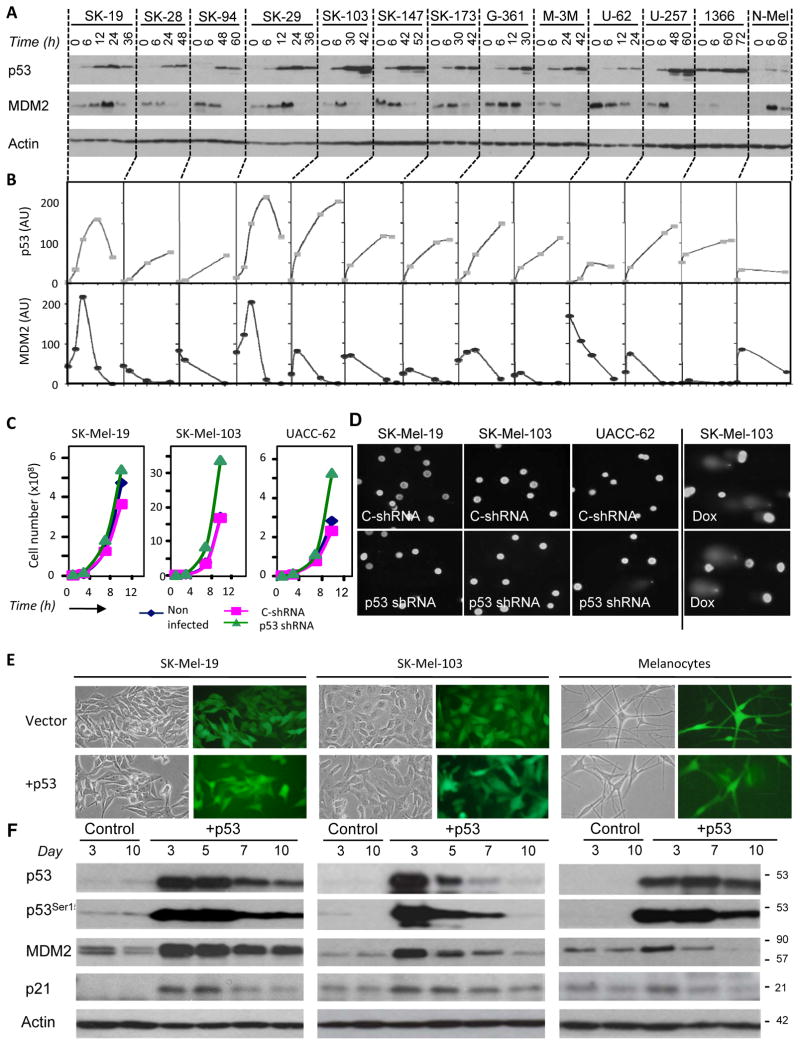
As shown in Fig. 1A, p53 levels were low to undetectable in the melanoma cells tested, but could be greatly induced by doxorubicin treatment (by 20–50 fold in p53 mutant cells, and up to 200 fold in cells expressing wild type 53). In melanocytes, however, doxorubicin-mediated p53 induction was considerably less efficient (Fig. 1A and quantification in Fig. 1B). Regarding MDM2, the expression of this protein was as expected (Polsky and Cordon-Cardo, 2003), 2 to10-fold higher levels in melanoma cells than in normal melanocytes (Fig. 1A,B). Interestingly, although the full length MDM2 90 kDa isoform declined as p53 levels increased upon doxorubicin treatment, there was not a direct correlation between these two proteins (Fig. 1B). These results indicated a differential regulation of the p53-MDM2 axis in melanocytes and melanoma cells.
FIGURE 1. Differential regulation and requirement of p53 in melanoma cells and normal melanocytes.
A, Protein immunoblots depicting p53 leves in basal conditions and upon treatment with doxorubicin (DOX, 0.5 μg/mL). B, Quantification of data in A as determined by densitometry. Shown are arbitrary units (AU) normalized to actin. C, Proliferative capacity of p53 shRNA-expressing melanoma cells versus their non-infected or control shRNA counterparts. D, Global assessment of DNA damage comet assay. Doxorubicin is shown as a control for DNA damage (far right panel). E, Microphotographs of indicated melanoma lines following ectopic overexpression of p53 (day 7). F, Protein immunoblots to visualize total p53, activated p53 (phosphorylated on Ser 15) and the p53 targets MDM2 and p21.
Next previously validated shRNAs (see Fig S1A) were used to define the relative dependency of melanocytes and melanoma cell lines on p53. Interestingly, p53-shRNA transduced melanoma cells maintained their morphology (Fig. S1B), proliferated at higher rates than their uninfected or control shRNA-counterparts (p>0.05; Fig. 1C; Fig. S1C) and showed no signs of global DNA damage (by comet assays or accumulation of phosphorylated histone H2AX; see Fig. 1D, Fig. S1A). Therefore, these results argue against p53 actively maintained in melanoma cells as a protective response against DNA damage or other forms of intrinsic cellular stress, as described in other systems (Bartkova et al., 2005; Murga et al., 2009).
As shown in Fig. S1C, melanocytes showed a transient increase in S-phase content after p53 depletion, but cell cycle progression eventually declined, with an accumulation of cells at the G2/M phase. Comet assays were negative (not shown), but a marked increase in multinucleated cells was observed upon p53 downregulation in melanocytes (Fig S1B). To follow are studies to determine whether it is feasible to exploit the retention of p53 by melanoma cells without compromising the viability of normal melanocytes.
High efficiency of melanoma cells to dampen abnormal p53 levels
We hypothesized that melanoma cells retain p53 but may be particularly efficient shutting down its induction and/or function. To test this end, normal melanocytes and two representative melanoma cell lines were infected with bicistronic lentiviral vectors coding for p53 and GFP (the later to monitor infection efficiencies by fluorescence microscopy, Fig. 1E). In all cells, p53 levels were induced by >50 fold by day 3 after lentiviral infection (Fig. 1F). Of note, this ectopic p53 became phosphorylated at Ser15 in the absence of external stimuli (Fig. 1F), indicating the presence of latent stress-activating signals (Vousden and Prives, 2009) in both normal and tumoral melanocytic cells. Interestingly high p53 levels could be sustained in normal melanocytes, but dampened in melanoma cells (Fig. 1F). The downregulation of p53 was associated with a transient, and more effective increase of MDM2 in melanoma cells than in normal melanocytes (Fig. 1F).
Tumor cell-selective senescence-like features by p53-MDM2 interaction inhibitors
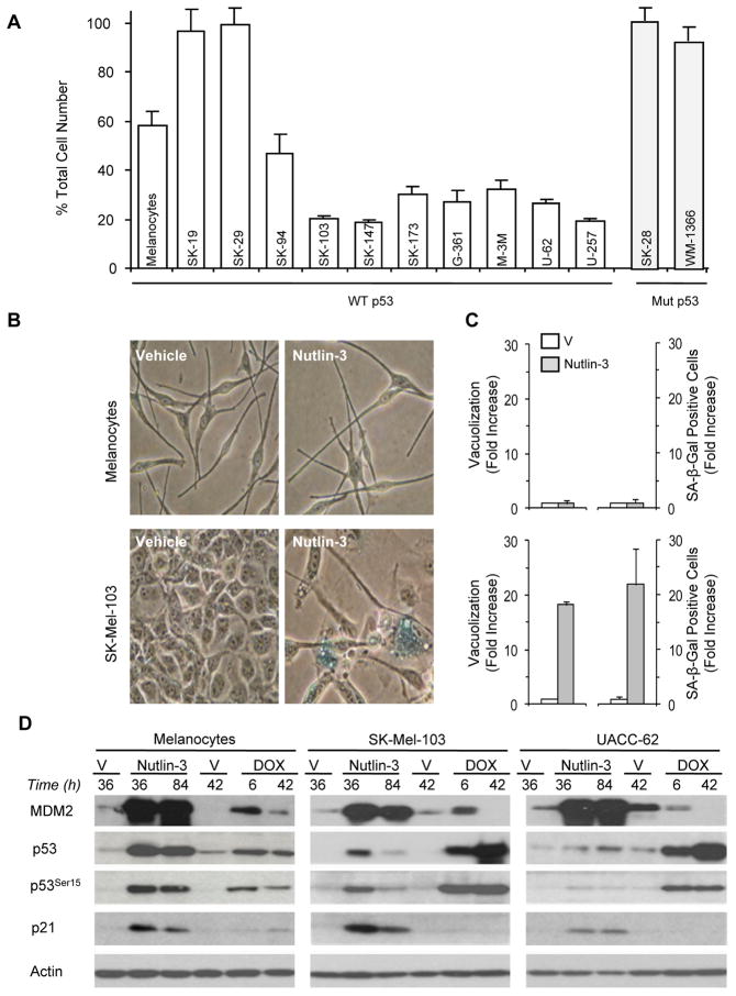
Next, we used Nutlin-3 to block p53-MDM2 interaction (Vassilev, 2007) and define the contribution of these proteins in melanoma cells. Despite Nutlin-3 being described to have antitumoral activity in some tumor cell types with mutant p53 (Ambrosini et al., 2007), this was not the case in SK-Mel-28 and WM-1366 (Fig. 2A and results not shown). However, most lines with wild type p53 responded to Nutlin-3 by a marked (although not absolute) arrest in cell proliferation (see Fig. 2A), as described in other tumor cells (Huang et al., 2009). By 3–4 days after treatment, melanoma cells became flattened, vacuolized and positive for senescence-associated β galactosidase activity (SA-β-Gal; Figs. 2B,C). The inhibitory effects of Nutlin-3 on melanoma cell proliferation could be observed even upon removal of this drug (Fig. S2A,B) and without significant induction of apoptosis (Fig. S3A–C). Normal melanocytes slowed their proliferation (albeit with delayed kinetics), but importantly with no obvious changes in cell morphology or acquisition of senescent-like features (see Fig. 2B,C and Fig. S3D, E).
FIGURE 2. Tumor cell-selective growth arrest by p53-MDM2 interaction inhibition.
A, Effects on cellular proliferation following treatment of the indicated cells with Nutlin-3 (5 μmol/L, 60 h). B, Microphotographs showing SA-β-Gal staining of normal melanocytes and SK-Mel-103 melanoma cells treated as indicated. C, Vacuolization and positive blue staining for SA-β-Gal of cells in B. Data correspond to the mean percentage ± SEM of cells from three separate visual fields. D, Protein immunoblots comparing effects of Nutlin-3 (5 μmol/L) and doxorubicin (DOX, 0.5 μg/mL) on the p53-MDM2 pathway in normal melanocytes and the indicated melanoma lines.
Differential impact of DNA damaging agents and p53-MDM2 interaction inhibitors on p53 levels
A possibility to account for the lack of Nutlin-3 induced senescence in melanocytes could be related to a lower induction and activation of p53-dependent signals than in melanoma cells. However, protein immunoblots indicated that MDM2 and the p53 target p21 were induced by Nutlin-3 in both cell types (see Fig. 2D). Interestingly, total and active (Ser-15-phosphorylated) p53 accumulated at higher levels in melanocytes. In fact, the induction of p53 upon treatment of melanoma cells with Nutlin-3 was modest when compared to doxorubicin (see particularly UACC-62 in Fig. 2D). Altogether, these results further emphasize the differential wiring of the p53-MDM2 axis in melanocytes and melanoma cells. Moreover, these data suggest additional roles of the oncogene MDM2 beyond the simple modulation of p53 expression.
Transient cell cycle arrest in normal melanocytes vs stable senescence-like phenotype in melanoma cells upon genetic depletion of MDM2
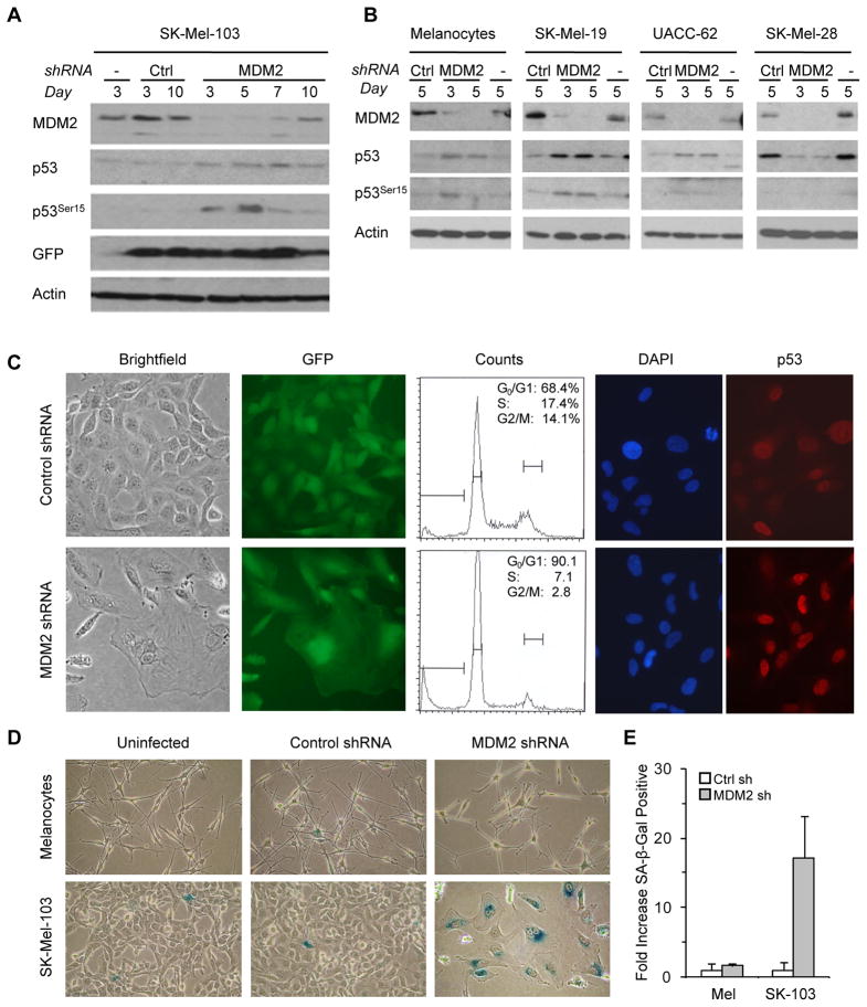
Nutlin-3 binds MDM2 specifically at the p53-binding pocket but does not block the interaction of MDM2 with other targets (Vassilev, 2007). To abrogate all MDM2-associated functions, melanocytes and melanoma cells were transduced with GFP-expressing lentiviruses that code for highly selective interfering shRNAs. This approach allowed for >90% inhibition of MDM2 within 4 days after infection (see Fig. 3A for detailed kinetics in SK-Mel-103, and Fig. 3B for representative examples in melanocytes and other melanoma cells). As the case for Nutlin-3, MDM2 shRNA led to a very modest increase in p53 in cells (SK-Mel-19, SK-Mel-103, UACC-62) in which this protein is not mutated, and even diminished the expression of p53 loss of function mutants (i.e. in SK-Mel-28). Immunophotographs showed an equivalent localization of p53 in control or MDM2-shRNA transduced cells (Fig. 3C). Therefore, MDM2 may be controlling the function, rather than the localization and levels of active p53. Melanoma cell lines that responded to Nutlin-3, also arrested when MDM2 was depleted (not shown). In fact, the resulting phenotype of MDM2 shRNA expressing melanomas was even more marked than for Nutlin-3 (Fig. 3C and Fig. S2A,D). Flow cytometry analyses indicated a G0/G1 cell cycle arrest (Fig. 3C). In addition, an intense SA-β-Gal staining reinforced the concept of a senescence-like phenotype after MDM2 depletion (Fig. 3D,E). Depletion of MDM2 in normal melanocytes (Fig. 3B) delayed cell division, but without signs of premature senescence (Fig. 3D,E). These results further emphasize the differential wiring of MDM2 in normal and tumor cells, offering a window for therapeutic intervention.
FIGURE 3. MDM2 genetic inactivation triggers melanoma growth arrest and senescence.
A,B, Protein immunoblots of the indicated cell lines that were either uninfected (−) or infected for GFP-lentiviruses coding for Control shRNA (Ctrl) or MDM2 shRNA. C, (Left panels) Representative brightfield and corresponding GFP fluorescent microphotographs (day 7) of SK-Mel-103 transduced with the indicated lentiviral vectors. (Middle panels) Cell cycle distribution as determined by flow cytometry (day 3). (Right panels) Visualization of p53 induction by immunofluorescence. D, SA-β-Gal staining in MDM2 shRNA-expressing SK-Mel-103 cells as compared to uninfected and Control shRNA cells. E, Corresponding quantification of cells exhibiting positive blue staining for SA-β-Gal. Data correspond to the mean ± SEM of cells from three experiments.
p16-independent fail-safe mechanism driven by modest changes in p53 in the absence of MDM2
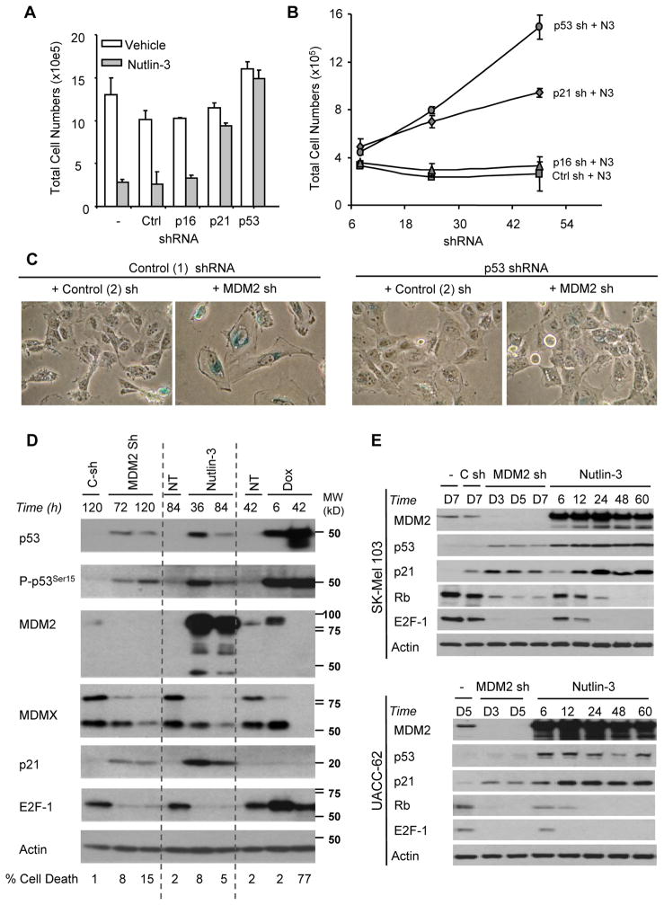
Given the modest induction of p53 by Nutlin-3 or MDM2 depletion, it was not obvious whether these agents activated senescence via p53 or as consequence of other deregulated MDM2 targets. For example, MDM2 can inhibit p21 in a p53 independent manner (Zhang et al., 2004). In addition, we questioned the role of the p16INK4a tumor suppressor since it is a key mediator of cellular senescence, and is retained in a proportion of spontaneous melanomas (Gray-Schopfer et al., 2007). Moreover, the tumor suppressor activities of p16INK4a can also be modulated by MDM2 (Xiao et al., 1995). To assess these different scenarios, SK-Mel-103 cells were infected with lentiviruses coding for validated shRNAs against p53, p21 or p16INK4a. Transduced cells were then treated with Nutlin-3 (Fig. 4A,B; Fig. S2) or infected with lentivirus coding for MDM2 shRNA (Fig 4C, see also Fig. 5E below). p53 shRNA but not p16INK4a depletion abrogated cell cycle arrest by both treatments (Fig. 4A), avoided cell flattening and the acquisition of SA-β-Gal positivity (Fig. 4C; Fig. S4). Inhibitory effects on Nutlin-3 and MDM2shRNA-driven senescence were also found for p21shRNA, although less efficiently than p53 depletion (Fig. 4A,B; Fig S4). These data are consistent with cDNA arrays that predicted a key role of p21 opposing melanoma cell proliferation (Terzian et al., 2010). Of note, p21 induction, which was obvious as early as 6 hours upon Nutlin-3 treatment (Fig. 4D, E), was completely abrogated by p53shRNA (see Fig. 5E). Altogether, the data presented above demonstrates that melanoma cells are “addicted” to MDM2 to blunt the pro-senescence function of p53, an activity that is modulated by p21 in a p16 independent manner.
FIGURE 4. Growth arrest effects of MDM2 inhibition are dependent upon p53 and the cyclin-dependent kinase (CDK) inhibitor p21.
A, Effects of Nutlin-3 (5 μmol/L, 48 h) on total cell numbers following introduction of Control (Ctrl), p16, p21, or p53 shRNA lentiviral constructs. Validation of genetic knockdown is shown in Figure 5E. B, Time-dependent effects of Nutlin-3 on cellular proliferation of SK-Mel-103 as described in A. C, Microphotographs depicting SA-β-Gal activity at day 7 following double infections with the indicated shRNA constructs in SK-Mel-103. D, Protein immunoblots illustrate differing effects on the cell cycle regulator E2F-1 following MDM2 inactivation by shRNA or Nutlin-3 (5 μmol/L) as compared to Doxorubicin (DOX; 0.5 μg/mL). E, Time-dependent effects on the p53-MDM2 pathway and S-phase cell cycle regulators comparing MDM2 inactivation by RNA interference (Control shRNA, C-sh; or MDM2 shRNA, MDM2 sh) to Nutlin-3 (5 μmol/L) treatment.
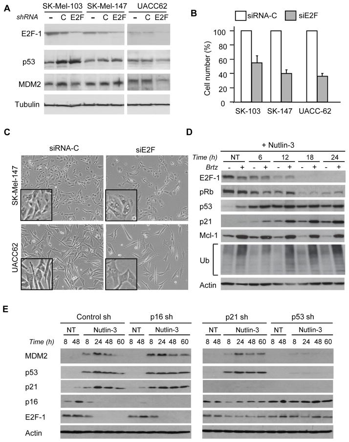
FIGURE 5. Proteasome independent but p53-dependent downregulation of E2F1 by functional loss of MDM2.
A, E2F-1 downregulation with siRNA pools. Scrambled siRNA pools are shown as reference controls. B, Comparative analysis of cell growth in indicated melanoma cell lines following introduction of the indicated siRNAs. C, Representative brightfield microphotographs (72 h) of control siRNA (siRNA-C) and E2F1 siRNA (siE2F1) in SK-Mel-147 and UACC62 cells. D, loss of E2F-1 is not dependent upon proteasomal degradation as shown by protein immunoblots following Nutlin-3 treatment in the presence or absence of the proteasome inhibitor Bortezomib (Brtz, 10 nM). MCL-1 and ubiquitin (Ub) are shown as controls for proteasomal inhibition. E, Protein immunoblots depicting time-dependent effects of Nutlin-3 (5 μmol/L) treatment following infections with Control, p16, p21 or p53 shRNA lentiviral constructs in SK-Mel-103.
DNA damage and MDM2 inactivation signal to p53 with differing effects on E2F1
In the context of defining the mechanism(s) underlying the induction of melanoma senescence by Nutlin-3 or MDM2shRNA, we considered of interest the MDMX and RB proteins. MDMX is a p53 repressor that can modulate the efficacy of Nutlin-3 (Hu et al., 2006). RB is a tumor suppressor that can be marked by MDM2 for ubiquitin proteasome degradation (Sdek et al., 2005). However, as shown in Fig. 4D, MDMX was regulated in a similar manner by Nutlin-3 (that blocks proliferation) and doxorubicin (which primes cells for apoptosis). Moreover, no RB induction was observed in the response to Nutlin-3 or MDM2 shRNA. In fact, both agents downregulated total and phosphorylated RB by 70–95% (Fig. 4E).
We then focused on E2F1 based on previous reports indicating various positive and negative feedback loops between this transcription factor and MDM2 (Polager and Ginsberg, 2009). Interestingly, while E2F1 was upregulated by doxorubicin in melanoma cells, it was depleted by MDM2 shRNA and Nutlin-3 (Fig. 4D). Importantly, E2F1 downregulation preceded entry into senescence following MDM2 depletion by shRNA (which occurs at 4–5 days) or after Nutlin-3 treatment (which occurs at 3–4 days) (see Fig. 4E for immunoblots in SK-Mel-103 and UACC-62). In contrast, normal melanocytes, which do not acquire senescence-like features upon treatment with Nutlin-3, do also retain E2F1 levels (Fig. S5).
Altogether, these data argue for E2F1 downregulation being an active driver of senescence-like features in melanoma cells, instead of a simple byproduct of cell cycle arrest. Consistent with this hypothesis, depletion of endogenous E2F1 by highly efficient siRNA pools (Fig. 5A) was sufficient to halt melanoma cell proliferation and induce senescence features (Fig. 5B, C). Conversely, short-term ectopic expression of E2F1 in melanoma cells by adenoviral infection, blunted the ability of Nutlin-3 to induce cell vacuolization or acquire SA-β-Gal activity (Fig. S6A, B).
Proteasome independent but p53-dependent downregulation of E2F1 by functional loss of MDM2
The next pending question was to which extent E2F1 was controlled directly by p53/p21 or whether known roles of MDM2 on ubiquitin-mediated E2F1 degradation (Campanero and Flemington, 1997; Hofmann et al., 1996) also played a role. The impact of ubiquitin-dependent degradation was analyzed by pretreatments with the proteasome inhibitor bortezomib. As shown in Fig. 5D, doses of bortezomib that can effectively stabilize ubiquitylated proteins and the classical proteasome target MCL-1, could not prevent the downregulation of E2F1 by Nutlin-3.
To demonstrate that E2F1 acts downstream of p53 and p21, but not p16, treatments were performed in an isogenic series of the SK-Mel-103 line generated by transduction of the corresponding shRNAs. Representative examples of the results obtained are summarized in Fig. 5E. In short, p16 shRNA, p21 shRNA and p53 shRNA provided a 0%, 70% and 100% protection, respectively, to Nutlin-3 induced E2F1 depletion. Therefore, these results indicate that melanoma cells depend on MDM2 to halt inhibitory effects of p53 on E2F1 via (at least in part) the tumor suppressor p21.
Melanoma cells depend on MDM2 to prevent senescence-like phenotype in vivo
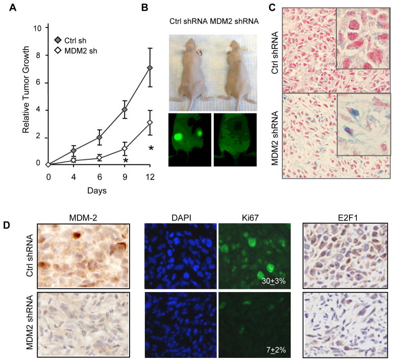
To assess the physiological relevance of MDM2 depletion, GFP-labeled SK-Mel-103 transduced with control or MDM2shRNA, were implanted into immunocompromised mice before any change in cell morphology or proliferation. Caliper measurements (Fig. 6A) and GFP-based fluorescence imaging (Fig. 6B) revealed a marked inability of MDM2 shRNA transduced cells to grow as xenografts. Consistent with a senescence-like arrest, MDM2 shRNA-derived tumors showed clear SA-β-Gal positive staining (Fig. 6C). Histological analyses further demonstrated the downregulation of MDM2 in the bulk of the tumors (Fig. 6D, left panels), as well as cell cycle arrest (visualized by a reduction in Ki67 staining from 30% to 7%, Fig. 6D, central panels), and downregulation of E2F1 (Fig. 6D, right panels). In conclusion, MDM2 depletion, when efficient, compromises the tumorigenic potential of otherwise aggressive melanoma cells. Moreover, these results also support the use of E2F1 to gauge the impact of MDM2 loss in vivo.
FIGURE 6. MDM2 blocks melanoma senescence-like arrest in vivo.
A, Tumor growth following implantation in nude mice of 0.5×10⁶ SK-Mel-103 cells expressing control (white diamonds) or MDM2 (black diamonds) shRNAs. Data represent final volume ± SEM (n=12 tumors). B, representative photographs and corresponding whole body fluorescent imaging of Control (left animal) or MDM2 shRNA-infected cells (right animal) at day 13 post transplantation. C, SA-β-Gal activity of tumors described above. D, Histological analyses of MDM2 inhibition as shown by immunohistochemical staining of formalin fixed paraffin-embedded tumor sections with MDM2 (left panels) and E2F-1 (right panels). Proliferative index is shown by staining with Ki67 (middle panels) and nuclear staining is shown by DAPI.
E2F1 marks the sensitivity of melanoma cells to Nutlin-3 and MDM2 shRNA
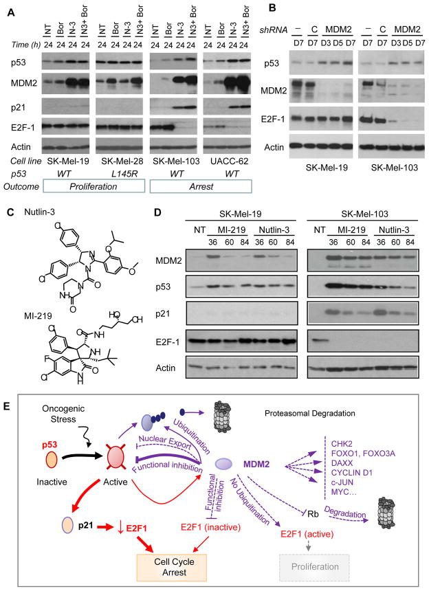
Active p53 is required for E2F1 downregulation and induction of senescence by Nutlin-3 and MDM2 shRNA (Fig. 4A,B; Fig. 5E). However, not all wild type p53-expressing melanomas were sensitive to Nutlin-3 and MDM2 shRNA (Fig. 2A). Given the impact of both treatments on E2F1, we questioned whether this protein could serve as a response indicator. To evaluate this hypothesis, we compared the response of cells that depend (SK-Mel-103, UACC-62) or are independent (SK-Mel-19, SK-Mel-29) on MDM2. Cells were treated with Nutlin-3 in the presence or absence of bortezomib (to assess putative effects of the proteasome on MDM2 and E2F1 in resistant cells). Resistant melanoma cells accumulated MDM2 efficiently upon Nutlin-3 treatment, but were markedly less potent at upregulating p21, and showed a complete maintenance of E2F-1 (Fig. 7A and results not shown). This similar maintenance in E2F-1 levels was also observed for cell lines resistant to MDM2 shRNA (Fig. 7B).
Figure 7. E2F1 marks the sensitivity of melanoma cells to MDM2 inactivation.
A–B, Expression of the indicated proteins following treatment with the proteasome inhibitor Bortezomib (Brtz, 10 nM), Nutlin-3 (N-3, 5 μmol/L) or the combination of both (N-3+Brtz) at 24 h (A), or following genetic inactivation of MDM2 by RNAi as a function of time (B). C, Molecular structure of small molecule inhibitors Nutlin-3 and MI-219 binding in the MDM2-p53 pocket. D, Depletion of E2F1 upon treatment of sensitive (SK-Mel-103) and resistant (SK-Mel-19) cells by Nutlin-3 (5 μmol/L) or MI-219 (5 μmol/L). E, Summary of described interactions between p53, MDM2, p21 and E2F1 (Polager and Ginsberg, 2009). The specific relationships found by this study in melanoma cells are indicated with thick and continuous lines.
To test the validity of E2F1 as a sensitivity marker of MDM2-based treatments, melanoma cells were treated with MI-219, a p53-MDM2 inhibitor structurally unrelated to Nutlin-3 (Fig. 7C). MI-219, which exhibits a higher binding affinity for MDM2 (Ki=5 nM) than Nutlin-3 (Ki=36 nM) (Shangary et al., 2008) was quantitatively more potent at inducing p53 and MDM2. Nevertheless, MI-219 and Nutlin-3 were qualitatively similar. The same sensitive cell lines showed E2F1 downregulation prior to the induction of cellular senescence (see Fig. 7D right panels). Conversely, the Nutlin-3 resistant cells, also failed to induce p21, downregulate E2F1 or alter cell cycle progression in response to MI-219 (see examples in Fig. 7D). In conclusion, our data identify E2F1 as a biomarker for tumors that become addicted to MDM2 to deactivate latent pro-senescence signals resulting from the maintenance of wild type p53.
DISCUSSION
The generation of bioavailable, and specific p53-MDM2 binding inhibitors is offering an alternative venue for the treatment of cancers that maintain active (or activatable) p53 (Shangary and Wang, 2009; Vassilev, 2007). Malignant melanomas, which usually present with wild type p53 (Gray-Schopfer et al., 2007), would therefore be ideal candidates for this therapeutic strategy. Nevertheless, the implementation of this concept is not straightforward. First, the traditional resistance chemoresistance of melanoma cells questions the functionality of this tumor suppressor (Pommier et al., 2004). Secondly, MDM2 is just one of many post-transcriptional regulators of p53 (Vousden and Prives, 2009). In turn, MDM2 can impinge on tumor cell physiology by diverse p53-independent mechanisms (Wade et al., 2010). Furthermore, no obvious outcome of Nutlin3 or MI-219 can be predicted from the genetic background of the cancer cells. Thus, a variety of effects of these compounds have been reported ranging from apoptosis (Vassilev, 2007), cytoskeletal reorganizations (Moran and Maki), endoduplication (Shen et al., 2008), DNA damage (Verma et al.), cellular quiescence (Korotchkina et al., 2009), senescence (Vassilev, 2007), and even treatment failure in tumors with wild type p53 (Long et al., 2010).
Here we have provided genetic and pharmacological evidence demonstrating that p53 can represent a point of vulnerability in selected melanoma cells. Specifically, we have identified an intrinsic repressive activity of p53, which in the absence of MDM2, would drive melanoma cells into a senescence-like program. These results are particularly relevant as melanoma is one of the prime examples of aggressive neoplasms in which senescence programs must be disengaged as a pre-requisite for tumor initiation. The p53/MDM2 wiring in melanoma cells and normal melanocytes is not equivalent, providing a window for therapeutic intervention. Importantly, we identified the transcription factor E2F1 as a biomarker to gauge the anti-melanoma activity of p53/MDM2 inhibitors.
The comparative analyses of the p53/MDM2 axis performed in melanocytes and melanoma cells allowed also to address key pending questions in the field: (i) is there a selective pressure for the maintenance of p53 in melanocytic cells?, (ii) is p53 active (or activatable) in melanoma cells?, and (iii) is MDM2 controlling stability, cellular localization and/or function of p53? The results of this study indicate that p53 is largely dispensable for melanoma cells but required to avoid mitotic defects in normal melanocytes. Moreover, we demonstrated that p53 depleted melanoma cells had no increased DNA damage and even showed an enhanced proliferative potential. These results are consistent with various animal models in which genetic depletion of p53 cooperates with HRAS, NRAS or BRAF-driven cell transformation (Bardeesy et al., 2001; Patton et al., 2005; Terzian et al.; Terzian et al., 2010). Instead, nuclear aberrations observed in melanocytes transduced with specific shRNAs indicated that p53 loss may in fact be detrimental as a very early event in the neoplastic transformation. The maintenance of viability and absence of senescence shown here in response to MDM2 shRNA, treatment with Nutlin3 or DNA damaging agents, may be the reflection of studies in mouse models, in which melanocyte senescence was found to be driven by ARF but not by p53 (Ha et al., 2007).
Interestingly, our data show that melanocytes may indeed be more suited than melanoma cells to sustain a transient deregulation of endogenous p53. In fact, perhaps one of the most unexpected results of this study is that melanoma cells can counteract large inductions of p53 (> 30 fold) when MDM2 is present, but succumb to relative minor changes of this protein if MDM2 is depleted. This form of cell cycle exit was found without obvious changes in the cellular localization of p53 (which remained nuclear). Consequently, although MDM2 can modulate p53 half life and cellular distribution (by its known roles as an E3 ubiquitin ligase and cytoplasmic-nuclear shuttle, respectively) (Wade et al., 2010), our data point to a direct control of p53 function as a central role of MDM2 in melanoma cells (see schematic in Fig. 7E).
Nutlin-3 and the interfering hairpins against MDM2 have also served as tools to uncover an intrinsic basal phosphorylation of p53 at Ser-15. This post-transcriptional modification is indicative of basal cellular stress (Wade et al., 2010). Therefore, these results a consistent with a scenario in which melanoma cells become addicted to MDM2 by having maintained p53 in an intrinsically stressful background without mutating or permanently halting its potential to tumor cell division.
From a mechanistic point of view, the interplay described here among MDM2, p53 and E2F1 is intriguing. In other systems, MDM2 can either activate or repress E2F1, by p53 dependent and independent pathways (Polager and Ginsberg, 2009). Furthermore, MDM2 can also activate E2F1 indirectly, by interacting and promoting the ubiquitin-dependent degradation of RB, an E2F1 inhibitor (Xiao et al., 1995). In turn, the crosstalk between E2F1 and p53 is not less intricate, with positive and negative feedback loops among them (see schematic in Fig. 7E). Our results indicate that in the melanoma cells tested, MDM2 does in fact have a positive effect on E2F1, but independent from proteasomal degradation and repression of RB. Instead, MDM2 was found to allow for sustained E2F1 expression, at least in part, by thwarting inhibitory effects on this protein elicited by p53 via the tumor suppressor p21. However, other p53-dependent events (i.e. miRNAs such as miR34a, 34b or 34c) may also contribute to the activation of pro-senescence signals as described in other tumor types (Kumamoto et al., 2008).
Finally, the identification of E2F1 as a putative biomarker for MDM2-based anticancer therapies also has important translational implications. As indicated above, Nutlin and MI-219 compounds are paving the way for pharmacological inhibitors of the p53-MDM2 interaction as a plausible anticancer therapy (Vassilev, 2007). It is not realistic, however, to expect a generalized efficacy of these treatments even if the target tumors follow the premise of wild type and responsive p53. The loss of E2F1 preceding the induction of premature senescence by Nutlin-3 or MDM2 shRNA provide a new strategy to gauge the efficacy of these agents in preclinical models.
In conclusion, the data presented here indicate that the bypass of premature senescence is not just a “one time event” at early stages of melanocyte transformation. The combination of interfering RNAs and structure-based pharmacological inhibitors identified an intrinsic addiction of melanoma cells to MDM2 tightly associated with the suppression of latent pro-senescence signals. This role of MDM2 was found linked not only to the mutational status of p53, but to the wiring of the transcription factor E2F1. These results provide new insights into molecular mechanisms of melanoma cell maintenance. In addition, they have practical implications for the selection of suitable genetic backgrounds for treatments based on p53/MDM2 interaction inhibition.
MATERIALS AND METHODS
Cells and reagents
Human melanocytes and melanoma lines were isolated and cultured as previously described (Verhaegen et al., 2006). Doxorubicin hydrochloride, was purchased from Sigma Chemical (St. Louis, MO), Bortezomib from Millennium Pharmaceuticals (Cambridge, MA) and Nutlin-3 from Cayman Chemical (Ann Arbor, MI). MI-219 was synthesized and purified as described before (Shangary and Wang, 2009).
Cell proliferation and viability assays
Total cell numbers were estimated by manual counting, and cell death rates were determined by standard typan blue exclusion assays. Experiments were analyzed in triplicate and data are presented as the mean ± SEM. Analyses of cell cycle proliferation were performed by flow cytometry using a FACS Calibur flow cytometer and the Cell Quest software (BD Biosciences, San Jose, CA). Senescence-associated β galactosidase (SA-β-Gal) activity was assessed and visualized as described using as positive controls melanocytes transduced with oncogenic BRAFV600E (Denoyelle et al., 2006).
RNA interference
Protocols and vectors for lentiviral-mediated expression of shRNAs against p53, p21 and p16INK4a have been reported before (Denoyelle et al., 2006; Nikiforov et al., 2007; Verhaegen et al., 2006). To deplete MDM2, a hairpin containing nt 362–380 (GenBank NM002392) was cloned into KH1-GFP and transduced following previously described protocols (Denoyelle et al., 2006). Parallel infections with KH1-GFP vectors coding for scrambled shRNA served as controls. E2F1 expression was inhibited with ON-Target plus SMARTpool siRNA E2F1 (J-003259-09) using as a reference control the ON-Target plus Non-targeting Pool (D-00181010-20, ThermoScientific, Epsom, UK).
Extract preparation and protein immunoblots
Total cell lysates were obtained by Laemmli extraction, separated by SDS-PAGE and transferred to Immobilon-P membranes (Millipore, Bedford, MA) for immunoblotting. Antibodies and protein quantification methods are listed in Supplementary Materials.
DNA damage (comet assay)
Single cell gel electrophoresis Comet™ assays (Trevigen, Gaithersburg, MD) were performed to evaluate the ability of damaged DNA fragments to migrate in an electric field. DNA was visualized with SYBR® Green.
Tumor growth
Athymic NCr-nu/nu mice (Charles River) were kept in pathogen-free conditions and animal care was provided in accordance with the procedures outlined in the Guide for the Care and Use of Laboratory Animals of the University of Michigan. To analyze the impact of MDM2 inactivation in vivo, 0.5×10⁶ SK-Mel-103 cells expressing MDM2 shRNA or Control shRNA were injected subcutaneously (s.c) in both rear flanks of 8 week old mice (n=12 tumors per condition). Tumors were imaged in vivo with an Illumatool TLS LT-9500 fluorescence light system (Lightools Research, Encinitas, CA). Tumor volume was estimated as V=L×W2/2, where L and W stand for tumor length and width, respectively. Frozen tumors were processed for SA-β-Gal staining and subsequently paraffin embedded as described elsewhere (Michaloglou et al., 2005). Statistical analyses of tumor growth were performed with the nonparametric Mann-Whitney U test for two-group comparisons. Two-tailed P < 0.05 was considered statistically significant.
Supplementary Material
Acknowledgments
Financial Support. M.S.S was supported by R01CA107237 from the NIH, SAF2008-01950 from the Spanish Ministry of Science and Innovation, and a development grant from the Spanish Association Against Cancer (AECC). S.W. was funded by R01CA121279 and M.V. is the recipient of a Career Development Award from the Dermatology Foundation.
We thank Joshua A. Bauer, J. Chadwick Brenner and Thomas E. Carey for shRNA constructs against p53 and MDM2, Andrzej Dlugosz for support and Mikhail Nikiforov for helpful suggestions at early stages of this study.
Footnotes
CONFLICT OF INTEREST
The p53-MDM2 binding inhibitor MI-219 was licensed by the University of Michigan to Ascenta Therapeutics Inc., and has been subsequently sub-licensed to sanofi-aventis. Shaomeng Wang owns stocks and serves as a consultant in Ascenta Therapeutics. The University of Michigan also owns stocks in Ascenta Therapeutics and receives milestone and royalty payments from Ascenta and sanofi-aventis.
Supplementary information is available at Oncogene’s website.
References
- Ambrosini G, Sambol EB, Carvajal D, Vassilev LT, Singer S, Schwartz GK. Mouse double minute antagonist Nutlin-3a enhances chemotherapy-induced apoptosis in cancer cells with mutant p53 by activating E2F1. Oncogene. 2007;26:3473–81. doi: 10.1038/sj.onc.1210136. [DOI] [PubMed] [Google Scholar]
- Avery-Kiejda KA, Zhang XD, Adams LJ, Scott RJ, Vojtesek B, Lane DP, et al. Small molecular weight variants of p53 are expressed in human melanoma cells and are induced by the DNA-damaging agent cisplatin. Clin Cancer Res. 2008;14:1659–68. doi: 10.1158/1078-0432.CCR-07-1422. [DOI] [PubMed] [Google Scholar]
- Bardeesy N, Bastian BC, Hezel A, Pinkel D, DePinho RA, Chin L. Dual inactivation of RB and p53 pathways in RAS-induced melanomas. Mol Cell Biol. 2001;21:2144–53. doi: 10.1128/MCB.21.6.2144-2153.2001. [DOI] [PMC free article] [PubMed] [Google Scholar]
- Bartkova J, Horejsi Z, Koed K, Kramer A, Tort F, Zieger K, et al. DNA damage response as a candidate anti-cancer barrier in early human tumorigenesis. Nature. 2005;434:864–70. doi: 10.1038/nature03482. [DOI] [PubMed] [Google Scholar]
- Bennett DC. How to make a melanoma: what do we know of the primary clonal events? Pigment Cell Melanoma Res. 2008;21:27–38. doi: 10.1111/j.1755-148X.2007.00433.x. [DOI] [PubMed] [Google Scholar]
- Campanero MR, Flemington EK. Regulation of E2F through ubiquitin-proteasome-dependent degradation: stabilization by the pRB tumor suppressor protein. Proc Natl Acad Sci U S A. 1997;94:2221–6. doi: 10.1073/pnas.94.6.2221. [DOI] [PMC free article] [PubMed] [Google Scholar]
- Castresana JS, Rubio MP, Vazquez JJ, Idoate M, Sober AJ, Seizinger BR, et al. Lack of allelic deletion and point mutation as mechanisms of p53 activation in human malignant melanoma. Int J Cancer. 1993;55:562–5. doi: 10.1002/ijc.2910550407. [DOI] [PubMed] [Google Scholar]
- Denoyelle C, Abou-Rjaily G, Bezrookove V, Verhaegen M, Johnson TM, Fullen DR, et al. Anti-oncogenic role of the endoplasmic reticulum differentially activated by mutations in the MAPK pathway. Nat Cell Biol. 2006;8:1053–63. doi: 10.1038/ncb1471. [DOI] [PubMed] [Google Scholar]
- Fernandez Y, Verhaegen M, Miller TP, Rush JL, Steiner P, Opipari AW, Jr, et al. Differential regulation of noxa in normal melanocytes and melanoma cells by proteasome inhibition: therapeutic implications. Cancer Res. 2005;65:6294–304. doi: 10.1158/0008-5472.CAN-05-0686. [DOI] [PubMed] [Google Scholar]
- Fu W, Ma Q, Chen L, Li P, Zhang M, Ramamoorthy S, et al. MDM2 acts downstream of p53 as an E3 ligase to promote FOXO ubiquitination and degradation. J Biol Chem. 2009;284:13987–4000. doi: 10.1074/jbc.M901758200. [DOI] [PMC free article] [PubMed] [Google Scholar]
- Gray-Schopfer V, Wellbrock C, Marais R. Melanoma biology and new targeted therapy. Nature. 2007;445:851–7. doi: 10.1038/nature05661. [DOI] [PubMed] [Google Scholar]
- Ha L, Ichikawa T, Anver M, Dickins R, Lowe S, Sharpless NE, et al. ARF functions as a melanoma tumor suppressor by inducing p53-independent senescence. Proc Natl Acad Sci U S A. 2007;104:10968–73. doi: 10.1073/pnas.0611638104. [DOI] [PMC free article] [PubMed] [Google Scholar]
- Hoek KS. DNA microarray analyses of melanoma gene expression: a decade in the mines. Pigment Cell Res. 2007;20:466–84. doi: 10.1111/j.1600-0749.2007.00412.x. [DOI] [PubMed] [Google Scholar]
- Hofmann F, Martelli F, Livingston DM, Wang Z. The retinoblastoma gene product protects E2F-1 from degradation by the ubiquitin-proteasome pathway. Genes Dev. 1996;10:2949–59. doi: 10.1101/gad.10.23.2949. [DOI] [PubMed] [Google Scholar]
- Hu B, Gilkes DM, Farooqi B, Sebti SM, Chen J. MDMX overexpression prevents p53 activation by the MDM2 inhibitor Nutlin. J Biol Chem. 2006;281:33030–5. doi: 10.1074/jbc.C600147200. [DOI] [PubMed] [Google Scholar]
- Huang B, Deo D, Xia M, Vassilev LT. Pharmacologic p53 activation blocks cell cycle progression but fails to induce senescence in epithelial cancer cells. Mol Cancer Res. 2009;7:1497–509. doi: 10.1158/1541-7786.MCR-09-0144. [DOI] [PubMed] [Google Scholar]
- Jemal A, Siegel R, Xu J, Ward E. Cancer statistics, 2010. CA Cancer J Clin. 2010;60:277–300. doi: 10.3322/caac.20073. [DOI] [PubMed] [Google Scholar]
- Kass EM, Poyurovsky MV, Zhu Y, Prives C. Mdm2 and PCAF increase Chk2 ubiquitination and degradation independently of their intrinsic E3 ligase activities. Cell Cycle. 2009;8:430–7. doi: 10.4161/cc.8.3.7624. [DOI] [PubMed] [Google Scholar]
- Korotchkina LG, Demidenko ZN, Gudkov AV, Blagosklonny MV. Cellular quiescence caused by the Mdm2 inhibitor nutlin-3A. Cell Cycle. 2009;8:3777–81. doi: 10.4161/cc.8.22.10121. [DOI] [PubMed] [Google Scholar]
- Kumamoto K, Spillare EA, Fujita K, Horikawa I, Yamashita T, Appella E, et al. Nutlin-3a activates p53 to both down-regulate inhibitor of growth 2 and up-regulate mir-34a, mir-34b, and mir-34c expression, and induce senescence. Cancer Res. 2008;68:3193–203. doi: 10.1158/0008-5472.CAN-07-2780. [DOI] [PMC free article] [PubMed] [Google Scholar]
- Long J, Parkin B, Ouillette P, Bixby D, Shedden K, Erba H, et al. Multiple distinct molecular mechanisms influence sensitivity and resistance to MDM2 inhibitors in adult acute myelogenous leukemia. Blood. 2010;116:71–80. doi: 10.1182/blood-2010-01-261628. [DOI] [PMC free article] [PubMed] [Google Scholar]
- Michaloglou C, Vredeveld LC, Soengas MS, Denoyelle C, Kuilman T, van der Horst CM, et al. BRAFE600-associated senescence-like cell cycle arrest of human naevi. Nature. 2005;436:720–4. doi: 10.1038/nature03890. [DOI] [PubMed] [Google Scholar]
- Moran DM, Maki CG. Nutlin-3a induces cytoskeletal rearrangement and inhibits the migration and invasion capacity of p53 wild-type cancer cells. Mol Cancer Ther. 9:895–905. doi: 10.1158/1535-7163.MCT-09-1220. [DOI] [PMC free article] [PubMed] [Google Scholar]
- Murga M, Bunting S, Montana MF, Soria R, Mulero F, Canamero M, et al. A mouse model of ATR-Seckel shows embryonic replicative stress and accelerated aging. Nat Genet. 2009;41:891–8. doi: 10.1038/ng.420. [DOI] [PMC free article] [PubMed] [Google Scholar]
- Nikiforov MA, Riblett M, Tang WH, Gratchouck V, Zhuang D, Fernandez Y, et al. Tumor cell-selective regulation of NOXA by c-MYC in response to proteasome inhibition. Proc Natl Acad Sci U S A. 2007;104:19488–93. doi: 10.1073/pnas.0708380104. [DOI] [PMC free article] [PubMed] [Google Scholar]
- Patton EE, Widlund HR, Kutok JL, Kopani KR, Amatruda JF, Murphey RD, et al. BRAF mutations are sufficient to promote nevi formation and cooperate with p53 in the genesis of melanoma. Curr Biol. 2005;15:249–54. doi: 10.1016/j.cub.2005.01.031. [DOI] [PubMed] [Google Scholar]
- Polager S, Ginsberg D. p53 and E2f: partners in life and death. Nat Rev Cancer. 2009;9:738–48. doi: 10.1038/nrc2718. [DOI] [PubMed] [Google Scholar]
- Polsky D, Cordon-Cardo C. Oncogenes in melanoma. Oncogene. 2003;22:3087–91. doi: 10.1038/sj.onc.1206449. [DOI] [PubMed] [Google Scholar]
- Pommier Y, Sordet O, Antony S, Hayward RL, Kohn KW. Apoptosis defects and chemotherapy resistance: molecular interaction maps and networks. Oncogene. 2004;23:2934–49. doi: 10.1038/sj.onc.1207515. [DOI] [PubMed] [Google Scholar]
- Prieur A, Peeper DS. Cellular senescence in vivo: a barrier to tumorigenesis. Curr Opin Cell Biol. 2008;20:150–5. doi: 10.1016/j.ceb.2008.01.007. [DOI] [PubMed] [Google Scholar]
- Ryan KM, Phillips AC, Vousden KH. Regulation and function of the p53 tumor suppressor protein. Curr Opin Cell Biol. 2001;13:332–7. doi: 10.1016/s0955-0674(00)00216-7. [DOI] [PubMed] [Google Scholar]
- Satyamoorthy K, Chehab NH, Waterman MJ, Lien MC, El-Deiry WS, Herlyn M, et al. Aberrant regulation and function of wild-type p53 in radioresistant melanoma cells. Cell Growth Differ. 2000;11:467–74. [PubMed] [Google Scholar]
- Sdek P, Ying H, Chang DL, Qiu W, Zheng H, Touitou R, et al. MDM2 promotes proteasome-dependent ubiquitin-independent degradation of retinoblastoma protein. Mol Cell. 2005;20:699–708. doi: 10.1016/j.molcel.2005.10.017. [DOI] [PubMed] [Google Scholar]
- Shangary S, Qin D, McEachern D, Liu M, Miller RS, Qiu S, et al. Temporal activation of p53 by a specific MDM2 inhibitor is selectively toxic to tumors and leads to complete tumor growth inhibition. Proc Natl Acad Sci U S A. 2008;105:3933–8. doi: 10.1073/pnas.0708917105. [DOI] [PMC free article] [PubMed] [Google Scholar]
- Shangary S, Wang S. Small-molecule inhibitors of the MDM2-p53 protein-protein interaction to reactivate p53 function: a novel approach for cancer therapy. Annu Rev Pharmacol Toxicol. 2009;49:223–41. doi: 10.1146/annurev.pharmtox.48.113006.094723. [DOI] [PMC free article] [PubMed] [Google Scholar]
- Shen H, Moran DM, Maki CG. Transient nutlin-3a treatment promotes endoreduplication and the generation of therapy-resistant tetraploid cells. Cancer Res. 2008;68:8260–8. doi: 10.1158/0008-5472.CAN-08-1901. [DOI] [PMC free article] [PubMed] [Google Scholar]
- Smalley KS, Contractor R, Haass NK, Kulp AN, Atilla-Gokcumen GE, Williams DS, et al. An organometallic protein kinase inhibitor pharmacologically activates p53 and induces apoptosis in human melanoma cells. Cancer Res. 2007;67:209–17. doi: 10.1158/0008-5472.CAN-06-1538. [DOI] [PubMed] [Google Scholar]
- Soengas MS, Capodieci P, Polsky D, Mora J, Esteller M, Opitz-Araya X, et al. Inactivation of the apoptosis effector Apaf-1 in malignant melanoma. Nature. 2001;409:207–11. doi: 10.1038/35051606. [DOI] [PubMed] [Google Scholar]
- Soengas MS, Lowe SW. Apoptosis and melanoma chemoresistance. Oncogene. 2003;22:3138–3151. doi: 10.1038/sj.onc.1206454. [DOI] [PubMed] [Google Scholar]
- Terzian T, Torchia EC, Dai D, Robinson SE, Murao K, Stiegmann RA, et al. p53 prevents progression of nevi to melanoma predominantly through cell cycle regulation. Pigment Cell Melanoma Res. doi: 10.1111/j.1755-148X.2010.00773.x. [DOI] [PMC free article] [PubMed] [Google Scholar]
- Terzian T, Torchia EC, Dai D, Robinson SE, Murao K, Stiegmann RA, et al. p53 prevents progression of nevi to melanoma predominantly through cell cycle regulation. Pigment Cell Melanoma Res. 2010 doi: 10.1111/j.1755-148X.2010.00773.x. [DOI] [PMC free article] [PubMed] [Google Scholar]
- Vassilev LT. MDM2 inhibitors for cancer therapy. Trends Mol Med. 2007;13:23–31. doi: 10.1016/j.molmed.2006.11.002. [DOI] [PubMed] [Google Scholar]
- Vazquez A, Bond EE, Levine AJ, Bond GL. The genetics of the p53 pathway, apoptosis and cancer therapy. Nat Rev Drug Discov. 2008;7:979–87. doi: 10.1038/nrd2656. [DOI] [PubMed] [Google Scholar]
- Verhaegen M, Bauer JA, Martin de la Vega C, Wang G, Wolter KG, Brenner JC, et al. A novel BH3 mimetic reveals a mitogen-activated protein kinase-dependent mechanism of melanoma cell death controlled by p53 and reactive oxygen species. Cancer Res. 2006;66:11348–59. doi: 10.1158/0008-5472.CAN-06-1748. [DOI] [PubMed] [Google Scholar]
- Verma R, Rigatti MJ, Belinsky GS, Godman CA, Giardina C. DNA damage response to the Mdm2 inhibitor nutlin-3. Biochem Pharmacol. 79:565–74. doi: 10.1016/j.bcp.2009.09.020. [DOI] [PMC free article] [PubMed] [Google Scholar]
- Vousden KH, Prives C. Blinded by the Light: The Growing Complexity of p53. Cell. 2009;137:413–31. doi: 10.1016/j.cell.2009.04.037. [DOI] [PubMed] [Google Scholar]
- Wade M, Wang YV, Wahl GM. The p53 orchestra: Mdm2 and Mdmx set the tone. Trends Cell Biol. 2010;20:299–309. doi: 10.1016/j.tcb.2010.01.009. [DOI] [PMC free article] [PubMed] [Google Scholar]
- Xiao ZX, Chen J, Levine AJ, Modjtahedi N, Xing J, Sellers WR, et al. Interaction between the retinoblastoma protein and the oncoprotein MDM2. Nature. 1995;375:694–8. doi: 10.1038/375694a0. [DOI] [PubMed] [Google Scholar]
- Yang G, Zhang G, Pittelkow MR, Ramoni M, Tsao H. Expression profiling of UVB response in melanocytes identifies a set of p53-target genes. J Invest Dermatol. 2006;126:2490–506. doi: 10.1038/sj.jid.5700470. [DOI] [PubMed] [Google Scholar]
- Zhang Z, Wang H, Li M, Agrawal S, Chen X, Zhang R. MDM2 is a negative regulator of p21WAF1/CIP1, independent of p53. J Biol Chem. 2004;279:16000–6. doi: 10.1074/jbc.M312264200. [DOI] [PubMed] [Google Scholar]
- Zhu Y, Poyurovsky MV, Li Y, Biderman L, Stahl J, Jacq X, et al. Ribosomal protein S7 is both a regulator and a substrate of MDM2. Mol Cell. 2009;35:316–26. doi: 10.1016/j.molcel.2009.07.014. [DOI] [PMC free article] [PubMed] [Google Scholar]
Associated Data
This section collects any data citations, data availability statements, or supplementary materials included in this article.


