Abstract
Background
Traumatic spondyloptosis is defined as greater than 100% of traumatic subluxation of one vertebral body in the coronal or sagittal plane which usually causes the complete transaction of spinal cord. It is a rare but severe injury of the vertebral column. We present four unusual cases of traumatic spondyloptosis causing complete spinal cord transaction, which were operated upon successfully.
Methods
We reviewed the imaging findings of four patients with traumatic thoraco-lumbar spondyloptosis from our radiology database, who presented to our trauma centre from August 2008 to September 2008. Four patients were identified with ages ranging from ten to 27 years. All patients had sustained high-energy closed spinal injuries. All patients underwent plain radiography, CT and MR imaging.
Results
Three patients had sagittal plane spondyloptosis and one patient had coronal plane spondyloptosis. Complete cord/cauda eqina transection was present in all patients. One patient had low lying cord with complete cord transection. All patients underwent surgery. Reduction of displacement with pedicle screw and rod fixation was carried out to realign the vertebral column. None of the patients recovered neurological function postoperatively.
Conclusions
To conclude, traumatic thoraco-lumbar spondyloptosis is very rare and radiology plays an important role in the diagnosis and management of traumatic spondyloptosis. Surgical reconstruction and stabilisation allows for rehabilitation.
Introduction
Spinal cord damage is caused either by trauma or disease. Traumatic spondyloptosis is defined as greater than 100% of traumatic subluxation of one vertebral body in the coronal or sagittal plane, which usually causes the complete transaction of spinal cord [5, 11]. It is a rare but severe injury of the vertebral column. We present four unusual cases of traumatic spondyloptosis causing complete spinal cord transaction, which were operated upon successfully.
Case reports
Patient 1
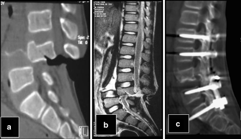
A ten-year-old boy fell from a running truck and was run over by the tractor-plough assembly, after which he developed paraplegia. The patient was haemodynamically stable and had developed bladder and rectal incontinence. The patient was brought to our hospital where various investigations were instigated. FAST (focused assessment with sonography in trauma) did not show any fluid in the dependent parts of the peritoneal cavity or pelvis. Plane radiographs showed grade 4 retrolisthesis of L3 over L4 with complete disruption of spinal alignment. CT corroborated the radiographic findings and also showed fracture of L3, misalignment of posterior elements, disruption of articular facets and transection of thecal sac at this level (Fig. 1a). However, paraplegia along with bowel bladder involvement was not totally clear, and MRI was planned. MRI showed the child to have low lying cord with complete transection of the spinal cord and total disruption of vertebral alignment (Fig. 1b). The patient was subsequently operated upon and the vertebrae were fixed with implants restoring vertebral alignment, but the cord still showed the sequelae of injury.
Fig. 1.
a, b CT and MRI of patient 1 showing the complete anterior dislocation of L4 over L5 causing complete disruption of low lying spinal cord. c Proper alignment of spinal column after pedicle screw fixation
Patient 2
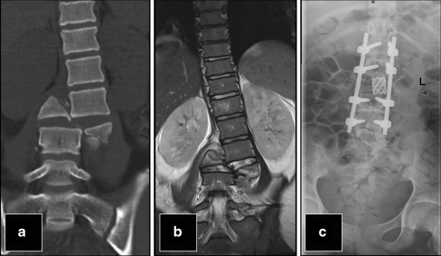
A 16-year-old male was admitted to our hospital with paraplegia following a motor vehicle crash. After basic resuscitation, a plain radiograph of the lumbosacral spine was obtained which showed complete collapse of the L3 vertebra with lateral displacement of the superior segment relative to the inferior segment. CT showed comminuted fracture of L3 vertebra with complete misalignment of vertebral column and the proximal segment was lying laterally in relation to the distal segment (Fig. 2). MRI showed additional findings of complete spinal cord transaction along with injury to the cord at the level of conus medullaris and cauda equina nerve roots. The patient was subsequently operated upon and vertebral alignment was restored.
Fig. 2.
a The lateral dislocation of L3 over L4 in patient 2 causing complete spinal cord injury. b Good alignment of spinal column following pedicle screw fixation
Patient 3
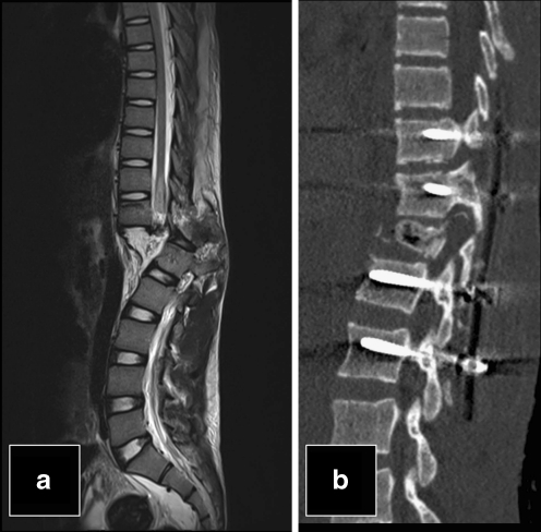
A 20-year-old male, who had fallen from height, had transient loss of consciousness and developed paraplegia subsequently. He had inability to move either lower limb with muscle power 0/5 in lower limbs and loss of sensation below L1. CT spine showed posterior spondyloptosis of T12 –L1 level with complete transaction of spinal cord. MRI dorso-lumbar spine showed complete disruption of terminal spinal cord at T12-L1 level (Fig. 3). The patient was subsequently operated upon and vertebral alignment was restored.
Fig. 3.
a The complete anterior dislocation of D12 over L1 in patient 3 causing complete disruption of spinal cord. b Proper alignment of spinal column after pedicle screw fixation
Patient 4
A 27-year-old male had fallen from height while working on the railing on the second floor of a multistoried building. He had transient loss of consciousness and developed paraplegia and severe back pain. Plain radiographs of spine showed a fracture of T12 vertebra with anterior and left lateral displacement of proximal segment relative to the lower segment and causing complete disruption of vertebral alignment. Contrast enhanced CT of the chest and abdomen showed fracture of the upper end plate, adjacent body, lamina and spinous process of T12 vertebra with disruption of T11-12 facet joint leading to complete misalignment of the vertebral column as seen on plain radiograph. MRI dorso-lumbar spine showed complete disruption of the terminal spinal cord at T11-12 level with injury to conus medullaris (Fig. 4). The patient was subsequently operated upon and vertebral alignment was restored.
Fig. 4.
MRI T2W image showing the complete anterior dislocation of D11 over D12 causing complete disruption of spinal cord
Discussion
Spondyloptosis is another form of spinal dislocation or advanced spondylolisthesis, in which one spinal segment is lodged in the anterior or posterior space of another [2, 6]. High thoracic lesions lead to paraparesis instead of quadriparesis, but autonomic symptoms are still marked. In the lower thoracic and lumbar lesions, urinary and bowel retention occur.
The injury mechanism of the patient would be compatible with the shear type of fracture dislocation according to the report of Denis [4]. According to this report, all three columns were disrupted. The spondyloptosis in our cases resulted from the concurrent mechanism of shearing and axial compression. Traumatic impact causes either a flexion-rotation stress or a shearing force that fractures the facets and ruptures ligaments, resulting in spinal disarticulation. These injuries have the highest association with spinal cord injury of all fracture types. The posterior reduction and fixation using pedicle screws in the thoraco-lumbar spondyloptsis is sufficient to provide good alignment and fixation [7, 10].
Spondylolisthesis treatment protocols are based on age, symptomatology, and slippage degree [6]. Fractures have been classified as stable or unstable according to the three-column concept [4]. Stable fractures can be treated by bed rest or brace, but unstable fractures are generally treated by open reduction and stabilisation with the rigid instrumentation.
This series describes four patients with spondyloptosis of the thoraco-lumbar spine including the first case with complete transection of a low lying cord. Traumatic spondyloptosis results from high energy trauma (Table 1). Due to three column involvement, the fracture is highly unstable.
Table 1.
Patient details
| Patient number | Age (y), Gender | Mechanism of injury | Spinal level | Plane of spondyloptosis | Cord status | Operative procedure | Post-operative improvement |
|---|---|---|---|---|---|---|---|
| 1 | 10, Male | Run over by tractor wheel | L4-L5 | Sagittal | Low lying cord with complete cord transaction | L2, L3, L4, L5 pedicle screw and rod fixation | Mild improvement |
| 2 | 16, Male | Road traffic accident | L3-L4 | Coronal | Complete cauda eqina and nerve roots transaction | Pedicle screw fixation at L1-L2 and L4-L5 | None |
| 3 | 20, Male | Fall from height | D12-L1 | Sagittal | Complete cord transaction | D11 and L2 pedicle screw with rod fixation | None |
| 4 | 27, Male | Fall from height | D11-D12 | Sagittal | Complete cord transaction | D10, D11 & D12 pedicle screw and rod fixation and L2 pedicle screw with rod fixation | None |
In our series, all four patients had sustained high-energy closed spinal injuries: two were involved in motor vehicle accidents and two patients had falls from height. All four patients presented with paraplegia (muscle power 0/5 in both lower limbs) and complete loss of sensation in lower limbs. All patients underwent plain radiography, CT and MR imaging. Three patients had sagittal plane spondyloptosis and one patient had coronal plane spondyloptosis. Complete cord/cauda eqina transection was present in all patients. One patient had a low lying cord with complete cord transection. All patients underwent surgery with reduction of displacement by pedicle screw and rod fixation to realign the vertebral column. None of the patients had full neurological function recovery as the injury was complete in all cases.
In one of our cases, the patient unfortunately had a low lying spinal cord which was transected and led to the catastrophic events. Usually the spinal cord ends at the lower margin of L1 or upper margin of L2, but in our patient, the spinal cord was lying at the L4 level, which resulted in the complete paralysis. Surprisingly the patient showed significant recovery following surgery. A review of previous cases of lumbar spondyloptosis suggests that the degree of anatomical injury found at surgery is a better predictor of patient outcome than fracture severity assessed radiologically [1]. In the other cases, patients developed urinary, rectal and sensory symptoms because of transected cauda equine nerve roots.
Yadla et al. undertook a retrospective review of a tertiary care regional spinal cord injury patient population in which they described five cases of traumatic spondyloptosis [11]. Four patients, all with sagittal-plane spondyloptosis, had a complete neurological deficit, and one with coronal-plane spondyloptosis presented with an incomplete neurological deficit. Other than this case series, very few case reports are described in literature [3, 8, 9]. The case with low lying spinal cord transection is not described in literature.
To conclude, traumatic thoraco-lumbar spondyloptosis is very rare. Radiology plays an important role in the diagnosis and management of complete spinal cord transections and helps in planning surgery. Surgical reconstruction and stabilisation allows for rehabilitation.
References
- 1.Bellew MP, Bartholomew BJ. Dramatic neurological recovery with delayed correction of traumatic lumbar spondyloptosis. Case report and review of the literature. J Neurosurg Spine. 2007;6(6):606–610. doi: 10.3171/spi.2007.6.6.16. [DOI] [PubMed] [Google Scholar]
- 2.Curylo LZ, Edwards C, DeWald R. Radiographic markers in spondyloptosis. Spine. 2002;27:2021–2025. doi: 10.1097/00007632-200209150-00010. [DOI] [PubMed] [Google Scholar]
- 3.Chul Woo Lee, Sun Chul Hwang, Soo Bin Im, Bun Tae Kim, Won Han Shin (2004) Traumatic thoracic spondyloptosis: a case report. J Korean Neurosurg Soc 622–624
- 4.Denis F. The three column spine and its significance in the classification of acute thoracolumbar spinal injuries. Spine. 1983;8:817–831. doi: 10.1097/00007632-198311000-00003. [DOI] [PubMed] [Google Scholar]
- 5.Denis F, Burkus JK. Shear fracture-dislocations of the thoracic and lumbar spine associated with forceful hyperextension (lumberjack paraplegia) Spine. 1992;17:156–161. doi: 10.1097/00007632-199202000-00007. [DOI] [PubMed] [Google Scholar]
- 6.El-Khoury GY, Whitten CG. Trauma to the upper thoracic spine: anatomy, biomechanics and unique imaging features. Am J Roentgenol. 1993;160:95–102. doi: 10.2214/ajr.160.1.8416656. [DOI] [PubMed] [Google Scholar]
- 7.Han IH, Song GS. Thoracic pedicle screw fixation and fusion in unstable thoracic spine fractures. J Korean Neurosurg Soc. 2002;32:334–340. [Google Scholar]
- 8.Hanley EN, Jr, Knox BD, Ramasastry S, Moossy JJ. Traumatic lumbopelvic spondyloptosis. A case report. J Bone Joint Surg Am. 1993;75(11):1695–1698. doi: 10.2106/00004623-199311000-00016. [DOI] [PubMed] [Google Scholar]
- 9.Meneghini RM, DeWald CJ. Traumatic posterior spondyloptosis at the lumbosacral junction. A case report. J Bone Joint Surg Am. 2003;85-A(2):346–350. doi: 10.2106/00004623-200302000-00026. [DOI] [PubMed] [Google Scholar]
- 10.Park YK, Lee JK, Lim DJ, Chung HS, Lee HK. Pedicle screw fixation of the thoracic spine. J Korean Neurosurg Soc. 1999;28:190–195. [Google Scholar]
- 11.Yadla S, Lebude B, Tender GC, Sharan AD, Harrop JS, Hilibrand AS, Vaccaro AR, Ratliff JK. Traumatic spondyloptosis of the thoracolumbar spine. J Neurosurg Spine. 2008;9(2):145–151. doi: 10.3171/SPI/2008/9/8/145. [DOI] [PubMed] [Google Scholar]


