Abstract
Mucocutaneous manifestations can be the first markers of HIV. We are reporting the case of an adolescent girl who presented with chronic, recurrent, intensely pruritic papular lichenified eruptions over extremities, face and trunk, which were exudative and crusted at places. She had delayed milestones with growth failure and no pubertal features. She did not have any risk factors to be suspected for HIV. The lesions were refractory to treatment, so she was tested for HIV and she came out to be HIV positive. This case reports pruritic papular eruptions as presenting illness of HIV.
Keywords: HIV/AIDS, pruritic papular eruptions, presenting illness
INTRODUCTION
Pruritic papular eruptions (PPEs) are the most common cutaneous manifestation of HIV disease, with a prevalence ranging from 11 to 46%, more so in less developed countries.[1] PPE can be the first marker of HIV infection.[2]
We present a case of a 13-year-old adolescent girl who had recurrent, intensely pruritic, and refractory to treatment papular lesions of no identifiable cause as the presenting illness of HIV.
CASE REPORT
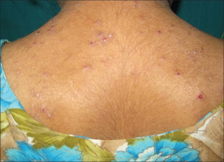
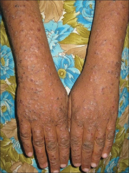
A 13-year-old female presented to Government Medical College, Vadodara, in March 2010, with complaints of severe generalized itching and skin lesions over extremities, face, neck, upper back and chest since 3 years [Figures 1 and 2]. She also had history of recurrent fever. There was no family history suggestive of atopic diathesis. The parents and siblings (one elder brother and one sister) were healthy, with no history of similar lesions. There was no past history of tuberculosis or blood transfusion in the patient or parents. There were no other risk factors for HIV in them.
Figure 1.

A 13-year-old female with pruritic lichenified papules over back with exudation
Figure 2.

Multiple excoriated lichenified papules over both upper limbs
She had not started menstruating and her secondary sexual characters were not developed.
On general examination, the patient had pallor. Her height was 143 cm [less than 3rd percentile height for age; z score < – 1 standard deviation (SD)][3] and weight was 25 kg with body mass index (BMI) 12.23 kg/m2 (z score for BMI < – 3 SD).[3]
On cutaneous examination, she had multiple excoriated lichenified papules over face, extremities, neck and upper back, with exudation and crusting at places and pediculosis over scalp. Her hymen was intact and external genitalia and perianal region were normal. There was no clinical evidence of any sexually transmitted disease (STD) and no inguinal lymphadenopathy.
Routine investigations including chest X-ray were normal except hemoglobin (9.5 g%). On the basis of history and clinical examination, the differential diagnoses kept were chronic papular urticaria, exudative discoid and lichenoid chronic dermatoses (EDLD), and mosquito bite hypersensitivity. The patient was given short-course oral prednisolone, starting with 15 mg and tapered weekly by 5 mg, and treated for pediculosis. She was referred to a pediatrician for her short stature for which she was clinically assessed and investigated for thyroid function tests which were within normal limits. Detailed hormonal analysis could not be done due to cost constraints.
Despite regular follow-up, the patient was not improving; hence, she was subjected to biopsy which showed hyperkeratosis with parakeratosis with neutophilic abscess in keratin layer. Epidermis showed pseudoepitheliomatous hyperplasia with non-specific inflammation in the underlying dermis. The histopathologic features were suggestive of eczematous dermatitis.
The patient was advised oral steroids intermittently, along with maintenance on topical steroids during periods of less activity. In July 2010, the patient was started on oral diamino-diphenyl sulfone (DDS) 50 mg, 1 tablet at night.
Recurrent, severely pruritic lichenified and exudative papular eruptions, which were refractory to treatment, along with delayed milestones led to get her investigated for HIV in February 2011. The enzyme-linked immunosorbent assay (ELISA) for HIV came out to be positive. Other investigations including serum venereal disease research laboratory (VDRL) test, serum hepatitis-B surface antigen (HBsAG), X-ray chest, and ultrasonogram (USG) abdomen were normal. Patient's parents and siblings were subjected to HIV testing but they were non-reactive. Both patient and her parents were enquired for sexual abuse, but no such history was available.
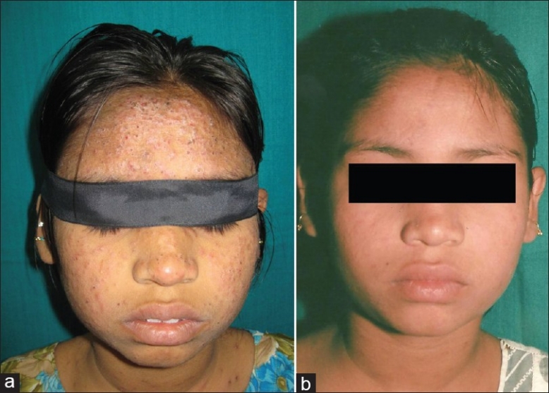
Patient's skin lesions were fitting into the criteria of PPE and hence she was categorized as WHO clinical stage IIa. Patient's CD4 count came out to be 431 cells/mm3. Her hemoglobin was 6.8 g%. She was started on D4T + 3TC + EFV regimen. The lesions started healing and pruritus minimized on initiation of antiretroviral therapy (ART) [Figures 3a and b].
Figure 3.

(a) Lichenified, crusted papular eruptions over face; (b) lesions over face healed after initiation of ART
DISCUSSION
The present case was initially not suspected of HIV because of absence of risk factors for HIV in the patient as well as her parents and siblings. The recalcitrant and persistent nature of her disease with delayed milestones prompted us to get her tested for HIV which came out to be positive. Subsequently, PPE related to HIV disease was considered as the diagnosis.
PPE is characterized by chronic bilaterally symmetric pruritic papules and sterile pustules on the trunk and extremities in an HIV-infected patient, with other definable causes of itching being ruled out.[1] The exact etiology of PPE is unknown, but might be an altered and exaggerated immune response to arthropod antigens.[4]
PPEs are regarded as WHO clinical stage II for infants and children.[5] PPEs manifest in advanced immunosuppressive stage in majority of the cases, but they may appear as an initial cutaneous disease with high CD4 count.[1] The present case also had CD4 count of 431 cells/mm3 when diagnosed. Liautaud et al. had observed in their study intensely pruritic eruptions as the first markers of HIV in 79% patients, and the eruptions appeared a mean of 8 months before the diagnosis of either Kaposi's sarcoma or opportunistic infection,[2] thus acting as the indicators of advancing immunosuppression.
PPEs are the most common cutaneous non-infectious manifestations of HIV, as observed by Sharma et al. who reported PPE in 35.8% cases[6] and by Lowe et al. who studied 139 HIV-positive adolescents and found PPE to be the most common HIV-related skin condition (42%).[7]
In the present report, the patient's presenting illness for HIV was PPE since 3 years, with low BMI. Colebunders et al. observed that 51% of HIV-positive patients had generalized PPE of unknown etiology of at least 1 month duration as their initial disease manifestation. Also, 95% patients with PPE and severe weight loss (greater than 10% of normal body weight) were seropositive.[8]
Differential diagnoses of PPE are follicular (eosinophilic, Demodex, staphylococcal, and pityrosporum folliculitis), and non-follicular pruritic eruptions, including scabies, insect bites, prurigo nodularis and eczematous eruptions.[9]
Topical steroids, emollients, and oral antihistaminics form the first line of management. But the pruritus is usually refractory to them.[4] Castelnuovo et al. studied 53 AIDS patients with PPE which were responsive to ART.[10]
HIV is known to cause growth failure as is also evident in the present case. A study by Aurpibul et al. showed that NNRTI-based ART leads to marked improvement in the growth of HIV-infected children. Timely initiation of ART before the children developed growth failure should be encouraged.
CONCLUSION
This case is reported as the diagnosis of HIV was missed because of no apparent risk factors in the patient or her family. HIV should be considered in the differential diagnosis when there is presence of refractory eczematous dermatitis with severely pruritic papular lesions and evidence of growth failure and delayed milestones in an adolescent.
PPE is a marker of advancing immunosuppression in HIV and has a major impact on the quality of life of the patient and should thus be recognized early and promptly treated.
Footnotes
Source of Support: Nil.
Conflict of Interest: None declared.
REFERENCES
- 1.Lakshmi SJ, Rao GR, Ramalakshmi, Satyasree, Rao KA, Prasad PG, et al. Pruritic papular eruptions of HIV: A clinicopathologic and therapeutic study. Indian J Dermatol Venereol Leprol. 2008;74:501–3. doi: 10.4103/0378-6323.44318. [DOI] [PubMed] [Google Scholar]
- 2.Liautaud B, Pape JW, DeHovitz JA, Thomas F, LaRoche AC, Verdier RI, et al. Pruritic skin lesions. A common initial presentation of acquired immunodeficiency syndrome. Arch Dermatol. 1989;125:629–32. doi: 10.1001/archderm.125.5.629. [DOI] [PubMed] [Google Scholar]
- 3.Growth reference data for 5-19 years. [Last accessed on 2011 July 10]. Available from: http://www.who.int/growthref/en/
- 4.Resneck JS, Jr, Van Beek M, Furmanski L, Oyugi J, LeBoit PE, Katabira E, et al. Etiology of Pruritic Papular Eruption with HIV Infection in Uganda. JAMA. 2004;292:2614–21. doi: 10.1001/jama.292.21.2614. [DOI] [PubMed] [Google Scholar]
- 5.Guidelines for HIV care and treatment in infants and children. IAP and NACO with support from Clinton Foundation, UNICEF, WHO. 2006. Nov, [Last accessed on 2011 June 26]. Available from: http://www.nacoonline.org/upload/Policies%20and%20Guidelines/4-%20Guidelines%20for%20HIV%20care%20and%20treatment%20in%20Infants%20and%20children.pdf .
- 6.Sharma A, Chaudhary D, Modi M, Mistry D, Marfatia YS. Noninfectious cutaneous manifestations of HIV/AIDS. Indian J Sex Transm Dis. 2007;28:19–22. [Google Scholar]
- 7.Lowe S, Ferrand RA, Morris-Jones R, Salisbury J, Mangeya N, Dimairo M, et al. Skin Disease Among Human Immunodeficiency Virus-Infected Adolescents in Zimbabwe: A Strong Indicator of Underlying HIV Infection. Pediatr Infect Dis J. 2010;29:346–51. doi: 10.1097/INF.0b013e3181c15da4. [DOI] [PMC free article] [PubMed] [Google Scholar]
- 8.Colebunders R, Mann JM, Francis H, Bila K, Izaley L, Kakonde N, et al. Generalized papular pruritic eruption in African patients with human immunodeficiency virus infection. AIDS. 1987;1:117–21. [PubMed] [Google Scholar]
- 9.James WD, Berger TG, Elston DM. 10th ed. Canada: Elsevier; 2006. Andrew's Diseases of the skin – clinical dermatology; p. 417. [Google Scholar]
- 10.Aurpibul L, Puthanakit T, Taecharoenkul S, Sirisanthana T, Sirisanthana V. Reversal of growth failure in HIV-infected Thai children treated with non-nucleoside reverse transcriptase inhibitor-based antiretroviral therapy. AIDS Patient Care STDS. 2009;23:1067–71. doi: 10.1089/apc.2009.0093. [DOI] [PubMed] [Google Scholar]


