

Abstract
Introduction:
Diagnosis of HIV infection in infants is difficult due to the presence of maternal antibodies; only nucleic acid assays are very helpful in early detection. Filter papers are especially useful for blood collection in resource-poor settings with limited access to diagnostic facilities.
Materials & Methods:
DBS samples were collected from the infants born to HIV seropositive mothers who had received single dose nevirapine at onset of labor. The samples were directly spotted onto the Whatman 903 cards from heel, big toe or finger prick depending on the age of the infants. A total of 766 infant samples were collected on dried blood spots (DBS) and transported to the Department of Experimental Medicine (DEM), Chennai, for testing from different government hospitals of rural and urban parts of Tamil Nadu, South India. According to National AIDS Control Organization's (NACO) protocol DNA was extracted from all these DBS and PCR was performed using the Roche kit version 1.5.
Results:
Fifteen infants were found to be HIV positive and 751 were HIV negative; all these 15 positive infants and 49 negative infants who were in the age group between 10 and 18 months were repeated with another DBS and compared with whole blood. The DBS results were concordant with the whole blood method and the sensitivity and specificity were 100%.
Keywords: Dried blood spots, HIV, Polymerase chain reaction
INTRODUCTION
Most HIV infection in children results from mother-to-child transmission (MTCT) of HIV, which can occur during pregnancy, labor and delivery, or the breast-feeding period. Despite the great advances that have been made in developing and implementing effective interventions to prevent HIV transmission from infected mothers to their infants, almost 2,000 infants in resource-poor countries are infected with HIV every day through MTCT.[1] Early diagnosis of human immunodeficiency virus type 1 (HIV-1) infection in infants born to HIV-1-seropositive mothers has proved difficult with conventional antibody tests.[2] The diagnosis of HIV infection among young infants now relies exclusively on virologic assays because serologic assays lack adequate sensitivity and specificity in differentiating maternal-derived versus infant-derived antibodies. Virologic assays are also helpful to confirm infection in patients with an advanced stage of disease who have inadequate specific antibody production.[3] In 1989, Science selected the polymerase chain reaction (PCR) as the major scientific development of the year.[4]
OBJECTIVE
The purpose of the study was to identify the HIV-1 status of the infants using DBS. The infants were born to HIV seropositive mothers who had received single dose nevirapine at the onset of labor. The mothers brought their infants for HIV testing to various government hospitals in Tamil Nadu, South India. All these infants received nevirapine syrup within 72 hours of age.
MATERIALS AND METHODS
This study was conducted in all the government Hospitals of rural and urban parts of the Tamil Nadu, South India. All the infants enrolled were received Single dose Nevirapine. The DBS samples were collected between October 2007 and September 2008 (11 months) after completion of Training of Trainers (TOT) program to all the Health Care Workers for the collection of DBS from the infants. Informed Consent for testing was obtained from infant's Parent/guardian. An inclusion criterion was HIV seropositive mother's infants only and all the infant's mothers received Single dose Nevirapine at the onset of labor.
According to NACO there are specific recommendations for sites from where specimens are to be collected from the infants: up to 4 months: heel prick, 4+ to 10 months: big toe and 10+ to 18 months: 3rd or 4th finger prick.
A total of 766 infant samples were collected from skin prick and coated onto filter paper (Whatman No. 903). The spotted filter papers were allowed to dry for at least 4 hours at room temperature and placed in individual zip locked bags containing a silica desiccant. All these samples were transported to Department of Experimental Medicine(DEM), The Tamil Nadu Dr. MGR Medical University, Chennai, Tamil Nadu. The Positive infant samples were repeated with another DBS and whole blood collected in EDTA vaccutainer tubes.
DNA extraction from DBS
One single spot of DBS was taken by 6 mm punches and was washed with 500 μl of Rnase free water (Invitrogen) for 3 times by vortexing for 10 seconds and centrifugation at a minimum speed. After removing the red-tinged liquid the DBS was treated in a 100 μl of 10% Chelax-100 resin (Biotechnology Grade, BioRad, Belgium) with this add a Roche internal control (IC) for each test 3.3 μl and kept it for 56°C (dry heat block) for 3 hours and 100°C (water bath) for 10 minutes. The extracted DNA was removed and stored at –20°C for further PCR amplification using Roche Amplicor HIV-1 DNA Test, version 1.5.[12]
DNA extraction from whole blood
In a screw caped tube 1.0 ml of blood wash was added and 500 μl of whole blood was added with this, incubated for 5 minutes at room temperature, and the tubes were centrifuged for 3 minutes at a maximum speed; the supernatant was slowly removed and again 1.0 ml of blood wash was added. The steps were repeated for 3 times. Working extraction reagent was prepared and Roche internal control (IC) was added with this; 200 μl of working reagent was added into all the tubes which contain the pellets. The tubes were incubated at 60°C for 30 minutes and 100°C for 30 minutes. After incubation the tubes were vortexed and centrifuged and 50 μl of the DNA was used for PCR amplification.
PCR amplification
Master mix was prepared by adding 100 μl of HIV-1 Mn2+ to HIV-1 MMX vial.
A total of 50 μl of the master mix was added into all the tubes and with this 50 μl of extracted DNA was added along the positive and negative controls. The reaction conditions for the program were HOLD Program: 2 minutes 50°C, CYCLE Program (5 cycles): 10 seconds 95°C, 10 seconds 52°C, 10 seconds 72°C, CYCLE Program (35 cycles): 10 seconds 90°C, 10 seconds 55°C, 10 seconds 72°C, HOLD Program: 15 minutes 72°C.
After amplification 100 μl of denaturation solution (DN) was added into all tubes, it should not be kept for more than 2 hours at room temperature.
Detection
A total of 100 μl of HIV-1 hybridization buffer is added to the micro well plate (MWP) and simultaneously 100 μl of the same buffer should be added to the internal control micro well plate (CT MWP). A total of 25 μl of amplicons is added to the appropriate wells of the MWP and CT MWP. Plates are covered and incubated for 1 hour at 37°C. The plates are washed with a wash buffer 5 times and dried with paper towels. One hundred microliters of Avidin-Horse radish peroxide conjugate (AV–HRP) is added to each well and incubated for 15 minutes at 37°C. After incubation the plates are washed with a wash buffer 5 times and are dried with paper towels. One hundred microliters of working substrate is added to the wells and kept for 10 minutes in dark. Finally 100 μl of stop solution is added and the absorbance is measured at 450 nm.
RESULTS
Results of HIV-1 DNA PCR using DBS
This study was carried out over a period of 11 months from October 2007 and September 2008. A total of 766 samples were tested for qualitative HIV-1 DNA PCR. Fourteen samples were found to be HIV positive and 752 samples were found to be HIV negative, but 2 sample results were equivocal, i.e., inconclusive – the OD reading of one sample was 0.259 and of other was 0.734; these 2 samples were retested using the same DBS and with another DBS samples. One was found to be positive and another was negative consistent with the reference standards in all cases.
After the repeated testing of equivocal samples the total number of positive samples was 15 (1.95%) and of negative samples was 751 (98.04%).
Results of HIV-1 DNA PCR using whole blood
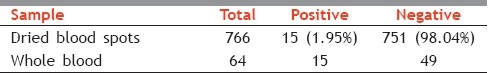
Whole blood samples were collected in EDTA tubes from 48 negative infants by DBS whose age range was between 10 months and 18 months including 14 HIV-positive samples and 2 equivocal samples. A total of 64 samples were tested by whole blood using a Roche kit. Fifteen samples were positive which were already tested positivebyDBS and 49 negative samples were negative.
The sensitivity was 100% and the specificity was 100% when DBS was compared with the whole blood. Table 1 shows the results of DBS and whole blood.
Table 1.
Results of dried blood spots and whole blood
DISCUSSION
In this 11-month study period a total of 766 DBS were received to perform the diagnosis of HIV-1 infection in infants by a DNA PCR test. Fifteen infants were found to be positive for HIV-1 infection and 751 infants were found to be negative by DBS.
Whole blood of all the 15 positive samples and 49 negative samples of the infants between 10 and 18 months of age group was also tested for HIV-1 DNA PCR and both the results were concordant.
Many centers continue to follow up children beyond 4--6 months of age to confirm the loss of maternal antibody, which usually occurs between 9 and 15 months of age. A positive DNA PCR at any time requires a repeat test on a second blood sample as soon as possible for confirmation. Two positive DNA PCRs at any time confirm infection.
According to the Roche Amplicorkit ,the whole blood has to be processed within 4 days of collection. This is difficult in our settings as the sample has to come from long distance to the testing centre (DEM). Thereforein this study we found it easy to perform the qualitative HIV-1 DNA PCR test with DBS than with whole blood.
The elution with Chelex-100 should eliminate the potential PCRinhibitors, since Chelex is a cation-chelating resin: the positivelycharged ions are captured by the resin, whereas the DNA, negativelycharged, remains free in the solution.[13]
The DBS format greatly facilitates the logistics of sample collection, processing, and shipping for limited resource settings. Whole blood saved as DBS can be transported or mailed to reference laboratories without refrigeration and has low biohazard risk.[14] Whole-blood can easily be coated on the filter paper from heel stick or finger punctures in infants; thus avoiding the use of syringes and vaccutainer tubes. Blood coated on filter paper lyses the cells and binds the DNA. Therefore, the sample centrifugation and extraction procedures are reduced. Dried blood on filter paper appears biologically stableand can be stored at room temperature. It can be transported easily and therefore it is convenient to use the DBS in resource-limited settings.[15]
The small amount of blood required, theease of collection, storage, and transport of samples, and thelow cost of the test make this assay ideal for HIV-1 testing ofinfants in the field or where resources arelimited.[16]
Dried blood specimens areideal for the diagnosis and evaluation of HIV-1 infection ininfants in resource-limited regions; however, previous reportshave shown a decline in the ability to detect HIV-1 antibodiesin samples stored under hot, humid conditions.[17]
Although HIV-1 DNA is stable in whole blood samples stored ina refrigerator for up to 10 days, testing of whole bloodcollected on filter paper offers an opportunity to simplifysample collection and the transport of specimens to laboratoriesfor diagnostic testing.[18] In conclusion using DBS as a sample source is the best method to perform the diagnosis of HIV infection in infants born to HIV seropositive mothers. Blood collection is easy for the phlebotomist and other healthcare workers; a little formal training is enough for them. A small quantity of blood, i.e., 50 μl, is enough to make a dried blood spot (DBS) whereas for the whole blood 500 μl is required to perform the test.
The positive infants are referred to the ART centers if they are less than 18 months of age. Future studies should be done of the dried blood spots with different types of filter papers with different volumes of the samples.
ACKNOWLEDGEMENTS
The authors are very grateful to all the HIV seropositive mothers and their infants participating in this study. The authors also wish to thank Tamil Nadu State AIDS Control Society and Clinton Foundation for their support to this study.
Footnotes
Source of Support: Nil.
Conflict of Interest: None declared.
REFERENCES
- 1.Guidelines on care, Treatment and support for women living with HIV/AIDS and their children in Resource-Constrained Settings. Geneva: World Health Organization; 2004. Antiretroviral for treating pregnant women and preventing HIV Infection in Infants. [Google Scholar]
- 2.Rakusan TA, Parrott RH, Sever JL. Limitations in the laboratory diagnosis of vertically acquired HIV infection. J Acquir Immune DeficSyndr. 1991;4:116–21. [PubMed] [Google Scholar]
- 3.Nielsen K, Bryson YJ. Diagnosis of HIV infection in children. PediatrClin North Am. 2000;47:39–63. doi: 10.1016/s0031-3955(05)70194-2. [DOI] [PubMed] [Google Scholar]
- 4.Guyer RL, Koshland DE. The molecule of the year. Science. 1989;246:1543–6. doi: 10.1126/science.2688087. [DOI] [PubMed] [Google Scholar]
- 5.Chadwick EG, Yogev R, Kwok S, Sninsky JJ, Kellogg DE, Wolinsky SM. Enzymatic amplification of the human immunodeficiency virus in peripheral blood mononuclear cells from pediatric patients. J Infect Dis. 1989;160:954–9. doi: 10.1093/infdis/160.6.954. [DOI] [PubMed] [Google Scholar]
- 6.Rogers MF, Ou CY, Rayfield M, Thomas PA, Schoenbaum EE, Abrams E, et al. Use of the polymerase chain reaction for early detection of the proviral sequences of human immunodeficiency virus in infants born to seropositive mothers. N Engl J Med. 1989;320:1649–54. doi: 10.1056/NEJM198906223202503. [DOI] [PubMed] [Google Scholar]
- 7.Williams P, Simmonds P, Yap PL, Balfe P, Bishop J, Brettle R, et al. The polymerase chain reaction in the diagnosis of vertically transmitted HIV infection. AIDS. 1990;4:393–8. doi: 10.1097/00002030-199005000-00003. [DOI] [PubMed] [Google Scholar]
- 8.Creek TL, Sherman GG, Nkengasong J, Lu L, Finkbeiner T, Fowler MG, et al. Infant human immunodeficiency virus diagnosis in resource-limited settings: Issues, technologies, and country experiences. Am J ObstetGynecol. 2007;197(Suppl 3):S64–71. doi: 10.1016/j.ajog.2007.03.002. [DOI] [PubMed] [Google Scholar]
- 9.Germer JJ, Gerads TM, Mandrekar JN, Mitchell PS, Yao JD. Detection of HIV-1 DNA proviral DNA with the AMPLICOR HIV-1 DNA test, version 1.5, following sample processing by the MagNa Pure LC instrument. J ClinVirol. 2006;37:195–8. doi: 10.1016/j.jcv.2006.08.001. [DOI] [PubMed] [Google Scholar]
- 10.Patton JC, Akkers E, Coovadia AH, Meyers TM, Stevens WS, Sherman GG. Evaluation of dried whole blood spots obtained by heel or finger stick as an alternative to venous blood for diagnosis of human immunodeficiency virus type 1 infection in vertically exposed infants in the routine diagnostic laboratory. Clin Vaccine Immunol. 2007;14:201–3. doi: 10.1128/CVI.00223-06. [DOI] [PMC free article] [PubMed] [Google Scholar]
- 11.McCabe ER, Huang S, Selzer WK, Law ML. DNA micro extraction from dried blood spots on filter paper blotters, Potential applications to new born screening. Hum Genet. 1987;75:213–6. doi: 10.1007/BF00281061. [DOI] [PubMed] [Google Scholar]
- 12.Zhang Q, Wang L, Jiang Y, Fang L, Pan P, Gong S, et al. Early infant human immunodeficiency virus type 1 detection suitable for resource-limited settings with multiple circulating subtypes by use of nested three-monoplex DNA PCR and dried blood spots. J ClinMicrobiol. 2008;46:721–6. doi: 10.1128/JCM.01539-07. [DOI] [PMC free article] [PubMed] [Google Scholar]
- 13.Fischer A, Lejczak C, Lambert C, Servais J, Makombe N, Rusine J, et al. Simple DNA extraction method for dried blood spots and comparison of two PCR assays for diagnosis of vertical human immunodeficiency virus type 1 transmission in Rwanda. J ClinMicrobiol. 2004;42:16–20. doi: 10.1128/JCM.42.1.16-20.2004. [DOI] [PMC free article] [PubMed] [Google Scholar]
- 14.Mehta N, Trzmielina S, Nonyane BA, Eliot MN, Lin R, Foulkes AS, et al. Low-cost HIV-1 diagnosis and quantification in dried blood spots by real time PCR. PLoS One. 2009;4:e5819. doi: 10.1371/journal.pone.0005819. [DOI] [PMC free article] [PubMed] [Google Scholar]
- 15.Jacob SM, Anitha D, Vishwanath R, Parameshwari S, Samuel NM. The use of dried blood spots on filter paper for the diagnosis of HIV-1 in infants born to HIV seropositive women. Indian J Med Microbiol. 2008;26:71–4. doi: 10.4103/0255-0857.38864. [DOI] [PubMed] [Google Scholar]
- 16.Beck IA, Drennan KD, Melvin AJ, Mohan KM, Herz AM, Alarcón J, et al. Simple, sensitive, and specific detection of human immunodeficiency virus type 1 subtype B DNA in dried blood samples for diagnosis in infants in the field. J ClinMicrobiol. 2001;39:29–33. doi: 10.1128/JCM.39.1.29-33.2001. [DOI] [PMC free article] [PubMed] [Google Scholar]
- 17.Behets F, Kashamuka M, Pappaioanou M, Green TA, Ryder RW, Batter V, et al. Stability of human immunodeficiency virus type 1 antibodies in whole blood dried on filter paper and stored under various tropical conditions in Kinshasa, Zaire. J ClinMicrobiol. 1992;30:1179–82. doi: 10.1128/jcm.30.5.1179-1182.1992. [DOI] [PMC free article] [PubMed] [Google Scholar]
- 18.Mitchell C, Jennings C, Brambilla D, Aldrovandi G, Amedee AM, Beck I, et al. Dried Blood Spot Working Group of the Infant Maternal Pediatric Adolescent AIDS Clinical Trials Network.Diminished human immunodeficiency virus type 1 DNA Yield from dried blood spots after storage in a humid incubator at 37°C compared to -20°C. J ClinMicrobiol. 2008;46:2945–9. doi: 10.1128/JCM.00359-08. [DOI] [PMC free article] [PubMed] [Google Scholar]