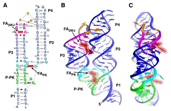
Figure 2.
Global structure of the aptamer domain of the THF riboswitch. (A) Secondary structure of the RNA used in the structure determination, redrawn to reflect the tertiary structure and include non-canonical base pairs, denoted using the Leontis-Westhof notation (Leontis and Westhof, 2001). The two molecules of folinic acid (FAPK and FA3WJ) observed in the crystal structure are denoted in red. (B) Cartoon representation of the global architecture of the THF aptamer. Coloring of the RNA is consistent with panel (a). (C) 90° rotation of the perspective shown in panel (b); note the placement of both THF binding sites on the same face of the RNA. See also Figure S1.

