Abstract
To explore the genetic basis of the pathogenesis and adaptation of avian influenza viruses (AIVs) to chickens, the A/duck/Yokohama/aq10/2003 (H5N1) (DkYK10) virus was passaged five times in the brains of chickens. The brain-passaged DkYK10-B5 caused quick death of chickens through rapid and efficient replication in tissues, accompanied by severe apoptosis. Genome sequence comparison of two viruses identified a single amino acid substitution at position 109 in NP from isoleucine to threonine (NP I109T). By analyzing viruses constructed by the reverse-genetic method, we established that the NP I109T substitution also contributed to increased viral replication and polymerase activity in chicken embryo fibroblasts, but not in duck embryo fibroblasts. Real-time RT-PCR analysis demonstrated that the NP I109T substitution enhances mRNA synthesis quickly and then cRNA and viral RNA (vRNA) synthesis slowly. Next, to determine the mechanism underlying the appearance of the NP I109T substitution during passages, four H5N1 highly pathogenic AIVs (HPAIVs) were passaged in the lungs and brains of chicken embryos. Single-nucleotide polymorphism analysis, together with a database search, suggests that the NP I109T mutation would be induced frequently during replication of HPAIVs in brains, but not in lungs. These results demonstrate that the amino acid at position 109 in NP enhances viral RNA synthesis and the pathogenicity of highly pathogenic avian influenza viruses in chickens and that the NP mutation emerges quickly during replication of the viruses in chicken brains.
INTRODUCTION
Avian influenza viruses (AIVs) belong to the genus Influenzavirus A of the family Orthomyxoviridae and contain eight genome segments of single-stranded, negative-sense RNA. AIVs are divided into low pathogenic (LP) and highly pathogenic (HP) phenotypes in chickens (44); pathogenicity depends on the cleavability of hemagglutinin (HA), which allows systemic replication in chickens (3, 11). The H5N1 HPAIVs may have been circulating in poultry farms since 1996 and have caused devastating damage to the poultry industry in Asia and Africa. The H5N1 HPAIVs also possess the unique capability of crossing host species barriers from birds to mammals, causing high mortality in mice (5, 10, 22), ferrets (8, 21), monkeys (32), and humans (6, 41, 47), and are considered a candidate for a new pandemic virus (24).
HA spike protein mediates binding to the cell surface sialic acid receptor to initiate virus infection, which plays an important role in overcoming the interspecies barrier. It is known that amino acids at the receptor binding site can alter the host range (1, 34, 57), and those in the vicinity of the receptor binding site also influence adaptation to new hosts (12, 29). Recently, the polymerase (PA, PB1, and PB2) genes have also been implicated in the adaptation of AIVs to mammals. PB1 may have contributed to the virulence of previous pandemic viruses, the H2N2 Asian flu and H3N2 Hong Kong flu viruses, in mammals (16, 28). Amino acids in the polymerase subunit PB2 (lysine at position 591, lysine at position 627, and asparagine at position 701) are critical for the adaptation of influenza A viruses to mammals, facilitating efficient replication of AIVs in human cells (17, 26, 55) and mammalian hosts (10, 25, 40). The amino acid mutation at position 627 in PB2 has been shown to appear quickly after passages of H5N1 HPAIV in mice (23). Moreover, PA is involved in high virulence in mammalian hosts (13, 18, 33). Thus, evidence of the importance of polymerase subunit proteins of AIVs in crossing the species barrier from birds to mammals is increasing.
In contrast, the molecular mechanisms that govern how duck origin AIVs acquire virulence in chickens are still poorly understood. Among HPAIVs, the Asian H5N1 HPAIV has extremely high pathogenicity in chickens; this high pathogenicity is associated with quick and high replication of the virus in chickens, accompanied by severe apoptosis of macrophages and early suppression of cytokine mRNA synthesis (42). The high pathogenicity of the H5N1 virus in chickens is related to PB2 and the amino acid at position 105 in NP (45). Additionally, Wasilenko et al. reported that NP, PB1, and PB2 contribute to the enhanced replication of H5N1 virus in chickens (53, 54). The approach of comparing two genetically similar viruses using reverse genetics suggests that both polymerase subunits and NP may play roles in the adaptation of AIVs from ducks to chickens. Nevertheless, this sophisticated approach carries the risk of detecting different genes or amino acids, depending on which virus strains are compared.
Passaging of AIVs in a new host may be a safe experimental approach to study the molecular basis underlying the adaptation of AIVs to a new host. Amino acid changes that are involved in the increased pathogenicity of AIVs after passaging in chickens can be identified. Ito et al. reported that an avirulent duck strain becomes highly pathogenic after being passaged in chickens via the air sac route because of a mutation from R-E-T-R to R-R-K-K-R at the HA cleavage site (14). It has also been reported that neuropathogenicity increased following the passaging of AIV in chicken brains, although the critical gene was not determined (38). The A/duck/Yokohama/aq10/2003 (H5N1) virus (DkYK10) belongs to the HP category but replicates relatively slowly and inefficiently in chickens, inducing high fever and severe gross lesions with a long time to death, while the A/chicken/Yamaguchi/7/2004 virus replicates quickly in chickens and causes peracute death before the onset of clinical signs or gross lesions (42). The duck origin DkYK10 may be an appropriate virus to study the genetic basis for the adaptation of HPAIVs from ducks to chickens.
To better understand the molecular mechanisms underlying the ability of duck origin HPAIVs to acquire pathogenicity in chickens, we passaged DkYK10 in chicken brains and obtained the more virulent DkYK10-B5. DkYK10-B5 quickly increased its pathogenicity in chickens due to a single amino acid substitution at position 109 in NP from isoleucine to threonine (NP I109T). The increased pathogenicity of DkYK10 in chickens was mediated by the enhanced activity of AIV transcription. Single-nucleotide polymorphism (SNP) analysis of the passaged samples indicates that replication in brains is crucial for the emergence of HPAIVs possessing this NP I109T substitution. SNP analysis, together with a sequence database search, suggests that the NP 109I residue could be mutated to 109T during many HPAIV replications in chicken brains.
MATERIALS AND METHODS
Viruses.
The HPAIV A/duck/Yokohama/aq10/2003 (H5N1) (DkYK10) (clade 6), with the amino acid sequence PQRERRRKKR at an HA cleavage site, was isolated from duck meat imported from China at the quarantine service in Japan (22). DkYK10 induces severe clinical signs and gross lesions in chickens, with a mean death time (MDT) of approximately 4 days (22, 42, 45). The following HP viruses were used for the embryo passage experiment: A/chicken/Yamaguchi/7/2004 (H5N1) (CkYM7) (clade 2.5) (24), A/chicken/Miyazaki/K11/2007 (H5N1) (CkMZ11) (clade 2.2), and A/whooper swan/Akita/1/2008 (H5N1) (WsAK1) (clade 2.3.2) (50).
Passage history of virus and comparison of pathogenicities.
Five 4- to 10-week-old specific-pathogen-free (SPF) White Leghorn (Gallus gallus domesticus) chickens were purchased from the Nippon Institute of Biological Science (Kobuchizawa, Yamanashi, Japan) and reared in a negative-pressure isolator with HEPA filters in the biosafety level 3 (BSL3) animal experimentation facility at the National Institute of Animal Health. A lung (L1) was obtained from a dead chicken after intranasal inoculation with DkYK10 at 106.0 50% egg infective doses (EID50)/0.1 ml. A 10% lung homogenate was prepared with phosphate-buffered saline (PBS), and the lung supernatant was intranasally inoculated into 5 new chickens for passage. The first dying chicken was anesthetized with diethyl ether 72 h postinoculation (p.i.), and the brain (B1) was collected. The supernatant of the 10% brain homogenate was inoculated intranasally into another 5 chickens, the first dying chicken was anesthetized 72 h later, and the brain (B2) was obtained. This brain passage of DkYK10 in 4- to 6-week-old chickens was repeated five times to obtain the virus that was passaged five times in brains, which was termed DkYK10-B5. The B1 to B5 viruses were collected from the first dying chicken 72, 72, 72, 72, and 48 h p.i., respectively. DkYK10-B5 was passaged another five times in brains, and the virus that was passaged 10 times in brains was termed DkYK10-B10. The B6 to B10 viruses were harvested from the first dying chicken 72, 54, 65, 71, and 78 h p.i., respectively.
Fever and mean death time.
A wireless thermosensor was used to record the fever and MDT of each chicken, as described previously (42). The thermosensor, which was designed for this experiment, was attached to the abdominal skin surface of each chicken with no-stretch sports tape. The body temperature signal of each chicken was continuously recorded in a computer every 5 min. The death time of each chicken was defined as the time when the body temperature dropped below 30°C after virus inoculation (42).
Sequence analysis.
RNA purification, cDNA synthesis, gene amplification by PCR, and sequencing were performed as described previously (48). Viral RNAs (vRNAs) were purified with the QIAamp Viral RNA Mini Kit (Qiagen, Germany) and transcribed into cDNA using random primers and PrimeScript reverse transcriptase (Takara, Shiga, Japan). All eight gene segments of DkYK10-B5 and DkYK10-B10 were amplified by PCR, and the PCR products were purified and sequenced using an automatic ABI Prism 3100 genetic analyzer (Applied Biosystems).
Reverse genetics.
All eight gene segments of DkYK10 and the NP gene segment of DkYK10-B5 were amplified by PCR, cloned into the pCR2.1 Topo (Invitrogen) vector, and then transferred into the pHW2000 expression vector (kindly provided by R. G. Webster, St. Jude Children's Research Hospital, Memphis, TN). To generate reassortant viruses, MDCK and 293T cells were cocultured and transfected with 1 μg of each of the eight plasmids and 16 μl TransIT-LT1 (Mirus Bio) in a total volume of 200 μl of Opti-MEM (Gibco) (45). After 48 h of cultivation, the culture supernatant was injected into the allantoic cavities of embryonated chicken eggs and harvested after 24 h of cultivation. The resulting reassortant viruses were named rgDKYK10 and rgDkYK10-B5.
Virus replication kinetics in chickens.
To compare virus replication kinetics in chickens, host animals were intranasally inoculated with 106.0 EID50/0.1 ml of rgDkYK10 (n = 12) or rgDKYK10-B5 (n = 9) and reared in negative-pressure isolators. At each sampling point, three chickens from each inoculation group were euthanized with ether and sacrificed, and the lungs, spleens, and kidneys were stored at −80°C. For virus titration, 10% tissue homogenate was prepared, and the supernatant was inoculated into 10-day-old embryonated chicken eggs; the EID50/g of tissue was calculated as described previously (31).
Detection of virus antigen and apoptotic cells in tissues.
Lungs, spleens, and brains were harvested sequentially from the same chickens that were used for the virus replication experiment and processed for immunohistochemical analysis to detect the AIV matrix protein (M) with anti-M monoclonal antibody, as described previously (46).
To compare apoptotic-cell numbers in tissues of chickens infected with HPAIVs, the lungs and livers from three chickens per group were fixed with 10% neutral buffered formalin. Within 7 days, these paraffin sections of tissue were stained using the ApopTag peroxidase in situ apoptosis detection kit (Chemicon International, Inc.), as described previously (45).
Growth kinetics of virus in cultured cells.
Primary chicken embryo fibroblasts (CEFs) and duck embryo fibroblasts (DEFs) (2.5 × 10⁵ cells/well) were cultured in a 6-well tissue culture plate, and the monolayer was inoculated with rgDkYK10 or rgDkYK10-B5 at a multiplicity of infection (MOI) of 0.001. Following adsorption for 1 h at 37°C, nonadsorbed viruses were removed and the medium was replaced. At 0, 12, 24, 36, and 48 h p.i., the culture supernatants were collected and stored at −80°C. For virus titration, serial 10-fold dilutions of viral samples were inoculated onto CEFs and cultivated at 37°C for 7 days to determine cytopathic effect and hemagglutination with chicken red blood cells. Fifty percent tissue culture infective dose (TCID50) assay endpoints were calculated using the method described by Reed and Muench (31).
Polymerase activity assay.
The polymerase activity assay was conducted as described previously (35, 45). Sixty percent confluent CEFs or DEFs cultured in 6-well tissue plates (Falcon) were transfected with 2 μg of pHH21-lucR, 1 μg of pHW2000-PB2, 1 μg of pHW2000-PB1, 1 μg of pHW2000-PA, and 2 μg of pHW2000-NP, using TransIT-LT1 (Mirus Bio). The pHH21-lucR vectors were constructed by inserting the luciferase gene, flanked by the noncoding region of the M segment, downstream of the RNA polymerase I promoter of pHH21 (kindly provided by Y. Kawaoka, University of Wisconsin). At 24 h after transfection, the cells were stored at −80°C. The cell extracts were prepared in 250 μl of passive lysis buffer, and luciferase levels were assayed with a luciferase assay system (Promega) and Arvo MX/Light (PerkinElmer, Japan) instrument according to the manufacturer's protocol. The experiment was performed in triplicate wells.
Real-time RT-PCR assay for vRNA, cRNA, and mRNA synthesis.
To compare the transcription and replication activities of rgDkYK10 and rgDkYK10-B5, CEFs were inoculated with viruses at an MOI of 10. After cultivation at 37°C for 2 days, the cell extracts were harvested at 0, 2, 4, 6, and 8 h p.i., and total cellular RNAs were purified with the RNeasy minikit (Qiagen). Three micrograms of total RNA was used for cDNA preparation with Primescript reverse transcriptase (Takara). The primers for cDNA synthesis of vRNA, cRNA, or mRNA for NP genes were 5′-TTGCKTCAAATGAGAACATG-3′, 5′-AGTAGAAACAAGGGTA-3′, and oligo(dT), respectively; those for H5 genes were 5′-ACATCAACACTAAAYCAGAG-3′, 5′-AGTAGAAACAAGGGTG-3′, and oligo(dT), respectively. After removing the primers with the High Pure PCR Cleanup Micro kit (Roche), the cDNAs were used to determine the quantity of mRNA, cRNA, and vRNA for the NP or H5 genes by real-time reverse transcription (RT)-PCR (49). The average threshold cycle (CT) value of triple wells was determined, and the relative viral RNA concentration (40 − CT) at each sampling point was determined after the RNA concentrations of two virus strains, rgDkYK10 and rgDkYK10-B5, at 0 h p.i. (immediately after virus inoculation) were adjusted to be the same. The 28S rRNA was used as a control to monitor cDNA synthesis (42).
SNP analysis.
To determine the mechanisms underlying the emergence of the NP 109T mutation in H5N1 HPAIVs, DkYK10 was passaged in CEFs and primary chicken kidney (CK) cells at an MOI of 0.01 and incubated for 2 days. In addition, DkYK10, CkYM7, CkMZ11, and WsAK1 (106 EID50) were inoculated in the allantoic cavities of 17-day-old chicken embryos, and the lungs and brains were harvested 48 h later. The lung or brain homogenates were then passaged in the allantoic cavities of embryonated eggs to obtain the respective lung or brain passage viruses. Viral RNAs were purified from the culture supernatants or supernatants of tissue homogenates with the High Pure Viral RNA kit (Roche), and 0.3 μg was transcribed into cDNA using random primers and PrimeScript reverse transcriptase (Takara, Shiga, Japan). Real-time PCR was performed to detect NP 109T using the Takara CycleavePCR Core Kit (Takara, Shiga, Japan), as well as a forward primer (5′-GCATAACAATAGAGAGAATG-3′), reverse primer (5′-TTGGAATGCCAKATCATAC-3′), and probe (5′-6-carboxyfluorescein [FAM]-GAGCTGAcTCTGTA-Eclipse [quencher]-3′). The reaction mixture consisted of 10 μl of CycleavePCR reaction mixture (2×; Takara), 1 μl of cDNA, 0.8 μl of forward and reverse primers (0.2 μM each), 0.8 μl of the probe (0.2 μM), and 0.4 μl of Rox reference dye II (50×; Takara). The PCR protocol was as follows: 1 cycle at 95°C for 10 s, followed by 45 cycles of 95°C for 5 s, 55°C for 10 s, and 72°C for 25 s. The average CT of triple wells per sample was determined, and the relative quantity of NP 109T RNA in samples was determined as 45 − CT. Then, 106 EID50 of DkYK10 was mixed with 106, 105, or 104 EID50 of DkYK10-B5, and their 45 − CT values were determined and used as a standard to estimate the titers of viruses with the NP 109T mutation.
Molecular epidemiological analysis.
To determine the frequency of NPs possessing 109T in the NCBI Influenza Virus Resource (http://www.ncbi.nlm.nih.gov/genomes/FLU/FLU.html), 1,597 NPs derived from H5N1 HPAIVs were analyzed. In addition, to determine the NP population that might participate in NP I109T substitution during passages, NP proteins with more than 98% and 99% homology to NPs of DkYK10, CkYM7, CkMZ11, or WsAK1 were searched for from among 1,464 NP proteins of Asian AIVs in GenBank.
Statistical analysis.
A single paired Student's t test was used to determine the statistical significance of fever, MDT, virus titers, polymerase activity, and viral RNA synthesis between DkYK10 and DkYK10-B5 or rgDkYK5 and rgDkYK10-B5. Statistical significance was set at a P value of <0.05.
RESULTS
Comparison of pathogenicities of DkYK10 and brain passage viruses in chickens.
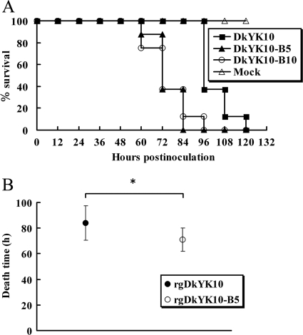
To compare the pathogenicities of DkYK10-B5 and DkYK10-B10 with that of the parental DkYK10, each of 8 chickens was inoculated intranasally with DkYK10, DkYK10-B5, or DkYK10-B10, and clinical signs, gross lesions, and mortality were observed every 12 h. Chickens infected with DkYK10 showed ruffled feathers and depression from 48 h p.i., and gross lesions were observed from 60 h p.i. In contrast, chickens infected with DkYK10-B5 or DkYK10-B10 showed ruffled feathers and depression from 36 h p.i., and gross lesions were observed as early as 48 h p.i.; these birds were dead 1 day before those infected with DkYK10 (Fig. 1 A). The body temperature kinetics of chickens infected with DkYK10-B5 or DkYK10-B10 were similar to those infected with DkYK10 until 55 h p.i.; however, then the kinetics of the DkYK10-B5 or DkYK10-B10 group dropped markedly to death (data not shown). The MDTs of chickens infected with DkYK10-B5 (70.1 ± 12.2 h p.i.) or DkYK10-B10 (69.1 ± 10.4 h p.i.) were shorter than those of chickens infected with parental DkYK10 (95.9 ± 9.2 h p.i.) (Fig. 1B).
Fig. 1.
Kinetics of the survival rate and MDT of chickens after intranasal inoculation with HPAIVs. (A) Chickens were inoculated with 106 EID50 of DkYK10, DkYK10-B5, or DkYK10-B10 (n = 8 for each inoculation group), and the kinetics of the survival rate were determined. The values represent the means ± standard deviations of 8 chickens in each group. (B) Comparison of the MDTs of chickens inoculated intranasally with 106 EID50 of rgDkYK10 or rgDkYK10-B5. The time of death was defined as the time when body temperature fell below 30°C after inoculation, and the mean MDT ± standard deviation for each group is shown. *, significant difference (P < 0.05).
The average virus titers in the lungs and spleens of chickens infected with DkYK10-B5 or DkYK10-B10 just before death were 10 times higher than in those infected with DkYK10, while there was no difference among the three strains in virus titers in brains (Table 1 ). Additionally, influenza virus M antigen was detected more frequently in the lungs and spleens of chickens infected with DkYK10-B5 or DkYK10-B10 than in those infected with DkYK10, although large amounts of M antigen were detected in the brains of all three virus-inoculated groups (Table 1).
Table 1.
Virus infectivity and distribution of virus M antigen in tissues from chickens inoculated with DkYK10, DkYK10-B5, or DkYK10-B10
| Virus | Infectivitya,b (log10 EID50/g) |
M antigena,c |
||||
|---|---|---|---|---|---|---|
| Lung | Spleen | Brain | Lung | Spleen | Brain | |
| DkYK10 | 6.8 ± 0.6 | 5.9 ± 0.6 | 7.6 ± 0.5 | +, +, +, + | −, −, −, + | +++, +++, +++, +++ |
| DkYK10-B5 | 7.8 ± 0.3 | 7.1 ± 0.3 | 7.9 ± 0.5 | +++, +++, +++, +++ | +++, ++, +, ++ | ++, ++, +++, +++ |
| DkYK10-B10 | 7.3 ± 0.3 | 6.9 ± 0.4 | 7.5 ± 0 | +++, +++, ++, +++ | +++, +, ++, ++ | ++, ++, +++, +++ |
Four chickens per group were inoculated intranasally with 106.0 EID50 of DkYK10, DkYK10-B5, or DkYK10-B10 and sacrificed just before natural death. Tissues were obtained for virus infectivity and M antigen distribution analysis.
Titers of individual lungs, spleens, and brains were determined with embryonated chicken eggs; the mean virus titers ± standard deviations of four chickens are shown.
Grading of M antigen is as follows: −, none; +, mild; ++, moderate; +++, severe.
Sequence comparison between DkYK10 and brain passage viruses.
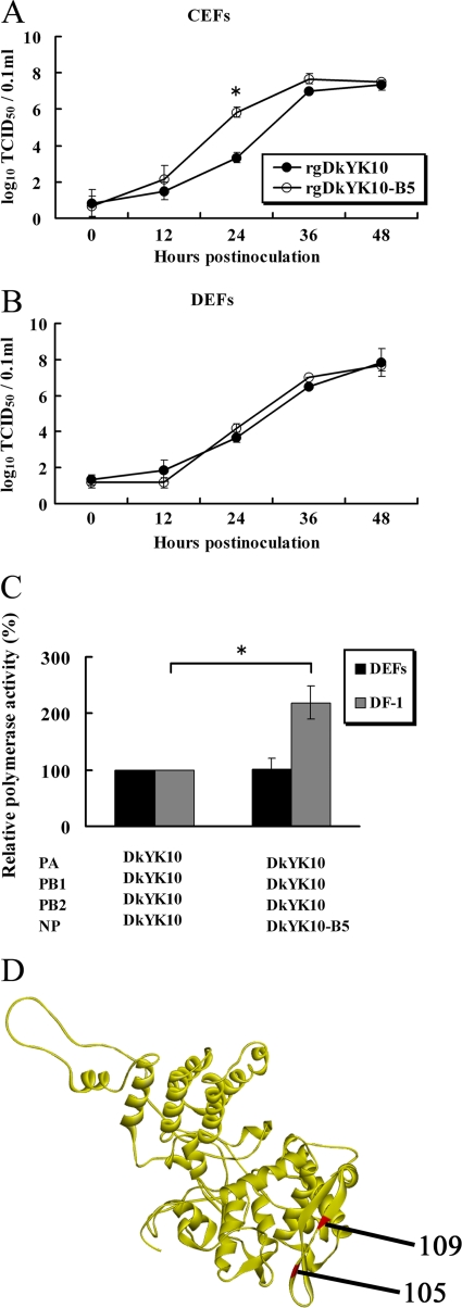
The whole-genome sequences of DkYK10 and two brain-passaged viruses were compared; we found that DkYK10-B5 had a single amino acid substitution at position 109 in NP (from isoleucine to threonine, NP I109T), while DkYK10-B10 had an additional mutation at position 188 in NA (from threonine to isoleucine). The NP 109T amino acid was located in the body domain (see Fig. 4D). This result, together with the virological and pathological data described above, suggested that the NP I109T mutation might be associated with the high pathogenicity of the two brain-passaged viruses.
Fig. 4.
Comparison of the viral growth kinetics of DkYK10 and DkYK1-B5 in CEFs and DEFs and their polymerase activities in DF-1 cells and DEFs. (A and B) CEFs (A) and DEFs (B) were inoculated with gDkYK10 and rgDkYK10-B5 at an MOI of 0.001, and virus titers of the culture supernatants at each time point were determined with CEFs and expressed as an average of three wells per time point. (C) Polymerase activity in DF-1 cells and DEFs was assayed by cotransfection with pHH21-lucR reporter plasmid and pHW2000 plasmids containing the PB2, PB1, PA, and NP genes. Cells cotransfected with pHH21-luc and pHW2000 were used as controls. At 24 h after transfection, luciferase activity in the cell lysates was determined, and the mean counts per second (CPS) ± standard deviations of triplicate experiments were determined. The relative polymerase activity versus parental DkYK10 is shown. (D) The amino acids at position 105 identified previously (45) and at position 109 detected in this study are shown in the crystal structure of NP. *, significant difference (P < 0.05).
High pathogenicity of brain passage virus in chickens.
To determine the effect of the NP mutation on the pathogenicity of DkYK10 in chickens, we generated two reassortant viruses using reverse genetics (rgDkYK10 and rgDkYK10-B5) and inoculated them intranasally into two groups of eight chickens each. The chicken pathogenicity test showed that the MDT of chickens infected with rgDkYK10-B5 was 71.1 ± 9.1 h p.i., which was significantly shorter than that of chickens infected with rgDKYK10 (84.0 ± 13.4 h p.i.) (P < 0.05) (Fig. 1B). In addition, fevers were slightly lower in chickens inoculated with rgDkYK10-B5 (2.0 ± 0.7°C) than in chickens inoculated with rgDkYK10 (2.6 ± 1.3°C) because of early death.
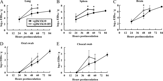
The replication kinetics of rgDkYK10-B5 in chickens' lungs, spleens, and brains were compared with those of rgDkYK10. Three birds were euthanized at each sampling point, and the average virus titers of tissues were determined (see Fig. 3). It was demonstrated that rgDkYK10-B5 replicated more quickly than rgDkYK10 in the chickens' lungs, spleens, and brains. At 48 h p.i., virus titers in the lungs (Fig. 2 A), spleens (Fig. 2B), and brains (Fig. 2C) of animals inoculated with rgDkYK10-B5 were approximately 100 times higher than those in animals inoculated with rgDkYK10. Significant differences in virus titers between two strains are shown in Fig. 2 (P < 0.05).
Fig. 3.
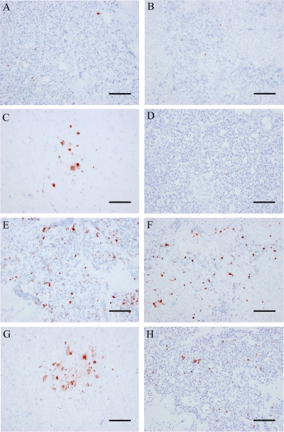
Immunohistochemical detection of AIV matrix protein and TUNEL staining of apoptotic cells in tissues from chickens infected with rgDkYK10 or rgDkYK10-B5. AIV M protein was detected in the lungs (A and E), spleens (B and F), and brains (C and G) from chickens intranasally inoculated with rgDkYK10 (A to C) or rgDkYK10-B5 (E to G) at 48 h p.i. The apoptotic cells were detected in the lung tissue of chickens infected with rgDkYK10 (D) or rgDkYK10-B5 (H) at 48 h p.i. Scale bars = 50 μm.
Fig. 2.
Kinetics of viral replication of rgDkYK10 and rgDkYK10-B5 in tissues from chickens and virus shedding in oral and cloacal swabs. Chickens were inoculated intranasally with 106 EID50 of rgDkYK10 or rgDkYK10-B5 (n = 3 for each inoculation group). Lungs (A), spleens (B), and brains (C), as well as oral (D) and cloacal (E) swabs, were collected sequentially at 24, 48, 60, and 84 h p.i., and their virus titers were determined with embryonated chicken eggs. The mean virus titers ± standard deviations from three birds at each point are shown (log10 EID50/g). *, significant difference (P < 0.05).
Virus titers in oral and cloacal swabs were higher in chickens infected with rgDkYK10-B5 than in those infected with rgDkYK10 around 48 to 60 h p.i. (Fig. 2D and E), although the final virus titers of rgDkYK10 in these swabs might be higher. Virus titers from oral swabs of chickens infected with rgDkYK10 or rgDkYK10-B5 at 60 h p.i. were 101.7 EID50/g and 102.5 EID50/g, respectively, while those from cloacal swabs at 48 h p.i. were 101.3 EID50/g and 102.5 EID50/g, respectively.
Detection of virus antigen in tissues.
The sequential distribution and intensities of AIV M antigens in the lungs, spleens, and brains of chickens infected with rgDkYK10 or rgDkYK10-B5 were compared immunohistochemically (Table 2). In the rgDkYK10 group, M antigen was detected at low levels in lungs and spleens from 48 to 84 h p.i. (Fig. 3 A to C) but was abundant in brains from 60 to 84 h p.i. (Table 2). In comparison, higher levels of M antigen were detected in the lungs, spleens, and brains of chickens infected with rgDkYK10-B5 at 48 and 60 h p.i. (Fig. 3E to G and Table 2).
Table 2.
Distribution of viral M antigen and apoptotic cells in tissues from 4-week-old SPF chickens inoculated intranasally with 106.0EID50 of rgDkYK10 or rgDkYK10-B5a
| Strain | Tissue | Detection of AIV matrix antigenb at (h p.i.) |
Tissue | Detection of apoptotic cellsc at (h p.i.) |
||||||
|---|---|---|---|---|---|---|---|---|---|---|
| 24 | 48 | 60 | 84 | 24 | 48 | 60 | 84 | |||
| rgDkYK10 | Lung | −, −, − | +, +, − | +, +, − | +, +, + | Lung | +, +, − | +, +, + | ++, +, + | +, ++, ++ |
| Spleen | −, −, − | +, +, − | +, −, + | +, +, − | Liver | +, +, + | +, +, + | +, +, + | ++, ++, +++ | |
| Brain | −, −, − | +, +, − | ++, +, ++ | +++, +++, +++ | ||||||
| rgDkYK10-B5 | Lung | −, −, − | +, +++, ++ | ++, ++, +++ | Lung | +, +, + | ++, ++, + | +++, +++, +++ | ||
| Spleen | −, −, − | +, ++, ++ | +, +, + | Liver | +, +, + | ++, +++, +++ | ++, ++, ++ | |||
| Brain | −, −, − | ++, ++, + | ++, ++, ++ | |||||||
Three chickens per group were killed at each sampling point (24, 48, 72, and 84 h p.i.). The lungs, spleens, and brains were examined for the virus replication kinetic analysis, and the lungs and livers were tested for the detection of apoptotic cells.
AIV matrix antigen was detected by immunohistochemistry. Grading by level of AI virus matrix protein or detected: −, none; +, mild; ++, moderate; +++, severe.
Apoptotic cells were detected with the ApopTag peroxidase in situ apoptosis detection kit (Chemicon International, Inc.) according to the manufacturer's instructions. Grading by level of apoptotic cells detected: −, none; +, mild; ++, moderate; +++, severe.
As shown in Fig. 3, M antigen was expressed in mononuclear cells from the lungs and spleens of chickens infected with rgDkYK10 or rgDkYK10-B5. In spleens, M antigen was detected in both the white and red pulp (Fig. 3B and F). In brains, M antigen was detected as small aggregations in both groups at 48 h p.i. and spread throughout the brain from 48 h p.i. onward (Table 3 and Fig. 3C and G).
Table 3.
SNP analysis at position 109 in NP after passaging four HPAIVs in chicken cells and organs
| Virus | Cells or tissue | Estimated titer of virus with NP 109T mutation at passage no.d: |
||
|---|---|---|---|---|
| 1 | 2 | 3 | ||
| DkYK10 | Chicken braina | >108 | >108 | Not tested |
| Kidney cellsb | ||||
| Embryoc lung | >107 | >106 | >107 | |
| Embryo brain | >108 | >108 | >107 | |
| CkYM7 | Embryo lung | >106 | ||
| Embryo brain | >105 | >106 | >107 | |
| CkMZ11 | Embryo lung | |||
| Embryo brain | >107 | >107 | >107 | |
| WsAK1 | Embryo lung | |||
| Embryo brain | >106 | >106 | ||
Ten percent lung homogenate (L1) was prepared from a dead chicken after inoculation with DkYK10 and intranasally inoculated into chickens. The brain (B1) was harvested from the first dying chicken, the supernatant of the 10% brain homogenate was inoculated intranasally into another 5 chickens, and B2 tissue was obtained.
The allantoic fluid infected with DkYK10 was inoculated onto primary chicken kidney cells at an MOI of 0.01 and incubated for 2 days at 37°C. The culture fluid was passaged another two times in the cells.
Seventeen-day-old embryonated chicken eggs were inoculated with HPAIV (106 EID50) in the allantoic cavity, and lung or brain tissues were harvested after 48 h of cultivation. The 10% lung or brain homogenates were then passaged into the allantoic cavities of the embryonated eggs to obtain the respective lung or brain passage viruses.
Virus RNA was purified from tissue samples and tested for the emergence of the NP-109T mutation using real-time PCR based on SNP analysis. The average SNP value (45 − CT) of 3 wells was determined. The 106 EID50 of DkYK10 was mixed with 103, 104, 105, or 106 EID50 of DkYK10-B5, and the SNP values of these mixtures were used as a standard to estimate titers of virus (EID50/g tissue) with the NP 109T mutation.
Detection of apoptotic cells.
To determine whether the efficient replication of rgDKYK10-B5 causes severe apoptosis in tissues, apoptotic cells were detected with the ApopTag peroxidase in situ apoptosis detection kit (terminal deoxynucleotidyltransferase-mediated dUTP-biotin nick end labeling [TUNEL] method). The analysis indicated that apoptotic cells in lungs and livers were detected quickly and frequently in chickens infected with rgDkYK10-B5 from 48 h p.i., while those induced by rgDkYK10 infection were sparse at 48 and 60 h p.i. and became frequent at 84 h p.i. (Table 2 and Fig. 3D and H). The majority of apoptotic cells may be mononuclear cells in the lungs and spleen and Kupffer cells in livers in both strains. Negligible numbers of apoptotic cells were detected in the lungs and livers of uninfected chickens.
Replication of virus in CEFs and DEFs.
We also determined whether the high virus replication in chickens was associated with high replication in culture cells. CEFs or DEFs were inoculated with DkYK10 or rgDkYK10-B5 at an MOI of 0.001, and the culture supernatants were sequentially harvested and titrated to compare growth kinetics. In CEFs, the titers of rgDkYK10-B5 at 24 h p.i. were significantly higher than those of rgDkYK10 (P < 0.05) (Fig. 4 A), although the final titers of both viruses reached the same level at 36 h p.i. To determine whether the NP I109T substitution was related to adaptation of the virus from ducks to chickens, the viral growth kinetics of rgDkYK10-B5 were compared with those of rgDkYK10 in DEFs. There was no difference in viral growth kinetics between the two viruses in DEFs (Fig. 4B).
Comparison of polymerase activities.
We investigated whether the NP I109T substitution enhanced the polymerase activity of DkYK10 in DF-1 cells by using a minigenome system with the luciferase reporter gene. The polymerase activities of rgDkYK10 and rgDkYK10-B5 were 4,367 ± 235 and 13,581 ± 362, respectively (Fig. 4C), indicating that the single amino acid substitution significantly enhanced the polymerase activity of DkYK10 in DF-1 cells, but not in DEFs (P < 0.05).
Comparison of transcription and replication activities.
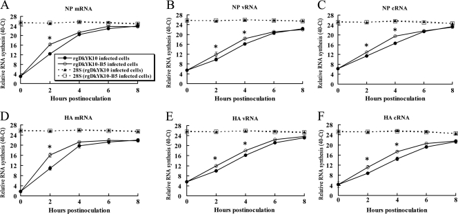
These findings indicated that the NP I109T substitution increased the polymerase activity of DkYK10 in CEFs. We further explored the mechanisms underlying enhanced RNA synthesis resulting from the NP mutation in CEFs. Real-time RT-PCR was performed to compare the kinetics of mRNA, vRNA, and cRNA synthesis of the NP gene in CEFs infected with rgDkYK10-B5 or rgDkYK10. In CEFs infected with rgDkYK10-B5, mRNA synthesis increased quickly, and the copy number at 2 h p.i. was 14.7 times greater than in CEFs infected with rgDkYK10 (Fig. 5A). The vRNA and cRNA syntheses in CEFs infected with rgDkYK10-B5 increased slowly at 2 to 4 h p.i.; the respective vRNA copy numbers at 2 h p.i. and 4 h p.i. were 4.1 and 4.8 times greater, and the respective cRNA copy numbers at 2 h p.i. and 4 h p.i. were 3.0 and 7.5 times greater, than those with rgDkYK10 (Fig. 5B and C). The H5 mRNA of rgDkYK10-B5 was also quickly synthesized in CEFs (Fig. 5D), while the cRNA and vRNA of rgDkYK10-B5 increased slowly (Fig. 5E and F). There was no difference in 28S RNA synthesis at any point between the rgDkYK10 and rgDkYK10-B5 groups. Significant differences (P < 0.05) in RNA synthesis between the two strains are shown in Fig. 5. These results indicate that the NP 109T mutation enhances mRNA synthesis quickly in CEFs, and cRNA and vRNA synthesis increased thereafter.
Fig. 5.
Kinetics of vRNA, cRNA, and mRNA synthesis of NP and H5 genes in CEFs inoculated with rgDkYK10 and rgDkYK10-B5. CEFs were inoculated at an MOI of 10 with rgDkYK10 or rgDkYK10-B5, and total cellular RNA was harvested at 0, 2, 4, 6, and 8 h p.i. Three micrograms of total RNA per sample was reverse transcribed into cDNA using Primescript reverse transcriptase (Takara) and specific primers for mRNA (A and D), vRNA (B and E), or cRNA (C and F) of the NP (A to C) and H5 (D to F) genes. The amounts of cDNA were used to determine the RNA amounts by real-time PCR for the NP and H5 genes (49). The average threshold cycle value (40 − CT) of triple wells was determined, and the relative viral RNA concentration (40 − CT) at each point is shown after the values at 0 h p.i. of the virus strains were adjusted. *, significant difference (P < 0.05).
Molecular epidemiological analysis of the amino acid at position 109 in NP.
To determine the epidemiological involvement of the NP I109T substitution in the field, 1,597 NPs of H5N1 HPAIVs registered in GenBank were surveyed. Only 5 NPs contained threonine (DkYK10-B5 type) at position 109 in NP, while 1,586 NPs contained isoleucine (DkYK10 type) and 6 NPs contained valine. The 5 NPs of DkYK10-B5 type were A/chicken/Nigeria/08RS848-59/2007(H5N1), A/chicken/Viet Nam/10/2005(H5N1), A/mallard/Manitoba/458/2005(H5N1), A/quail/Shantou/3846/2002(H5N1), and A/goose/Hong Kong/437-6/1999(H5N1). This analysis suggests that the participation of the NP I109T substitution in increased pathogenicity of H5N1 viruses in chickens may be low in the field.
Chicken embryo passages and SNP analysis.
To determine when the NP I109T substitution emerges during the passaging of DkYK10 in chicken brains, we compared the sequences of the NP genes of all brain passage samples. The NP I109T substitution was detected from the second brain passage sample onward. Although the NP I109T substitution was quickly induced by brain passaging, molecular epidemiological analysis suggests that the emergence frequency in the field may be limited. Next, we evaluated the significance of the NP I109T substitution in experimentally passaged samples using SNP analysis.
Before this experiment, we established SNP analysis for the detection of the NP 109T gene in samples. SNP analysis could quickly detect the NP 109T molecule in the first brain-passaged sample. SNP analysis also quantitatively detected the NP 109T molecule, even in the mixture of DkYK10 (106) and DkYK10-B5 (104) (Table 3), while the conventional plasmid cloning and sequencing method detected the NP 109T molecule only in the mixture of DkYK10 (106) and DkYK10-B5 (106).
DkYK10 was passaged five times in primary CEFs or CK cells, and the samples were tested for the presence of the NP 109T molecule by SNP analysis. However, the NP 109T molecule was not detected. Next, DkYK10 was inoculated into the allantoic cavities of 17-day-old embryos and cultivated for 48 h to obtain lung- or brain-passaged samples. Supernatants of the passaged tissue homogenates were inoculated into the cavities for tissue passaging. SNP analysis of the samples demonstrated that passage of HPAIVs in embryo brains is a suitable experimental system in which to evaluate NP 109T emergence.
Next, we determined whether the NP I109T substitution occurred in three other H5N1 HPAIVs by passaging them in embryonic lungs or brains. CkYM7, CkMZ11, and WsAK1 were individually passaged three times in chicken embryo lungs or brains and then tested for the appearance of the NP 109T molecule by SNP. The NP I109T substitution occurred in all three HPAIVs after brain passaging; the frequency of the substitution varied depending on the viral strain, but the substitution almost never occurred after lung passages.
Molecular epidemiological analysis of NP.
There was more than 97.8% amino acid homology among the four NPs of H5N1 HPAIVs, all of which could acquire the NP I109T substitution during brain passages. Next, we searched the NCBI Influenza Virus Resource to discern the population of NPs with over 98% amino acid identity to any of the four H5N1 HPAIVs. Sixty-four percent and 85% of 1,464 NP proteins derived from Asian AIVs had greater than 99% and 98% homology, respectively, to any of the four H5N1 HPAIVs. This analysis suggests that many NPs may acquire the NP I109T substitution during HPAIV replication in the brains of chickens.
DISCUSSION
The molecular basis underlying the high pathogenicity of HPAIVs in chickens remains largely unknown. The well-known pathogenicity enhancement of AIVs during passaging in chickens is caused by addition of multiple basic amino acids at the HA cleavage site (14, 20, 38). Recently, NP, PB1, and PB2 (53); NP and PB2 (45); and NS1 (19) have been shown to contribute to the pathogenicity of H5N1 HPAIVs in chickens, while PB1 plus PA (13) and PA (39) are associated with high pathogenicity in ducks. We further elucidated the molecular basis for HPAIV pathogenesis in chickens. The NP I109T substitution enhanced the replication of DkYK10 in chickens and CEFs, as well as polymerase activity in CEFs. A clear difference in pathogenicity in chickens between DkYK10 and DkYK10-B5 was replication speed in tissues and cells, although final virus titers were slightly higher. Quick replication of virus in tissue led to early spread of the virus to multiple organs, which quickly resulted in multiple organ failure and death in chickens. Our previous studies showed that the replication speed of virus in tissues and cells is correlated with pathogenicity in chickens (42, 43, 45). These results indicate that the increased pathogenicity of DkYK10-B5 is intrinsically related to the enhancement of polymerase activity in cells by the NP I109T substitution. Previous studies suggested that the brain is an important tissue for HPAIVs to acquire pathogenicity in chickens (14, 37, 38). Interestingly, this study directly indicated that the NP I109T substitution appears during replication of DkYK10 in chicken brains but does not appear or appears inefficiently in lungs and primary cells (CK cells and CEFs). In addition, the NP I109T substitution was generated in 4 different NP genes under the four different gene constellations. These results suggest that the increased pathogenicity of many HPAIVs may be caused by the NP I109T mutation during passages in the brains of chickens.
The high staining of M antigen was observed in the brains of chickens inoculated with the original DkYK10, but not in lungs, spleens, and kidneys. The SNP analysis showed that the NP 109T mutant was detected in the brains of the chickens (>106 EID50/g tissue), but not in other tissues (data not shown). These results suggest that the large amounts of M antigen in the brains of chickens infected with the parental DkYK10 may be due to high replication of the NP mutant. It is hypothesized that high mutation rates of RNA viruses create a cloud of mutations, which afford a great probability of evolving and adapting to new hosts or tissues, and selection of viruses in new hosts or tissues acts on clouds of mutants rather than individual mutants (the quasispecies theory) (51). According to the theory, it is considered that a virus stock may contain multiple mutations at the population level and that a minor population can be switched to a major population in some new tissues. In our case, it is possible that DkYK10 may contain a cloud of mutants, NP 109I and NP 109T, and the minor population (NP 109T) in the stock became major in brains while NP 109I or other populations may have become major in other tissues. Many clouds of mutants may be present in virus samples, but these mutations cannot be detected by the conventional cloning-sequencing method. In contrast, the SNP assay is more sensitive for identification of minor mutations and is useful to study the evolution and adaptation of AIVs to new hosts or tissues.
Surprisingly, the NP I109T mutation appeared in different NP genes under four different genetic backgrounds of HPAIVs. However, the frequency of appearance of the NP mutation varied among 4 HPAIVs, and the mutation appeared quickly in DkYK10, while it appeared slowly in 3 other viruses. The NP gene itself or the gene constellation of DkYK10 may influence the efficient replication of the NP 109T mutant virus in brains. Also, the NP I109T mutation was readily observed in the lungs of embryos infected with DkYK10 compared with 3 other H5N1 viruses. It is likely that the DkYK10 mutant replicated efficiently in brains and might be transmitted from brains to lungs. Although the NP 109T mutation was related to high replication in chicken tissues, the frequency of the NP mutation in the GenBank database was low. It seems likely that the NP mutant virus remains sequestered in the brain and is not transmitted to other chickens. Even so, that does not negate the possibility that the NP mutant virus will become prevalent in chicken populations for the following reasons. (i) The mutation emerged frequently and quickly in chicken brains. (ii) Some mutant viruses may transfer from brains to lungs or mucosal surfaces soon after emergence, because the mutant virus was detected in lungs at the first passage of DkYK10 in embryos. (iii) The mutant virus, DkYK10-B5, can replicate more quickly on mucosal surfaces than the parent virus. (iv) Virus mounts in swabs are correlated with transmissibility in chickens (43). Therefore, rather than relying solely on the data for the first decade, a long-term survey may be required to determine the epidemiological significance of the NP mutation.
NP is a polypeptide of 498 amino acids with a crescent-like structure; it can be divided into two domains, a head and a body (56). The head domain is formed by residues 150 to 272 and 438 to 452, and the body domain consists of polypeptide segments 21 to 149, 273 to 396, and 453 to 489 (56). The molecular mechanism of the NP I109T substitution underlying the increasing replication of AIVs in chicken cells is unknown. A crystal structure model of the NP protein was obtained from the SWISS-MODEL Workspace (http://swissmodel.expasy.org/workspace/) (36) and analyzed by using the MATRAS v.1.2 program (15). Although the I109T replacement did not cause any change in NP structure or static potential in this model (data not shown), it is possible that the replacement may change the conformation of NP, which may influence the interaction with viral polymerase subunits (PB1 and PB2) or cellular transcription factors, such as importin-α isoforms. Interaction of NP with PB2 regulates viral replication in mammalian cells (4, 17, 30), and interaction of NP with importin-α enhances nuclear transport of RNP and viral replication (7, 30). Interestingly, the NP I109T mutation increased the polymerase activity of DkYK10 in CEFs, but not in DEFs, and emergence of NP 109T may require replication in chicken brains, but not in chicken lungs. These results suggest that cellular proteins expressed in brain cells may be related to the emergence of the NP mutant virus. We previously identified another substitution (M105V) in the NP body domain that may also be related to adaptation from ducks to chickens (45). Thus, the NP body domain may play a critical role in the adaptation of AIVs from ducks to chickens. However, molecular mechanisms for enhancing polymerase activity differ between the two NP amino acids, 105V and 109T. NP 105V requires additional genes, such as M or NS genes (45), while NP 109T does not. To understand the molecular mechanisms underlying the increased pathogenicity of HPAIVs in chickens or adaptation of AIVs to the new host, the roles of the NP body domain in functional interactions with cellular or viral proteins remain to be determined.
The mechanisms regulating the synthesis of mRNA, cRNA, and vRNA of influenza A virus are not fully understood, and the roles of NP in viral RNA synthesis are in dispute. It is currently hypothesized that an interaction of NP with polymerase subunits or viral RNAs may regulate the switch from transcription (capped RNA-primed initiation) to replication (unprimed initiation and prevention of premature termination) (2, 27). Recently, Vreede et al. proposed a stabilization model for the role of NP in influenza virus RNA synthesis (52). Influenza virus synthesizes both mRNA and cRNA at an early stage after infection, and mRNA is protected from cellular degradation by the presence of a 5′ cap and a 3′ poly(A) tail, while cRNA may be rapidly degraded. At an early stage of infection, small amounts of NP protein may allow cRNA degradation and may delay vRNA synthesis. Our real-time RT-PCR analysis demonstrated that mRNAs of NP and H5 genes were synthesized quickly and efficiently throughout the experimental period in both viruses, while their cRNAs and vRNAs were synthesized slowly and constantly without further increase from 4 to 8 h p.i. This study suggests that NP 109T may at least enhance the transcription of AIVs. A previous study demonstrated that mRNA transcription can occur in the absence of NP and that NP is required for antitermination [readthrough at the mRNA poly(A) site], which is an essential step for replication (2). Although vRNA synthesis is considered to be 10 to 20 times higher than cRNA synthesis (9), real-time RT-PCR analysis revealed that the molecular ratio of vRNA to cRNA is almost equal in the NP and H5 genes. The roles of NP in AIV transcription and replication remain largely unknown.
ACKNOWLEDGMENTS
We thank Masaru Kobayashi and Makiko Shishido for excellent technical assistance.
This work was supported by a grant from the Core Research for Evolutional Science and Technology (CREST) program of the Japan Science and Technology Agency.
Footnotes
Published ahead of print on 27 July 2011.
REFERENCES
- 1. Aytay S., Schulze I. T. 1991. Single amino acid substitutions in the hemagglutinin can alter the host range and receptor binding properties of H1 strains of influenza A virus. J. Virol. 65:3022–3028 [DOI] [PMC free article] [PubMed] [Google Scholar]
- 2. Beaton A. R., Krug R. M. 1986. Transcription antitermination during influenza viral template RNA synthesis requires the nucleocapsid protein and the absence of a 5′ capped end. Proc. Natl. Acad. Sci. U. S. A. 83:6282–6286 [DOI] [PMC free article] [PubMed] [Google Scholar]
- 3. Bosch F. X., Orlich M., Klenk H. D., Rott R. 1979. The structure of the hemagglutinin, a determinant for the pathogenicity of influenza viruses. Virology 95:197–207 [DOI] [PubMed] [Google Scholar]
- 4. Boulo S., Akarsu H., Ruigrok R. W., Baudin F. 2007. Nuclear traffic of influenza virus proteins and ribonucleoprotein complexes. Virus Res. 124:12–21 [DOI] [PubMed] [Google Scholar]
- 5. Chen H., et al. 2004. The evolution of H5N1 influenza viruses in ducks in southern China. Proc. Natl. Acad. Sci. U. S. A. 101:10452–10457 [DOI] [PMC free article] [PubMed] [Google Scholar]
- 6. de Jong J. C., et al. 2005. The influenza season 2004/05 in the Netherlands with the largest epidemic of the last 5 years caused by the virus variant A/California and the composition of the vaccine for the season 2005/06. Ned. Tijdschr. Geneeskd. 149:2355–2361 [PubMed] [Google Scholar]
- 7. Gabriel G., Herwig A., Klenk H. D. 2008. Interaction of polymerase subunit PB2 and NP with importin alpha1 is a determinant of host range of influenza A virus. PLoS Pathog. 4:e11. [DOI] [PMC free article] [PubMed] [Google Scholar]
- 8. Govorkova E. A., et al. 2005. Lethality to ferrets of H5N1 influenza viruses isolated from humans and poultry in 2004. J. Virol. 79:2191–2198 [DOI] [PMC free article] [PubMed] [Google Scholar]
- 9. Hatada E., Hasegawa M., Mukaigawa J., Shimizu K., Fukuda R. 1989. Control of influenza virus gene expression: quantitative analysis of each viral RNA species in infected cells. J. Biochem. 105:537–546 [DOI] [PubMed] [Google Scholar]
- 10. Hatta M., Gao P., Halfmann P., Kawaoka Y. 2001. Molecular basis for high virulence of Hong Kong H5N1 influenza A viruses. Science 293:1840–1842 [DOI] [PubMed] [Google Scholar]
- 11. Horimoto T., Kawaoka Y. 1994. Reverse genetics provides direct evidence for a correlation of hemagglutinin cleavability and virulence of an avian influenza A virus. J. Virol. 68:3120–3128 [DOI] [PMC free article] [PubMed] [Google Scholar]
- 12. Hulse D. J., Webster R. G., Russell R. J., Perez D. R. 2004. Molecular determinants within the surface proteins involved in the pathogenicity of H5N1 influenza viruses in chickens. J. Virol. 78:9954–9964 [DOI] [PMC free article] [PubMed] [Google Scholar]
- 13. Hulse-Post D. J., et al. 2007. Molecular changes in the polymerase genes (PA and PB1) associated with high pathogenicity of H5N1 influenza virus in mallard ducks. J. Virol. 81:8515–8524 [DOI] [PMC free article] [PubMed] [Google Scholar]
- 14. Ito T., et al. 2001. Generation of a highly pathogenic avian influenza A virus from an avirulent field isolate by passaging in chickens. J. Virol. 75:4439–4443 [DOI] [PMC free article] [PubMed] [Google Scholar]
- 15. Kawabata T. 2003. MATRAS: a program for protein 3D structure comparison. Nucleic Acids Res. 31:3367–3369 [DOI] [PMC free article] [PubMed] [Google Scholar]
- 16. Kawaoka Y., Krauss S., Webster R. G. 1989. Avian-to-human transmission of the PB1 gene of influenza A viruses in the 1957 and 1968 pandemics. J. Virol. 63:4603–4608 [DOI] [PMC free article] [PubMed] [Google Scholar]
- 17. Labadie K., Dos Santos Afonso E., Rameix-Welti M. A., van der Werf S., Naffakh N. 2007. Host-range determinants on the PB2 protein of influenza A viruses control the interaction between the viral polymerase and nucleoprotein in human cells. Virology 362:271–282 [DOI] [PubMed] [Google Scholar]
- 18. Li Z., et al. 2005. Molecular basis of replication of duck H5N1 influenza viruses in a mammalian mouse model. J. Virol. 79:12058–12064 [DOI] [PMC free article] [PubMed] [Google Scholar]
- 19. Li Z., et al. 2006. The NS1 gene contributes to the virulence of H5N1 avian influenza viruses. J. Virol. 80:11115–11123 [DOI] [PMC free article] [PubMed] [Google Scholar]
- 20. Löndt B. Z., Banks J., Gardner R., Cox W. J., Brown I. H. 2007. Induced increase in virulence of low virulence highly pathogenic avian influenza by serial intracerebral passage in chickens. Avian Dis. 51:396–400 [DOI] [PubMed] [Google Scholar]
- 21. Maines T. R., et al. 2005. Avian influenza (H5N1) viruses isolated from humans in Asia in 2004 exhibit increased virulence in mammals. J. Virol. 79:11788–11800 [DOI] [PMC free article] [PubMed] [Google Scholar]
- 22. Mase M., et al. 2005. Isolation of a genotypically unique H5N1 influenza virus from duck meat imported into Japan from China. Virology 339:101–109 [DOI] [PubMed] [Google Scholar]
- 23. Mase M., et al. 2006. Recent H5N1 avian influenza A virus increases rapidly in virulence to mice after a single passage in mice. J. Gen. Virol. 87:3655–3659 [DOI] [PubMed] [Google Scholar]
- 24. Mase M., et al. 2005. Characterization of H5N1 influenza A viruses isolated during the 2003-2004 influenza outbreaks in Japan. Virology 332:167–176 [DOI] [PubMed] [Google Scholar]
- 25. Munster V. J., et al. 2007. The molecular basis of the pathogenicity of the Dutch highly pathogenic human influenza A H7N7 viruses. J. Infect. Dis. 196:258–265 [DOI] [PubMed] [Google Scholar]
- 26. Naffakh N., Massin P., Escriou N., Crescenzo-Chaigne B., van der Werf S. 2000. Genetic analysis of the compatibility between polymerase proteins from human and avian strains of influenza A viruses. J. Gen. Virol. 81:1283–1291 [DOI] [PubMed] [Google Scholar]
- 27. Palese P., Shaw M. L. 2007. Orthomyxoviridae: the viruses and their replication, p. 1647–1689 In Knipe D. M., Howley P. M. (ed.), Fields virology, 5th ed., vol. II. Lippincott Williams & Wilkins, Philadelphia, PA [Google Scholar]
- 28. Pappas C., et al. 2008. Single gene reassortants identify a critical role for PB1, HA, and NA in the high virulence of the 1918 pandemic influenza virus. Proc. Natl. Acad. Sci. U. S. A. 105:3064–3069 [DOI] [PMC free article] [PubMed] [Google Scholar]
- 29. Perez D. R., et al. 2003. Role of quail in the interspecies transmission of H9 influenza A viruses: molecular changes on HA that correspond to adaptation from ducks to chickens. J. Virol. 77:3148–3156 [DOI] [PMC free article] [PubMed] [Google Scholar]
- 30. Portela A., Digard P. 2002. The influenza virus nucleoprotein: a multifunctional RNA-binding protein pivotal to virus replication. J. Gen. Virol. 83:723–734 [DOI] [PubMed] [Google Scholar]
- 31. Reed L., Muench H. 1938. A simple method of estimating fifty percent endpoints. Am. J. Hyg. 27:493–497 [Google Scholar]
- 32. Rimmelzwaan G. F., et al. 2001. Pathogenesis of influenza A (H5N1) virus infection in a primate model. J. Virol. 75:6687–6691 [DOI] [PMC free article] [PubMed] [Google Scholar]
- 33. Rolling T., et al. 2009. Adaptive mutations resulting in enhanced polymerase activity contribute to high virulence of influenza A virus in mice. J. Virol. 83:6673–6680 [DOI] [PMC free article] [PubMed] [Google Scholar]
- 34. Romanova J., et al. 2003. Distinct host range of influenza H3N2 virus isolates in Vero and MDCK cells is determined by cell specific glycosylation pattern. Virology 307:90–97 [DOI] [PubMed] [Google Scholar]
- 35. Salomon R., et al. 2006. The polymerase complex genes contribute to the high virulence of the human H5N1 influenza virus isolate A/Vietnam/1203/04. J. Exp. Med. 203:689–697 [DOI] [PMC free article] [PubMed] [Google Scholar]
- 36. Schwede T., Kopp J., Guex N., Peitsch M. C. 2003. SWISS-MODEL: an automated protein homology-modeling server. Nucleic Acids Res. 31:3381–3385 [DOI] [PMC free article] [PubMed] [Google Scholar]
- 37. Shinya K., et al. 2009. Ostrich involvement in the selection of H5N1 influenza virus possessing mammalian-type amino acids in the PB2 protein. J. Virol. 83:13015–13018 [DOI] [PMC free article] [PubMed] [Google Scholar]
- 38. Silvano F. D., Yoshikawa M., Shimada A., Otsuki K., Umemura T. 1997. Enhanced neuropathogenicity of avian influenza A virus by passages through air sac and brain of chicks. J. Vet. Med. Sci. 59:143–148 [DOI] [PubMed] [Google Scholar]
- 39. Song J., et al. 2011. The PA protein directly contributes to the virulence of H5N1 avian influenza viruses in domestic ducks. J. Virol. 85:2180–2188 [DOI] [PMC free article] [PubMed] [Google Scholar]
- 40. Subbarao E. K., London W., Murphy B. R. 1993. A single amino acid in the PB2 gene of influenza A virus is a determinant of host range. J. Virol. 67:1761–1764 [DOI] [PMC free article] [PubMed] [Google Scholar]
- 41. Subbarao K., et al. 1998. Characterization of an avian influenza A (H5N1) virus isolated from a child with a fatal respiratory illness. Science 279:393–396 [DOI] [PubMed] [Google Scholar]
- 42. Suzuki K., et al. 2009. Association of increased pathogenicity of Asian H5N1 highly pathogenic avian influenza viruses in chickens with highly efficient viral replication accompanied by early destruction of innate immune responses. J. Virol. 83:7475–7486 [DOI] [PMC free article] [PubMed] [Google Scholar]
- 43. Suzuki K., Okada H., Itoh T., Tada T., Tsukamoto K. 2010. Phenotypes influencing the transmissibility of highly pathogenic avian influenza viruses in chickens. J. Gen. Virol. 91:2302–2306 [DOI] [PubMed] [Google Scholar]
- 44. Swayne D. E., Halvorso D. A. 2003. Influenza, p. 135–160 In Saif Y. M., Barnes J. H., Glisson J. R., Fadly A. M., McGougald L. R., Swayne D. E. (ed.), Diseases of poultry, 11th ed. Iowa State Press, Ames, IA [Google Scholar]
- 45. Tada T., et al. 2011. NP and PB2 contribute to increased virulence of H5N1 highly pathogenic avian influenza viruses in chickens. J. Virol. 85:1834–1846 [DOI] [PMC free article] [PubMed] [Google Scholar]
- 46. Tanimura N., et al. 2006. Pathology of fatal highly pathogenic H5N1 avian influenza virus infection in large-billed crows (Corvus macrorhynchos) during the 2004 outbreak in Japan. Vet. Pathol. 43:500–509 [DOI] [PubMed] [Google Scholar]
- 47. Tran T. H., et al. 2004. Avian influenza A (H5N1) in 10 patients in Vietnam. N. Engl. J. Med. 350:1179–1188 [DOI] [PubMed] [Google Scholar]
- 48. Tsukamoto K., et al. 2008. Subtyping of avian influenza viruses H1 to H15 on the basis of hemagglutinin genes by PCR assay and molecular determination of pathogenic potential. J. Clin. Microbiol. 46:3048–3055 [DOI] [PMC free article] [PubMed] [Google Scholar]
- 49. Tsukamoto K., et al. 2010. Broad detection of diverse H5 and H7 hemagglutinin genes of avian influenza viruses by real-time RT-PCR using primer/probe sets containing mixed bases. J. Clin. Microbiol. 48:4275–4278 [DOI] [PMC free article] [PubMed] [Google Scholar]
- 50. Uchida Y., et al. 2008. Highly pathogenic avian influenza virus (H5N1) isolated from whooper swans, Japan. Emerg. Infect. Dis. 14:1427–1429 [DOI] [PMC free article] [PubMed] [Google Scholar]
- 51. Vignuzzi M., Stone J. K., Arnold J. J., Cameron C. E., Andino R. 2006. Quasispecies diversity determines pathogenesis through cooperative interactions in a viral population. Nature 439:344–348 [DOI] [PMC free article] [PubMed] [Google Scholar]
- 52. Vreede F. T., Jung T. E., Brownlee G. G. 2004. Model suggesting that replication of influenza virus is regulated by stabilization of replicative intermediates. J. Virol. 78:9568–9572 [DOI] [PMC free article] [PubMed] [Google Scholar]
- 53. Wasilenko J. L., et al. 2008. NP, PB1, and PB2 viral genes contribute to altered replication of H5N1 avian influenza viruses in chickens. J. Virol. 82:4544–4553 [DOI] [PMC free article] [PubMed] [Google Scholar]
- 54. Wasilenko J. L., Sarmento L., Pantin-Jackwood M. J. 2009. A single substitution in amino acid 184 of the NP protein alters the replication and pathogenicity of H5N1 avian influenza viruses in chickens. Arch. Virol. 154:969–979 [DOI] [PubMed] [Google Scholar]
- 55. Yamada S., et al. 2010. Biological and structural characterization of a host-adapting amino acid in influenza virus. PLoS Pathog. 6:e1001034. [DOI] [PMC free article] [PubMed] [Google Scholar]
- 56. Ye Q., Krug R. M., Tao Y. J. 2006. The mechanism by which influenza A virus nucleoprotein forms oligomers and binds RNA. Nature 444:1078–1082 [DOI] [PubMed] [Google Scholar]
- 57. Yen H. L., et al. 2009. Changes in H5N1 influenza virus hemagglutinin receptor binding domain affect systemic spread. Proc. Natl. Acad. Sci. U. S. A. 106:286–291 [DOI] [PMC free article] [PubMed] [Google Scholar]


