Immunohistochemical distribution of desmin in the human fetal heart
Masahito Yamamoto, Shin-ichi Abe, Jose Francisco Rodríguez-Vázquez, Mineko Fujimiya, Gen Murakami and Yoshinobu Ide
Journal of Anatomy (2011) 219, pp. 253–258
doi: 10.1111/j.1469-7580.2011.01382.x
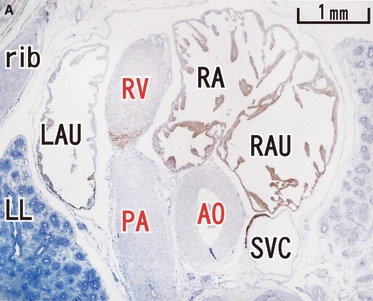
In the text and legends of our paper we used the term “cavernous sinus” instead of “coronary sinus”. In Figure 1, the arrows were described as indicating the atrio-ventricular node; this is incorrect, and unfortunately the quality of the appropriate sections does not allow us to discover whether the AV node is desmin-positive or not. In Figure 2, the caudalmost panel is E, not F, and three structures in panel A were mislabeled. The corrected text, legends to Figs 1 and 2, and relabeled Fig. 2A are shown below.
The authors apologise for these mistakes, and thank Professor R.H. Anderson for pointing them out. The Editor-in-chief regrets that these errors were not noticed before publication.
Results, first sentence
Slight variations in the results of immunostaining were observed among specimens even at the same stage. Desmin immunoreactivity was restricted to the atrial walls, proximal parts of the pulmonary vein and superior vena cava, coronary sinus, and the muscular sleeve around the developing sinuses and leaflets of the aortic valve (Figures 1–3).
Figure 1 Sagittal sections of a 9-week human fetus. Panels A–D show selected sections in left-to-right order. The right side of each panel corresponds to the ventral side of the body. Intervals between panels are 0.6 mm (A, B), 0.5 mm (B, C) and 0.2 mm (C, D), respectively. Panels A, B and D represent desmin expression while panel C represents MHC. Desmin reactivity is seen along the atrium (LA, RA), proximal parts of the pulmonary vein (LPV, RPV), superior vena cava (SVC), coronary sinus (CS) and in the muscular sleeve around the developing aortic valve (arrows in panel D). The auricle (RAU) is almost negative. MHC is positive in the ventricle as well as in the atrium. The asterisk in panel C indicates a protrusion of the left atrium. All panels were prepared at the same magnification (scale bar in panel A). Other labels for this and subsequent figures: AO, aorta; DA, ductus arteriosus; ES, esophagus; LMT, left coronary artery; PA, pulmonary artery/arteries; R/LAU, right/left auricle; R/LB, right/left bronchus; R/LL, right/left left lung; R/LV, right/left ventricle.TH, thymus.
Figure 2 Horizontal sections of a 12-week human fetus. The section illustrated in panel A is the most cranial and panel E the most caudal. The upper part of the panel corresponds to the ventral side of the body. Intervals between sections are 1.2 mm (A, B), 1.0 mm (B, C) and 1.2 mm (C–E), respectively. Panel D corresponds to a level 0.1 mm cranial to panel E, while panel F is 0.4 mm caudal to panel C. Panels A–D represent desmin, panel E is MHC and panel F is α-SMA. Desmin expression is seen along the atrium (LA, RA), proximal parts of the pulmonary vein (LPV, RPV), superior vena cava (SVC) and the coronary sinus (CS). The auricle is weakly positive. MHC (panel E) is positive in the ventricle as well as in the atrium. α-SMA (panel F) is positive along the vascular wall. Arrows in panel B indicate a valve-like structure between the atria. Scale bars in panels A and D represent magnifications for panels A–C and F, and panels D and E, respectively.
 |


