Abstract
Skeletal muscle contains multiple progenitor populations of distinct embryonic origins and developmental potential. Myogenic progenitors, usually residing in a "satellite cell position" between the myofiber plasma membrane and the laminin-rich basement membrane that ensheaths it, are self-renewing cells that are solely committed to the myogenic lineage1,2. We have recently described a second class of vessel associated progenitors that can generate myofibroblasts and white adipocytes, which responds to damage by efficiently entering proliferation and provides trophic support to myogenic cells during tissue regeneration3,4. One of the most trusted assays to determine the developmental and regenerative potential of a given cell population relies on their isolation and transplantation5-7. To this end we have optimized protocols for their purification by flow cytometry from enzymatically dissociated muscle, which we will outline in this article. The populations obtained using this method will contain either myogenic or fibro/adipogenic colony forming cells: no other cell types are capable of expanding in vitro or surviving in vivo delivery. However, when these populations are used immediately after the sort for molecular analysis (e.g qRT-PCR) one must keep in mind that the freshly sorted subsets may contain other contaminant cells that lack the ability of forming colonies or engrafting recipients.
Keywords: Cellular Biology, Issue 49, Muscle, white adipose, stem cells, flow cytometry, purification
Protocol
1. Tissue Collection:
Use mice between 7 and 12 weeks old. Usually, at least two animals are used: one is to set up the FACS single-color controls required for compensation and for fluorescence-minus-one isotype control samples. The other is the actual sample for sorting.
Euthanize the mice by CO2 inhalation or according to an ethics protocol of choice. Place mice on a paper towel, facing up, and spray with 70% ethanol thoroughly to prevent contamination with the mouse fur.
Lift the skin in the abdominal region with forceps, then make a small longitudinal cut with sharp scissors.
Peel the two flaps of skin in opposite directions (up and down) to completely expose the hind limb muscles underneath.
Excise the muscle tissue by inserting and extending the scissors along both sides of the tibia.
Repeat the same process for the femur to extract the muscle tissue.
Place all excised muscle tissue in 60mm petri-dishes, using one dish for the tissue from each mouse.
Repeat the above for all mice.
2. Dissociate Tissue Into Single Cells by Enzymatic Digestion:
Throughout this section of the protocol, use sterile reagents and tools and work under sterile conditions.
Add 2 mL of Collagenase II ( Sigma, cat#6885, 500u/ mL) and 20 μL of 250mM CaCl2 stock to each Petri-dish.
Tear the muscle tissue into small pieces ( about 1 mm3) with two forceps (one serrated, one with 2 teeth, from Fine Science Tools , cat#11051-10, 11154-10), paying attention to remove and discard as much contaminated non-muscle tissue as possible.
Cover the Petri-dishes and incubate them at 37 degrees Celsius for 30 minutes.
Retrieve the Petri-dishes and use a 3 mL syringe plunger to thoroughly mash the tissue pieces. Use one plunger for each sample to avoid contaminations.
Add some (~ 5 mL) sterile cold PBS to each Petri-dish and transfer the tissue sludge into a 50mL Falcon tube (rinse the Petri-dish if necessary to recover all the tissue)
Shake the Falcon tube vigorously, fill the tube with PBS, centrifuge @800rpm x 5 minutes ( Eppendorf bench top Model :5810R), and aspirate the supernatant (repeat 3 times)
Add 1mL mixture of Collagenase D (Roche, Cat# 11088882001, 1.5u/ mL) /Dispase II (Roche, Cat# :04942078001, 2.4u/ mL) and 10μL of CaCl2 (250mM) to each tube, incubate at 37 degrees Celsius with rotation for one hour.
Add 5-10 mL of cold, sterile PBS, vortex and pipette thoroughly to homogenize the tissue.
Fill up tubes with PBS and mix well.
Transfer the liquid to a 40μm cell strainer placed on top of a 50 mL Falcon tube to filter out the remaining large pieces of tissue. Divide each sample evenly into three 50mL Falcon tubes. Fill up each of the tubes with PBS then centrifuge @1600rpm for 5 minutes at 8 degrees Celsius.
Discard the supernatant and collect cells for staining.
3. Antibody Staining and Sorting:
Throughout this section of the protocol, use sterile FACS and collection buffers and work under sterile conditions. Commercial antibodies are generally considered sterile if properly handled.
- For single color and isotype control samples, resuspend the sample in 1mL FACS buffer and divide the suspended cells in nine 1.5mL pre-labelled Eppendorf tubes (100 μL each tube).
- Set up single color staining mixes according to the following table: ATTENTION: The final concentration of isotype control antibodies in the stain should match that of the primary antibody they are the control for. For example, if anti-Sca-1 is used for 1 μg/106 cells, its corresponding isotype must be used at the same concentration.
Tube # Name of tube Company Cat# clone isotype dilution 1 Non-stain 2 Hoechst33342 sigma B2261 2-5ug/ml 3 PI sigma P4864 1ug/ml 4 CD31-FITCCD45-FITC ebioscience 11-0311-85 390 Rat IgG2a 500 House made I3/2 Rat IgG2b 500 5 Sca1-PECY7 ebioscience 25-5981-82 D7 Rat IgG2a 5000 6 α-7 APC House made R2F2 Rat IgG2b 1000 - Set up isotype control staining mixes using the following table:
Tube # Nameof tube Hoechst PI CD31-FITC CD45-FITC Scal-PE-CY7 a-7-APC Rat-IgG2b-APC Rat-IgG2a-pecy7 Rat-IgG2a-FITC Rat-IgG2b-FITC 7 Iso-CD31/CD45 + + - - + + - - + + 8 Iso-Scal-pecy7 + + + + - + - + - - 9 Iso- α-7-APC + + + + + - + - - - - Pipet the single color and isotope control staining mixes (we usually adjust the concentration of antibodies so that between 5 and 10 μL of staining mix are added to each sample) into the appropriate Eppendorf tube from Step 20. Mix well and incubate on ice for 1 hour.
Stain cells from sample mouse with 800μL of the antibody cocktail (below) for FACS cell sorting. Mix well and incubate on ice for 1 hour. Antibody cocktail: Hoechst, PI, CD31-FITC, CD45-FITC, Sca1-PECY7, α-7 APC ATTENTION: The optimal ratio of antibodies to cells must be determined by individually titrating each antibody, as significant differences may exist between batches. The optimal amount of antibody to be used should be expressed in μgs of antibody per 106 target cells and kept constant when using the same batch of antibody. Clearly, if the number of target cells does not change significantly between experiments, or if the staining volume is scaled with the number of cells, a fixed dilution of each antibody can also be used.
Wash: Add approximately 1mL of FACS buffer to each of the 9 control tubes; add approximately 20mL of FACS buffer to the sample tube.
Centrifuge the Eppendorf tubes @ 3000rpm for 5 minutes ( Eppendorf bench top centrifuge, Model:5417C ). Centrifuge the sample tube @1600rpm for 5 minutes (Eppendorf bench top centrifuge, Model:5810R).
Aspirate and discard the supernatant from all the tubes.
Resuspend cells in the FACS buffer (1mL for each of the controls, 4mL for the sorting sample), Pipette cells into "FACS tubes" through the cell strainer cap (BD Falcon cat# 352235, 40μm pore size) to remove remaining clumps that may affect the cytometer.
Set up the FACS Sorter with single color controls and isotype controls.
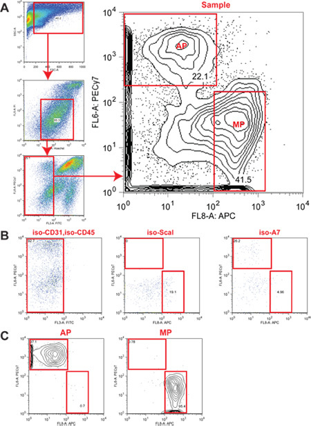
- Collect sorted cells according to the following table in a 3.5 mL collection buffer (95% DMEM, 5%FBS) separately. Normally, when harvesting both hind limbs, a single wild type mouse could yield about 150,000 myogenic progenitor cells or 100,000 adipogenic progenitor cells, with the viability above 95%, as judged by reanalyzing sorted cells by FACS at the end of the procedure (Figure 1) Definition of myogenic and adipogenic progenitors based on surface staining
Antibody/stain Myogenic progenitor (MP) Adipogenic progenitor (AP) Hoechst + + PI - - CD31-FITC - - CD45-FITC - - Sca1-PECY7 - + α-7 APC + -
4. Transplantation:
Centrifuge sorted cells @1500rpm for 5 minutes, remove the supernatant and transfer cells to autoclaved Eppendorf tubes (1.5 mL), rinse cells with 500 μL of sterile PBS (serum free!), then centrifuge @ 3000rpm for 5 minutes. Maintaining all reagents in sterile conditions and working in a tissue culture hood, resuspend myogenic progenitors in PBS, or adipogenic progenitor in Matrigel ( BD cat# 354234) at approximately 106 cells /mL.
- Transplant myogenic progenitors or adipogenic progenitor to recipient mice.
- Anaesthetize the mouse with isoflouran according to your institution policy.
- Shave the hair around the injection region, then sterilize the skin by rubbing with 70% ethanol
- Inject 20μL of myogenic progenitors or adipogenic progenitors (= 20k cells) using a 3/10 CC insulin syringe ( BD cat# 309300).
After an appropriate time (three weeks works well, but myofibers expressing donor-derived transgenic markers are usually evident within one week after transplant), anaesthetize the recipient mice from step 29 by intraperitoneal injection of 400 μL Avertin (Sigma Cat# T04840-2, 25 mg/ mL), then perfuse transcardially with 50mL PBS (contains 10 μM of EDTA) first, followed by 15mL of 4% PFA. Collect target tissue, post-fix with 4% PFA overnight if required, then transfer to 20% sucrose overnight. Embed in OCT, freeze and cryosection.
5. Representative Results:
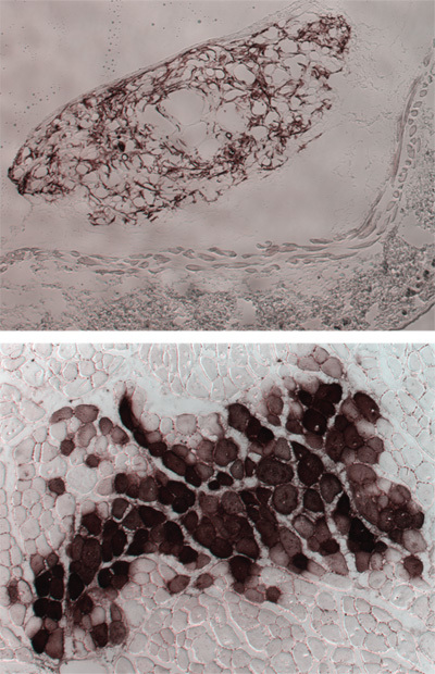
When MP are transplanted into mouse skeletal muscle, they readily fuse with pre-existing myofibers. Therefore any genetic label present in the donor cells will be easily detectable in the fibers that received them. Figure 2 shows an example where the transplanted cells expressed human alkaline phostphatase, revealed histochemically.
When AP are transplanted the result is highly dependent on the environment of the transplantation site: when transplanted subcutaneously these cells give origins to adipocytes and myofibroblasts (see figure 2). In many other sites, including muscle, they do not survive unless fatty degeneration has been induced3.
 Figure 1. Sorting strategy for the isolation of progenitor populations from skeletal muscle. (A) Sorting strategy: Viable cells were identified based on forward scatter and side scatter. Hoechst staining was used to exclude anuclear debris and propidium iodide (PI) staining to exclude dead cells. Hematopoietic (CD45) and endothelial (CD31) cells were excluded from the sorting gates. The α7+ Hoechst+ PI- CD45- CD31- Scal- subset contains all myogenic progenitors (MP). The Scal+ Hoechst+ PI- CD45- CD31- α7- population contains adipogenic progenitors (AP). (B) Fluorescence-minus-one (FMO) isotype controls confirm the specificity of the stain. (C) Purity checks of MP and AP subsets after sorting.
 Figure 2. Representative results following MP and AP transplantation. AP were transplanted subcutaneously (top panel) and MP intramuscularly (bottom panel). In both cases, donor cells originated from a mouse expressing transgenic human alkaline phosphatase, identified by the brown staining.
Discussion
The goal of this protocol is to strike a reasonable balance between high yields and high viability of the purified cells. The most critical step in ensuring that healthy cells are recovered is, predictably, the enzymatic dissociation of the starting tissue. The handling of the tissue should be particularly gentle, which is hard to demonstrate or appreciate even in an audiovisual format. Another key factor is the length of the procedure. The longer it takes to go from the donor to the recipient animal, the lower the viability and therefore, the engraftment efficiency. Should there be any question about viability that is not immediately answered by looking at the frequency of PI positive event in the purified cell samples after sorting, we suggest that a limiting dilution assay to measure clonogenicity is performed3. Typical sorts from undamaged muscle routinely yield about 1 in 15-20 cells capable of initiating myogenic or fibro/adipogenic colonies in vitro. While essentially 100% of the colonies initiated from MPs contain differentiated myotubes, only about one third of colonies obtained from FAPs contain adipocytes in addition to smooth muscle actin positive myofibroblasts.
Disclosures
No conflicts of interest declared.
References
- Buckingham M, Montarras D. Skeletal muscle stem cells. Curr Opin Genet Dev. 2008;18:330–336. doi: 10.1016/j.gde.2008.06.005. [DOI] [PubMed] [Google Scholar]
- Relaix F, Marcelle C. Muscle stem cells. Curr Opin Cell Biol. 2009;21:748–753. doi: 10.1016/j.ceb.2009.10.002. [DOI] [PubMed] [Google Scholar]
- Joe AW. Muscle injury activates resident fibro/adipogenic progenitors that facilitate myogenesis. Nat Cell Biol. 2010;12:153–163. doi: 10.1038/ncb2015. [DOI] [PMC free article] [PubMed] [Google Scholar]
- Natarajan A, Lemos DR, Rossi FM. Fibro/adipogenic progenitors: A double-edged sword in skeletal muscle regeneration. Cell Cycle. 2010;9 doi: 10.4161/cc.9.11.11854. [DOI] [PubMed] [Google Scholar]
- Sherwood RI. Isolation of adult mouse myogenic progenitors: functional heterogeneity of cells within and engrafting skeletal muscle. Cell. 2004;119:543–554. doi: 10.1016/j.cell.2004.10.021. [DOI] [PubMed] [Google Scholar]
- Sacco A, Doyonnas R, Kraft P, Vitorovic S, Blau HM. Self-renewal and expansion of single transplanted muscle stem cells. Nature. 2008. [DOI] [PMC free article] [PubMed]
- Montarras D. Direct isolation of satellite cells for skeletal muscle regeneration. Science. 2005;309:2064–2067. doi: 10.1126/science.1114758. [DOI] [PubMed] [Google Scholar]


