Abstract
Microarray expression profiling of the nervous system provides a powerful approach to identifying gene activities in different stages of development, different physiological or pathological states, response to therapy, and, in general, any condition that is being experimentally tested1. Expression profiling of neural tissues requires isolation of high quality RNA, amplification of the isolated RNA and hybridization to DNA microarrays. In this article we describe protocols for reproducible microarray experiments from brain tumor tissue2. We will start by performing a quality control analysis of isolated RNA samples with Agilent's 2100 Bioanalyzer "lab-on-a-chip" technology. High quality RNA samples are critical for the success of any microarray experiment, and the 2100 Bioanalyzer provides a quick, quantitative measurement of the sample quality. RNA samples are then amplified and labeled by performing reverse transcription to obtain cDNA, followed by in vitro transcription in the presence of labeled nucleotides to produce labeled cRNA. By using a dual-color labeling kit, we will label our experimental sample with Cy3 and a reference sample with Cy5. Both samples will then be combined and hybridized to Agilent's 4x44 K arrays. Dual-color arrays offer the advantage of a direct comparison between two RNA samples, thereby increasing the accuracy of the measurements, in particular for small changes in expression levels, because the two RNA samples are hybridized competitively to a single microarray. The arrays will be scanned at the two corresponding wavelengths, and the ratio of Cy3 to Cy5 signal for each feature will be used as a direct measurement of the relative abundance of the corresponding mRNA. This analysis identifies genes that are differentially expressed in response to the experimental conditions being tested.
Protocol
1. Quality control analysis of RNA sample with 2100 Bioanalyzer
Before you start,
Denature the ladder and your samples at 70 °C for 10 min. Chill immediately on ice.
Clean the instrument electrodes with RNaseZAP for 1 min, followed by RNase-free water for 10 sec. Allow the electrodes to air-dry.
Prepare the gel matrix by pipeting 550 μl of the gel matrix into a spin filter provided with the kit. Centrifuge at 1,500 RCF for 10 min at room temperature. Aliquot the filtered gel into 65 μl aliquots and store at 4°C for up to 1 month.
- To prepare the chip priming station,
- Insert a 1-ml syringe through the clip and into the luer lock adapter.
- Adjust the base plate to position C by loosening the screw under the base, lifting the plate and retightening the screw.
- Adjust the syringe clip to the top position.
Equilibrate the dye concentrate to room temperature. Vortex for 10 sec. Add 1 μl of dye to a 65 μl aliquot of filtered gel. Vortex well and centrifuge at 13,000 RCF for 10 min at room temperature. Use within one day. When you are ready to run your samples,
- Position a chip on the priming station. In this protocol we are using RNA 6000 nano chips. To load the gel,
- Pipette 9 μl of the gel-dye mix in the well marked with a white "G" against a black background. Make sure your tip is positioned at the very bottom of the well.
- Position the syringe plunger at 1 ml. Close the priming station. Make sure it click-locks.
- Press the plunger down slowly but steadily until it is held by the clip.
- Wait for 30 sec. Release the clip.
- Let the plunger go up by itself. After it stops moving, wait for a few seconds and pull the plunger back to the 1 ml position. Open the priming station.
Pipette 9 μl of the gel-dye mix in each of the 2 wells marked "G".
Pipette 5 μl of the marker in each of the 12 sample wells and in the ladder well.
Pipette 1 μl of the prepared ladder in ladder well.
Pipette 1 μl of each sample into the sample wells.
Pipette 1 μl of the marker in each unused sample well.
Vortex the chip for 1 min at 2,400 rpm.
- To run the chip,
- Start the 2100 Expert Software.
- Position the chip and close the lid. If the instrument is on-line, an icon will show whether the lid is open or closed and what type of chip is inserted. Make sure the correct port is selected.
- Run the assay within 5 min of loading the samples to prevent evaporation.
2. Amplification and labeling
To prepare for microarray analysis, RNA samples are amplified and labeled, usually in a reaction based on T7 RNA polymerase3. Double stranded cDNA is generated by reverse transcription. cDNA is used in an in vitro transcription reaction to generate what is known as cRNA. This reaction is performed in the presence of labeled ribonucleotides, producing microgram quantities of labeled RNA for array hybridization. The choice of amplification/labeling methods depends on the subsequent microarray platform to be used. In this section, we describe the generation of fluorescently labeled RNA using Agilent's Quick Amp labeling kit.
Pipette 50 ng to 5 μg of RNA in a 1.5 ml tube. The volume should not exceed 8.3 μl. If necessary, concentrate your RNA sample by vacuum centrifugation or precipitation with alcohol.
Add 1.2 μl of T7 primer.
Bring to a final volume of 11.5 μl with RNase-free water.
Incubate at 65°C for 10 min to denature the RNA. We recommend using a thermocycler with a hot lid to prevent condensation in the top of the tube.
During the 10 min incubation, warm 5X First Strand Buffer to 80°C for 5 min. Vortex and spin down. Keep at RT until ready to use.
After the 10 min incubation, quickly cool down the denatured RNA samples on ice.
- Prepare the cDNA master mix. Per reaction, add the following:
- 4 μl 5X First Strand Buffer
- 2 μl 0.1 M DTT
- 1 μl 10 mM dNTPs
- 1 μl MMLV-RT enzyme
- 0.5 μl RNase Out.
Mix components by inverting the tube 4 times. Do not vortex. Spin down to collect the contents at the bottom of the tube.
Add 8.5 μl of master mix per sample. Mix by pipeting up and down.
Incubate for 2 hr at 40°C, followed by 10 min at 65°C. We recommend using a thermocycler with a heated lid.
Transfer the tubes to ice for 5 min.
- Prepare the transcription master mix. Per reaction, add the following:
- 15.3 μl nuclease-free water
- 20 μl 4X transcription buffer
- 6 μl 0.1 M DTT
- 8 μl NTPs
- 6.4 μl 50% PEG (PEG should be prewarmed to 40°C and vortexed to ensure resuspension)
- 0.5 μl RNase Out
- 0.6 μl inorganic pyrophosphatase
- 0.8 μl T7 RNA polymerase
- 2.4 μl Cy3- or Cy5 CTP
Briefly spin the samples to collect the contents at the bottom of the tube.
Add 60 μl of Transcription Master Mix per sample.
Incubate at 40°C for 2 hr.
Add 20 μl of RNase-free water.
Purify the labeled cRNA to remove unincorporated nucleotides. We recommend using Qiagen's RNeasy mini columns.
Quantify the labeled cRNA. We recommend using a NanoDrop2000 spectrophotometer in Microarray Measurement mode. Make sure you select RNA-40 as the sample type.
Record sample concentration, yield (calculated as the concentration multiplied by the volume) and specific activity (calculated as 1000 multiplied by the ration of dye concentration over cRNA concentration). For microarray hybridization, it is necessary to achieve a yield of at least 825 ng and a specific activity of at least 8 pmol/μg.
3. Microarray hybridization
Turn on the hybridization station and set at 65°C at least 2 hours before hybridization begins.
- To prepare the hybridization sample, mix the following in a 1.5 ml tube:
- 825 ng Cy3-cRNA (this is usually your "treated" sample)
- 825 ng Cy5-cRNA (this is usually your "reference" sample)
- 11 μl 10X Blocking agent.
- Add RNase-free water to a final volume of 52.8 μl
- Add 2.2 μl fragmentation buffer.
Vortex at low speed to mix.
Incubate at 60°C for exactly 30 min. This is the cRNA fragmentation step, which generates labeled fragments for array hybridization. It is very important to incubate exactly as described. We recommend the use of a thermocycler with a heated lid. It is very important to incubate exactly as described to generate probes of the correct average length. Excessive or insufficient fragmentation will result in false negatives or false positives.
Add 55 μl of 2X Hybridization buffer to stop fragmentation. Mix by pipetting, taking special care not to introduce bubbles.
Centrifuge for 1 min at maximum speed to collect the content at the bottom of the tube. If bubbles are observed, centrifuge longer to remove them.
Place the tube on ice, protected from the light. Load the sample on the array as soon as possible.
- To load the array, first prepare the hybridization chamber. This protocol describes the use of Agilent's 4x44K arrays with the Roche-Nimblegen hybridization system and A4 mixer chambers.
- Place a microarray slide in the assembly/disassembly tool, barcode side first.
- Open an A4 mixer and expose the adhesive. Holding the array and assembly/disassembly tool to prevent any movement, place the A4 mixer on the slide, starting at the far end. Make sure it is correctly aligned to the tool. Stick the mixer all along the array slide.
- Pull from the end of the mixer to remove the slide/mixer assembly.
- Using the braying tool, press all along the adhesive gasket to make sure it is tightly glued to theslide. Small air bubbles trapped between the adhesive and the slide can be seen against a dark background. Take extra time to remove these bubbles with the braying tool. Your array is now ready to be loaded.
Use a positive displacement pipette to load 100 μl of the hybridization sample. Push the tip tightly inside the port hole; then dispense slowly and smoothly so that air is not trapped in the array area. Never try to suck the sample back up. Once the entire sample has been dispensed, keep the tip on the port hole without releasing the plunger. When the sample covers the whole array and liquid starts to come out the vent port, quickly remove the pipette. Only release the plunger once the tip is away from the slide.
Gently dab the excess liquid at both ports. Make sure you do not draw sample out of the chamber itself.
Cover the port holes with stickers by using tweezers.
Put the slide on the hybridization station, and check that the bladder holes are correctly positioned over the O-rings. This will ensure proper mixing during hybridization.
Close the slide cover and the station lid. Set the station to mixing mode B.
Hybridize the array at 65°C for 17 hours.
Prepare Wash Buffer 2 by adding Triton X-102 to a final concentration of 0.005%. Keep at 37°C overnight.
4. Microarray washing
Add Triton X-102 to Wash Buffer 1 to a final concentration of 0.005%. Keep at room temperature.
When hybridization is finished, fill a glass dish "A" with Wash Buffer 1. The dish should be big enough to hold the assembly/disassembly tool and allow some maneuvering as well. All washes should be done in glassware. Avoid plastic dishes, since they tend to leach compounds resulting in high background in the array.
Put a slide rack and a stir bar in a staining dish "B". Fill with Wash buffer 1, making sure it covers the rack. Place on a stir plate at room temperature.
Put a stir bar on an empty staining dish "C" and place on a stir plate.
Remove the array and put it in the assembly/disassembly tool. Submerge the whole assembly in the dish "A".
While holding the assembly/disassembly tool and the slide from opposite sides with one hand, carefully peel off the A4 mixer. Make sure you do not scratch the array area.
Quickly place the slide in the rack in staining dish "B". From now on, exposure to the air should be minimized since the Cy5 dye is sensitive to ozone. Do not wash more than 8 arrays at a time.
Wash for 1 minute stirring at medium speed.
Fill staining dish "C" with pre-warmed Wash buffer 2. Transfer the slides and wash for 1 minute.
Very slowly remove the slide rack from staining dish "C" to minimize droplets left behind on the slide.
Dry the slide by spinning for 2 minutes. If a compatible centrifuge is not available, centrifuge the slide in a 50-ml conical tube or blow argon gas over it.
Place slides in a 50-ml conical tube and fill it with argon gas. Scan immediately to avoid signal loss. We recommend use of the GenePix 4000B scanner from Molecular Devices.
5. Representative Results:
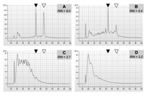
Good quality total RNA samples should produce only two major peaks when run in a Bioanalyzer for quality control, corresponding to the 2 major ribosomal RNA species. Some RNA degradation will show as a smear before the first ribosomal RNA peak. Severely degraded, low quality RNA will show a broad peak or a series of peaks at low retention times, while the 2 ribosomal RNA peaks will be of very low intensity or not identifiable at all. For examples of the 2100 Bioanalyzer output, see figure 1.
The Expert 2100 software will calculate an RNA Integrity Number, or RIN, as a quantitative measurement of RNA quality. High quality samples, with RIN values higher than 9, are obviously best for microarray applications. However, we have used samples with RIN values as low as 5.2 in generating microarray data of reasonable quality.
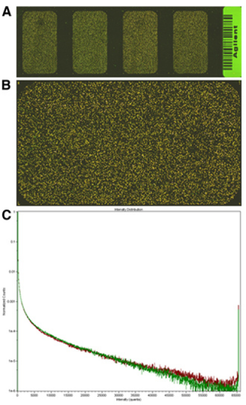
Good quality arrays should produce high signal at relatively low PMT values. In our experiment, most of the transcripts are expected to be present at similar levels in the experimental and reference sample; massive, widespread changes in gene expression will probably lack any biological significance. Therefore, most of the array should look yellow rather than green or red. Good quality signals should also be in a dynamic range such that the signal histograms fully overlap. For examples, see figure 2.
 Figure 1. Example of four RNA samples isolated from human brain tumor tissue. RNA was isolated using the protocol presented in 3.1.1. Sample quality was assessed using the 2100 Bioanalyzer on an RNA 6000 chip as described in 3.1.3. Samples are representative of 4 distinct RNA qualities: A, very good RNA quality (RIN>9); B, partially degraded RNA (RIN 5-6, note the significantly lower peak for 28S rRNA); C, highly degraded RNA (RIN<3); D, almost completely degraded RNA (RIN=2). We have successfully used samples with qualities similar to or better than B (RIN>5.2) for downstream microarray applications. Solid and open arrowheads indicate the position of the 18S and 28S rRNA species, respectively.
 Figure 2. Examples of array images and scatter plots. A, whole slide preview, showing the four arrays in the 4X44K Agilent format; B, high resolution scan of one of the array areas, note that neither of the signals (red or green) is predominant; C, scatter plot of the image in B, note that the signals fully overlap and no more than 1x10⁻⁶ features are at saturating intensity.
Discussion
Expression profiling with DNA microarrays provides a straightforward approach to identify differentially expressed genes between two biological samples. Successful expression profiling experiments require high quality RNA samples, robust labeling and hybridization. In our experience, the commercial arrays and labeling kits from Agilent provide high quality data at a reasonable cost. While Agilent also offers their own hybridization and scanning machinery, we favor the hybridization system from Roche-Nimblegen and the GenePix 4000B microarray scanner from Molecular Devices. Note that the Roche-Nimblegen hybridization unit was formerly known as the MAUI hybridization system. After the recent corporate acquisition by Roche, the A4 mixers have been discontinued. As of the moment of this writing, they can still be found through other sellers such as Kreatech Diagnostics (www.kreatech.com; this website also offers a convenient array compatibility tool), but long-term stock availability is uncertain. Other hybridization systems are available (for example, from Agilent); however, if compatible mixers can be found for the array being used, we recommend the Roche-Nimblegen's system for its quality and reproducibility.
Disclosures
No conflicts of interest declared.
Acknowledgments
This work was supported by grants to E. D. from the James S. McDonnell Foundation 21stCentury Award Program, and a NIH Director's New Innovator Award. G. A. B. was supported in part by a postdoctoral fellowship from the California Institute of Regenerative Medicine.
References
- Diaz E. One Decade Later: What has Gene Expression Profiling Told us About Neuronal Cell Types, Brain Function and Disease. Curr Genomics. 2009;10:318–318. doi: 10.2174/138920209788921029. [DOI] [PMC free article] [PubMed] [Google Scholar]
- Barisone GA. Expression profiling in Neuroscience, edited by Ioannis Karamanos. New York: SPRINGER SCIENCE+BUSINESS MEDIA, LLC; 2010. [Google Scholar]
- Gelder RNVan. Amplified RNA synthesized from limited quantities of heterogeneous cDNA. Proc Natl Acad Sci U S A. 1990;87:1663–1663. doi: 10.1073/pnas.87.5.1663. [DOI] [PMC free article] [PubMed] [Google Scholar]


