Abstract
Purpose
We prospectively evaluated the surgical outcomes of single scrotal incision orchiopexy in children with a palpable undescended testis compared with the traditional two incision orchiopexy.
Materials and Methods
A total of 398 orchiopexies (292 children) were included and randomly assigned to the single scrotal incision orchiopexy group (Group I, 147 children, 201 testes) or the traditional inguinal incision orchiopexy group (Group II, 145 children, 197 testes). The final number of patients enrolled (excluding those lost to follow-up) was 107 children (146 testes) in group I and 105 children (141 testes) in group II. Success was defined as no complications, postoperative intrascrotal location of the testis, and no conversion to the traditional inguinal approach. Surgical outcomes and complications were compared between the two groups. Testicular location, complications, and subjective satisfaction rate were assessed at the follow-up evaluation at least 12 months postoperatively.
Results
The overall success rate in group I was 92.5% in 135 of 146 testes; the remaining 9 testes required conversion to traditional two incision orchiopexy. In group II, orchiopexy was successful in 136 of 141 testes (96.5%). The operation time and hospital stay were significantly shorter in group I (40.5±25.9 minutes, 2.1±0.8 days) than in group II (62.3±35.6 minutes, 2.5±0.7 days), respectively (p<0.001, p=0.03). Postoperative complications were found in two cases (hematoma, wound dehiscence) in group I and in one case (wound dehiscence) in group II; all cases with complications recovered with conservative care. The subjective rate of satisfaction with the cosmetic result was 96.6% in group I and 96.5% in group II (p=0.97).
Conclusions
We conclude that single scrotal incision orchiopexy is a simple technique that is associated with a shorter operation time and hospital stay than the traditional method and that is more feasible cosmetically.
Keywords: Cryptorchidism, Orchiopexy, Scrotum, Testis
INTRODUCTION
Undescended testis is one of the most common disorders of childhood, with a rate of 3.68% among full-term infants managed by surgical correction [1]. Surgical intervention is warranted during early infancy to avoid secondary degeneration of the testis, to improve fertility later, to help with the detection of malignancy, and to reduce the chance of testicular torsion [2,3]. The inguinal approach is the traditional method for correcting undescended testis. In this approach, two incisions are made: one inguinal or groin incision to open the inguinal canal to visualize the cord structure and a second scrotal incision to fix the testes within the scrotum [4]. It was believed that inguinal incision is helpful for sufficient mobilization of the spermatic cord, separation of the processus vaginalis or hernia sac, high ligation of the hernia sac, and to achieve an adequate length for the testes to be relocated to the dependent portion of the scrotum.
However, Bianchi and Squire introduced the high scrotal incision orchiopexy technique for a palpable cryptorchid testis to decrease the potential morbidity of traditional inguinal incision orchiopexy [5]. Until now, few prospective studies have been reported regarding the success rate of this technique compared with traditional inguinal orchiopexy for undescended palpable testis [6]. In the present study, therefore, we evaluated a single scrotal incision technique for palpable undescended testis within the inguinal canal or distal to the external inguinal ring compared with traditional inguinal orchiopexy.
MATERIALS AND METHODS
1. Study design
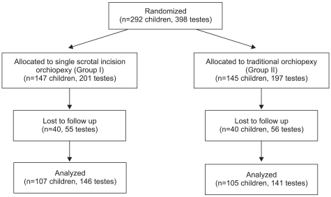
From January 2007 to December 2010, a total of 292 children (398 testes) with palpable undescended testes were randomly assigned to two groups: single scrotal incision orchiopexy (Group I, 147 children with 201 testes) or traditional inguinal incision orchiopexy (Group II, 145 children with 197 testes). Patients were assigned to the scrotal or inguinal group in a 1:1 ratio through a simple randomization procedure. A total of 80 patients were lost to follow-up (Fig. 1). A total of 107 children (146 testes) underwent single scrotal incision orchiopexy (group I) and 105 children (141 testes) underwent traditional inguinal incision orchiopexy (group II). They were followed up and evaluated until 12 months after the operation and included in the final data analysis. The patients' mean ages (months) at the time of operation in groups I and II were 40.1±10.3 and 41.8±11.4, respectively (Table 1). All patients were seen at least 1 week postoperatively to evaluate the possible occurrence of wound infection or skin problems and to assess for any other operation-related complications. At 3 months and 12 months after the operation, the patients were followed up for evaluation of long-term complications, overall success rate, and parents' satisfaction rate. Success was defined as no complications, postoperative intrascrotal location of the testis, and no conversion to the other method. The parents' satisfaction with the cosmetic results of the operation was assessed by use of a simple questionnaire that consisted of the answers 'satisfied,' 'not fully satisfied,' and 'unsatisfied.' This study was approved by the institutional review board of our hospital. All parents signed an informed consent form before participation in this study allowing the use of the patients' medical records for a scientific purpose.
FIG. 1.
Disposition of subjects assigned to the study.
TABLE 1.
Comparison of basal characteristics between the traditional (group II) and single scrotal incision orchiopexy (group I) patients included in the final analysis
Group I: single scrotal incision orchiopexy, Group II: traditional orchiopexy, a: Chi-square test, b: Student's t-test
2. Exclusion criteria
Children who had undergone a previous inguinal or pelvic surgery or who had a secondary ascending testis, ectopic testis, or undescended testis related to ambiguous genitalia or intersex condition were excluded from the study. Patients with primary and secondary hypogonadism and a detected hormonal abnormality or history of hormonal treatment were not included. All children were examined twice, preoperatively in the supine position by the primary surgeon and again after induction of general anesthesia to exclude retractile testis.
3. Surgical procedure
All operations were performed by 1 surgeon (Kim SO). The first surgical step of the single scrotal incision orchiopexy after induction of general anesthesia was a transverse skin incision that was commonly made along the high scrotal skin fold. The dartos pouch was adequately created through this incision for later relocation of the affected testis. The assistant manipulated the testis and held it between the thumb and index finger in a stable position, and in sequence the surgeon used blunt and sharp dissection of the subcutaneous tissues to approach the testis. The scrotal wound was retracted in an upward direction to facilitate easier dissection, and the surgeon divided the various covering and adhesive tissues of the cord. The dissection was carried out to the most cephalad to secure sufficient cord length and to possibly enter the lower half of the inguinal canal from below. The gubernacular attachments were released to enable identification of the testes within the cremasteric fibers, a patent processus vaginalis, and the cord structures. The cremasteric fibers and hernia sac were carefully separated from the cord structures, and the cranial sac was mobilized under traction into the canal and was ligated with a suture, as in traditional inguinal incision orchiopexy. When additional cord length was required, additional dissection was done through this incision by opening the external ring and canal, as necessary. Despite the additional dissection, if more length was required, the surgeon converted to the traditional inguinal orchiopexy method. The testis was then relocated into the dartos pouch, and two fixing sutures were made between the testicular tunica albuginea and inner scrotal wall medially and laterally to prevent ascent. The scrotal skin was closed with interrupted absorbable sutures. The operative time was recorded.
Traditional inguinal orchiopexy was performed by the existing method. After inguinal skin incision, the cord structure and testis were taken out of the inguinal incision site and then sufficiently dissected for mobilization. High ligation was done with the processus vaginalis, and the surgeon made an incision along the scrotal crease and relocated the testis in the scrotum.
4. Statistics
Statistical analysis was performed by using SPSS ver. 13.0 (SPSS Inc., Chicago, IL, USA). The Student's t-test and chi-square test were used for data analysis. p<0.05 were deemed to be statistically significant.
RESULTS
At the 12-month follow up, a total of 107 children (146 testes) who underwent single scrotal incision orchiopexy (group I) and 105 children (141 testes) who underwent traditional inguinal incision orchiopexy (group II) were included in the final data analysis. There were no significant differences in baseline characteristics between the groups. The mean follow-up time was 12.9±3.4 months in Group I and 12.7±3.3 months in Group II, and the evaluation is still ongoing (Table 1).
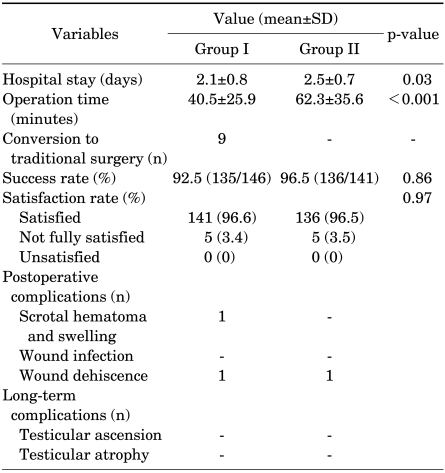
When the overall success rate was compared between the groups, it was not significantly different: 92.5% (135/146 testes) in group I and 96.5% (136/141 testes) in group II (p=0.86) at 12 months (Table 2). The results were not significantly different from those at 3 months. There was a significant difference between Groups I and II in terms of the period of hospitalization (days) (2.1±0.8 vs 2.5±0.7; p=0.03) and the operation time (minutes) (40.5±25.9 vs 62.3±35.6; p<0.001) (Table 2).
TABLE 2.
Comparison of surgical outcomes between traditional (group II) and single scrotal incision orchiopexy (group I)
Group I: single scrotal incision orchiopexy, Group II: traditional orchiopexy (In Group II, 4 cases were converted to other surgery: 2 cases to laparoscopic exploration and 2 cases to orchiectomy)
In nine cases in group I, the dissected cord length was insufficient because of severe adhesion despite a distal location in seven cases and a higher location in two cases, and we converted to the traditional inguinal approach in these cases. In the converted cases, the period of hospitalization (days) was 3.3±0.7 and the operation time (minutes) was 55±30.3. Thus, it seemed to take more time compared with the rest of group I, but we did not perform statistical analysis. All converted cases were unilateral. In four cases in group II, traditional incision failed because of the need for laparoscopic exploration of hidden testes in two cases and the need for orchiectomy due to already atrophied testes in two cases. Postoperative scrotal hematoma was found in one case in group I, and wound dehiscence was found in one case in each group. All cases of complications were unrelated to conversion and recovered with conservative care. Other complications including wound infection, testicular ascension, and testicular atrophy were not detected even after long-term follow-up. Both groups were pleased with the cosmetic result of the operation in terms of the parents' subjective satisfaction rate: 96.6% in group I and 96.5% in group (Table 2).
DISCUSSION
Single scrotal incision orchiopexy for a palpable undescended testis is well tolerated and has a cosmetically satisfactory result. Compared with traditional inguinal incision orchiopexy, single incision orchiopexy showed several benefits such as a shorter operation time and shorter hospital stay. The results of this study suggest that single incision orchiopexy is a useful method in terms of simplicity without significant surgical difficulties. Also, the success rate of single incision orchiopexy was as high as 92.5%; only 11 testes required conversion to traditional inguinal incision orchiopexy or had postoperative complications. There was no significant difference in the subjective satisfaction rate between the two groups.
Traditional inguinal incision orchiopexy was previously regarded as a mandatory procedure for obtaining adequate mobilization of the spermatic cord, but requires two standard skin incisions for direct visualization of the cord structures, and separation and high ligation of commonly associated inguinal hernia is not easy without opening the inguinal canal [4]. However, most undescended testes are palpable distal to the inguinal canal. Furthermore, in the pediatric population, there is good mobility of the skin incision and a relatively short distance from the external to the internal inguinal ring. These points led others to believe that one scrotal incision rather than two may be sufficient for orchiopexy in patients with a palpable, low-lying undescended testis.
The single incision transscrotal technique was introduced by Bianchi and Squire in the 1980s [5]. Bianchi and Squire proposed that moving the incision by retraction and the short distance from the internal to the external ring made it possible to dissect the hernia sac without opening the canal. Moreover, they suggested that achieving adequate palpable testis cord length was dependent more on releasing the hernial sac from the cord than on dissection around the spermatic cord vessels [5]. The suggested that the benefits of using one incision in the scrotal skin fold included decreased pain, improved cosmesis, and a shorter operative time with less incision needed to close the wound window [5,7]. Caruso et al evaluated the Bianchi single scrotal incision technique for orchiopexy in patients with a palpable undescended testis distal to the external inguinal ring and reported that this technique is simple and safe in such cases [8]. Only 1 of 42 testicles approached in this manner required a conversion to the traditional inguinal incision orchiopexy to gain adequate cord length. An important point in their report was that, after opening the inguinal canal when they converted to the inguinal approach, they found that the ligated hernia sac was retracted well into the low retroperitoneum above the level of the internal ring. Their findings support previous results showing that a palpable undescended testis may be surgically relocated into the dependent scrotum without sacrificing the traditional principles of orchiopexy [5,7].
The present study is distinct from previous descriptions of scrotal orchiopexy. In previous reports, single scrotal incision orchiopexy was rarely tried in patients with an undescended testis in the inguinal canal and the exact success rate and satisfaction rate were rarely calculated. Bianchi and Squire performed single scrotal incision orchiopexy in 120 patients with undescended testis and reported a 95.8% success rate [4]. They reported that the testicular locations of failed cases were in high areas such as the inguinal canal. Dayanç et al prospectively evaluated the success rate with or without inguinal hernia in patients with an undescended testis within the inguinal canal or beyond the external inguinal ring [9]. They divided the patients into two groups: one with the testis located within the inguinal canal and the other with the testis located beyond the external inguinal ring. Scrotal orchiopexy was performed successfully (97.6%) in 42 of 43 testes in the distal to the external inguinal ring group, and only 1 patient required conversion to a traditional inguinal incision. The average operating time was 18 minutes, and no hydrocele or hernia was noted. In the 29 testes that were located within the inguinal canal, 3 cases needed conversion to traditional inguinal orchiopexy, with an average operative time of 25 minutes and a success rate of 89.7%. In the present study, the overall success rate was 92.5% (135/146) in the single scrotal incision orchiopexy group and 96.5% (136/141) in the traditional inguinal incision orchiopexy group, thus showing no statistically significant difference between the two procedures. However, the operation time and hospital stay period were significantly shorter in the single scrotal incision group than in the traditional inguinal incision orchiopexy group, as in the previous study [10]. In our study, in two of the nine cases that were converted to traditional orchiopexy, the testes were located in the higher inguinal canal, and 7 were severely adhered with adjacent tissues. In those cases, the main reason for conversion was insufficient length of cord. In the failed orchiopexy cases in the traditional orchiopexy group, small and hidden testes were the main cause of failure. According to the results of our study, we suggest that possible conversion to traditional orchiopexy should be considered before the operation when the testis is located in the inguinal canal or higher. Our results show that single scrotal orchiopexy can be safely performed through a high scrotal incision. An additional inguinal incision is only required in a small number of subjects in whom the palpable testis has a high location and the vascular length is insufficient or the processus vaginalis is not sufficient.
A possible controversy regarding this scrotal approach technique is whether the dissection is high enough to easily allow for adequate lengthening of the cord and placement of the testis into the scrotum without tension. Also, there is concern that a single scrotal approach may not allow sufficient ligation of the processus vaginalis to avoid hernia or hydrocele formation after the operation. Several others have used the high scrotal incision technique to correct abnormalities of the patent processus vaginalis, such as hernia and hydrocele [5,7,11,12]. Moreover, many other studies have suggested that failed orchiopexy from a scrotal incision is not due to incomplete division of the hernia sac from the spermatic vessels [13,14]. Despite the controversy over the relationship between the success rate of single scrotal incision orchiopexy and ligation of the patent processus vaginalis [8,15], we successfully ligated the processus vaginalis in all cases.
In most cases, the scar was invisible at the follow-up visit. Thus, it could be said that the scrotal incision orchiopexy was cosmetically feasible. In the single scrotal incision group compared with the traditional inguinal incision group, objective indexes such as operation time and hospital stay without complications were good and the subjective satisfaction rate was also good. The results of the present study confirm our belief that single scrotal incision orchiopexy is a simple, safe, and cosmetically satisfactory technique. This study had some limitations, such as the relatively small number of patients included in the study. Also, important variables in this analysis, such as testis volume, were not considered in the follow-up evaluation.
CONCLUSIONS
The results of our study showed that single scrotal incision orchiopexy is a simple technique associated with a short operation time and hospital stay and is a cosmetically feasible method. Most palpable testes can be safely approached through a scrotal incision, but an additional inguinal incision should be considered in some cases of high inguinal testes where insufficient dissection of vascular length or the processus vaginalis is encountered.
Footnotes
The authors have nothing to disclose.
References
- 1.Berkowitz GS, Lapinski RH, Dolgin SE, Gazella JG, Bodian CA, Holzman IR. Prevalence and natural history of cryptorchidism. Pediatrics. 1993;92:44–49. [PubMed] [Google Scholar]
- 2.Clarnette TD, Rowe D, Hasthorpe S, Hutson JM. Incomplete disappearance of the processus vaginalis as a cause of ascending testis. J Urol. 1997;157:1889–1891. [PubMed] [Google Scholar]
- 3.Engeler DS, Hösli PO, John H, Bannwart F, Sulser T, Amin MB, et al. Early orchiopexy: prepubertal intratubular germ cell neoplasia and fertility outcome. Urology. 2000;56:144–148. doi: 10.1016/s0090-4295(00)00560-4. [DOI] [PubMed] [Google Scholar]
- 4.Ritchey ML, Bloom DA. Modified dartos pouch orchiopexy. Urology. 1995;45:136–138. doi: 10.1016/s0090-4295(95)97502-0. [DOI] [PubMed] [Google Scholar]
- 5.Bianchi A, Squire BR. Transscrotal orchidopexy: orchidopexy revised. Pediatr Surg Int. 1989;4:189–192. [Google Scholar]
- 6.Handa R, Kale R, Harjai M, Minocha A. Single scrotal incision orchiopexy for palpable undescended testis. Asian J Surg. 2006;29:25–27. doi: 10.1016/S1015-9584(09)60289-X. [DOI] [PubMed] [Google Scholar]
- 7.Iyer KR, Kumar V, Huddart SN, Bianchi A. The scrotal approach. Pediatr Surg Int. 1995;10:58–60. [Google Scholar]
- 8.Caruso AP, Walsh RA, Wolach JW, Koyle MA. Single scrotal incision orchiopexy for the palpable undescended testicle. J Urol. 2000;164:156–158. [PubMed] [Google Scholar]
- 9.Dayanç M, Kibar Y, Tahmaz L, Yildirim I, Peker AF. Scrotal incision orchiopexy for undescended testis. Urology. 2004;64:1216–1218. doi: 10.1016/j.urology.2004.06.069. [DOI] [PubMed] [Google Scholar]
- 10.Lee HR, Lee YS, Kim HS, Lee JY, Kim JC, Koh JS. A comparison between single scrotal incision orchiopexy and the inguinal approach in patients with palpable undescended testes distal to the external inguinal ring. Korean J Urol. 2009;50:1133–1137. [Google Scholar]
- 11.Lais A, Ferro F. Trans-scrotal approach for surgical correction of cryptorchidism and congenital anomalies of the processus vaginalis. Eur Urol. 1996;29:235–238. [PubMed] [Google Scholar]
- 12.Koyle MA, Walsh R, Caruso A, Wilson E. Scrotal (Bianchi) approach to patent processus vaginalis in children. Tech Urol. 1999;5:95–99. [PubMed] [Google Scholar]
- 13.Maizels M, Gomez F, Firlit CF. Surgical correction of the failed orchiopexy. J Urol. 1983;130:955–957. doi: 10.1016/s0022-5347(17)51594-3. [DOI] [PubMed] [Google Scholar]
- 14.Cartwright PC, Velagapudi S, Snyder HM, 3rd, Keating MA. A surgical approach to reoperative orchiopexy. J Urol. 1993;149:817–818. doi: 10.1016/s0022-5347(17)36218-3. [DOI] [PubMed] [Google Scholar]
- 15.Bassel YS, Scherz HC, Kirsch AJ. Scrotal incision orchiopexy for undescended testes with or without a patent processus vaginalis. J Urol. 2007;177:1516–1518. doi: 10.1016/j.juro.2006.11.075. [DOI] [PubMed] [Google Scholar]


