Abstract
Aim:
To evaluate and compare the microleakage of self-etch adhesives placed under different clinical techniques and to analyze the resin–dentin interfacial ultrastructure under scanning electron microscope (SEM).
Materials and Methods:
100 extracted human premolars were divided into two groups for different adhesives (Clearfil S3 and Xeno III). Class V cavities were prepared. Each group was further divided into four subgroups (n = 10) according to the placement technique of the adhesive, i.e. according to manufacturer's directions (Group 1), with phosphoric acid etching of enamel margins (Group 2), with hydrophobic resin coat application (Group 3), with techniques of both groups 2 and 3 (Group 4). The cavities were restored with composite. Ten samples from each group were subjected to microleakage study. Five samples each of both the adhesives from groups 1 and 3 were used for SEM examination of the micromorphology of the resin–dentin interface.
Results:
At enamel margins for both the adhesives tested, groups 2 and 4 showed significantly lesser leakage than groups 1 and 3. At dentin margins, groups 3 and 4 depicted significantly reduced leakage than groups 1 and 2 for Xeno III. SEM observation of the resin–dentin interfaces revealed generalized gap and poor resin tag formation in both the adhesives. Xeno III showed better interfacial adaptation when additional hydrophobic resin coat was applied.
Conclusions:
In enamel, prior phosphoric acid etching reduces microleakage of self-etch adhesives, while in dentin, hydrophobic resin coating over one-step self-etch adhesives decreases the microleakage.
Keywords: Enamel etching, hydrophobic resin coat, microleakage, resin–dentin interfacial micromorphology, self-etch adhesives
INTRODUCTION
One of the greatest challenges in restorative dentistry is to obtain an effective seal of the tooth/restoration interface. Composite restorations rely on adhesive systems which form a micromechanical bond with the tooth structure. Based upon the clinical approach, bonding to tooth hard tissues can be accomplished using one of the following adhesion strategies: the etch and rinse approach, self-etch approach and glass-ionomer approach.[1] Etch and rinse technique involves the use of 30–40% phosphoric acid etchant, while the self-etch approach is based on the use of non-rinse acidic monomers that simultaneously condition and prime dentin and enamel. The self-etch systems have been classified as two-step and one-step (all-in-one) adhesives. One-step self-etch systems incorporate all the components of an adhesive system (etchant, primer and bonding resin) into a single solution and combine all the three bonding steps into a single application. Clinically, self-etch systems not only simplify the bonding process by eliminating steps, but also eliminate some of the technique sensitivity associated with the use of etch and rinse systems.[2] Furthermore, as the moisture level of dentin is not a critical factor with these adhesives, the issue of wet bonding is not a concern. Moreover, the risk of incomplete resin infiltration is eliminated by simultaneous infiltration of the exposed collagen fibril scaffold with resin up to the same depth of demineralization.[2]
These one-step self-etch adhesives have been subdivided into three categories as strong, moderate (intermediary strong) and mild, based upon their pH value.[1] As self-etch adhesives use acidic monomers rather than the traditional phosphoric acid to condition tooth structure, they do not produce the same degree of porosity in enamel surfaces as that attained with phosphoric acid etching.[3] Bonding of one-step self-etch systems to enamel still remains critical and is controversially discussed by various authors.[4,5] There is a significant difference in enamel roughness with the self-etch adhesive systems when compared with the traditional phosphoric acid conditioning.[6] Barkmeier et al.[6] reported that extending the treatment time with the self-etch adhesives also does not significantly increase bond strengths and concluded that phosphoric acid treatment of enamel appears to be much more effective than acidic monomers for bonding resin-based materials. The insufficient etching of self-etch adhesives may also be attributed to the diminishing abilities of decalcification due to total inactivation of the acid in contact with the enamel surface.[7]
On the other hand, the poor bonding performance of one-step self-etch adhesives reported in dentin may be attributed to different factors.[8] These products create very thin coatings, which may be oxygen inhibited, resulting in a poorly polymerized adhesive layer.[9] They are prone to phase separation as the solvent evaporates from the solution and they behave as permeable membranes after polymerization.[9] These one-step self-etch adhesives are extremely hydrophilic as they contain high concentrations of both ionic and hydrophilic monomers.[10] It is difficult to evaporate water from these one-step self-etch adhesives, and even if evaporation is successful, water will rapidly diffuse back from the bonded dentin into the adhesive resin. Such hydrophilicity renders these adhesives very permeable and denies their ability to hermetically seal dentin surfaces. This water sorption plasticizes polymers and lowers their mechanical properties.[11] Although hydrophobic dimethacrylates are added to all-in-one adhesives to produce stronger cross-linked polymer networks, the hydrophilic monomers tend to cluster together before polymerization to create hydrophilic domains[12,13] and microscopic water-filled channels called water-trees.[14,15] These water-trees permit movement of water from the underlying dentin through the hybrid and adhesive layers to the adhesive–composite interfaces.[16] It has been suggested that the osmotic gradient responsible for the induction of this type of water transport is derived from the dissolved ions within the oxygen inhibition layer of these polymerized adhesives.[17] These ions osmotically attract water, which diffuses in from the outside through the hydrophilic adhesive layer to create the water blisters. Moreover, the collection of water droplets on the surface of a polymerized adhesive can result in a mode of polymerization of the resin composites, which is referred to in polymer chemistry as emulsion polymerization.[18] In such situations, the hydrophilic composite forms an emulsion in the presence of water, which results in the appearance of numerous resin beads along the interface instead of a continuous film of polymerized composite.
Therefore, it can be stated that the simplification of bonding steps has not improved the quality or the durability of bonding to dental tissues. Water sorption by hydrophilic and ionic resin monomers within both the hybrid layer and the adhesive layer may contribute to the degradation of resin–dentin bond strength over time as the hydrophilicity and hydrolytic stability of resin monomers are generally antagonistic. It has also been demonstrated that employing simplified self-etch adhesives in enamel can result in osmotic blistering and, consequently, bond failure when they are not covered by a hydrophobic resin layer.[19] By executing the rationale behind the use of two-step self-etch systems, the performance of one-step self-etch adhesives may also be improved by treating them as a primer and covering it with a less hydrophilic resin coating such as those employed in conventional three-step etch and rinse adhesives.[20] However, the effect of this alternative technique seems to vary with the brand of adhesive tested and requires further investigation.[20,21]
Different studies have reported the effect of additional acid etching on bonding of one-step self-etch adhesives to enamel or the effect of application of additional hydrophobic resin coat on bonding of one-step self-etch adhesives to dentin. However, most of these studies have been performed on flat surfaces which do not take into account the influence of C-factor on bonding. Therefore, the objective of this study was to analyze the effect of additional phosphoric acid etching of enamel and hydrophobic resin coat application on the microleakage of one-step self-etch adhesives at enamel and dentin margins of class V cavities and to observe the morphological characteristics of the resin–dentin interface after such treatment under scanning electron microscope (SEM) and to gain information on their penetration abilities. The null hypothesis tested was that additional acid etching of enamel and hydrophobic resin coat application on the prepared tooth surface will not affect the microleakage of one-step self-etch adhesives.
MATERIALS AND METHODS
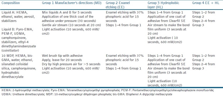
This study was performed in 100 intact caries-free human premolars extracted for orthodontic purpose. After debridement and disinfection in 1% thymol solution, teeth were stored in distilled water until use. Buccal class V cavities centered on the cementoenamel junction were prepared with a 3 mm mesiodistal width, 3 mm occlusogingival dimensions and 1.5 mm depth, using ISO 012 straight fissure diamond point (Dentsply Detrey, USA) in an air–water cooled high-speed handpiece. The bur was changed after every five preparations. The gingival margins on dentin were maintained as butt joint but in enamel a 45° bevel was given. The teeth were divided into two groups according to the one-step self-etch adhesives used: Clearfil S3 Bond (CS3) (Kuraray Medical, Tokyo, Japan) and Xeno III (Dentsply, Konstanz, Germany). Each group was further divided into four subgroups according to the four application modes [Table 1].
Table 1.
Adhesive systems: Composition and application modes of different groups

Group 1 (MD): Adhesive applied according to the manufacturer's directions.
Group 2 (EE): Enamel margins etched with 37% phosphoric acid (Etchant, 3M ESPE, St. Paul, MN, USA) for 15 seconds prior to adhesive application.
Group 3 (HL): The adhesive applied as in Group 1, followed by an additional application of hydrophobic resin coat (adhesive from Clearfil SE Bond, Kuraray, Japan).
Group 4 (EE + HL): Enamel margin was etched with 37% phosphoric acid for 15 seconds prior to adhesive application (as in Group 2) which was followed by application of an additional coat of hydrophobic resin layer (as in Group 3).
The cavities were bulk filled with resin composite (Z 250, 3M ESPE, St. Paul, MN, USA), light cured for 40 seconds at 600 mW/cm2 and polished. The restored teeth in each group were thermocycled for 500 cycles at 5°C and 55°C.
For microleakage test, 10 samples from each group were coated with two layers of sticky wax, leaving a 1 mm window around the cavity margins. The samples were then immersed in freshly prepared 2% methylene blue dye for 48 hours. The teeth were then rinsed with water, the sticky wax was removed and the teeth were left to air dry at room temperature for 24 hours. The teeth were sectioned longitudinally in a buccolingual direction by a cut through the center of the restoration. Dye penetration at the tooth restoration interface was assessed by a stereomicroscope at magnification 10× [Table 2].
Table 2.
Microleakage scores used for the study

Specimen preparation for scanning electron microscopy
Five samples each of both the adhesives from groups 1 and 3 were used for SEM examination of the resin–dentin interfaces. For SEM evaluation, the specimens were sectioned vertically in a buccolingual plane through the center of the restoration and polished. The sections were fixed in 10% formalin for 24 hours and decalcified in 6 N HCl for 30 seconds, rinsed in distilled water and deproteinized by 10-minute immersion in 1% NaOCl, and then rinsed in distilled water. After acid base treatment, the specimens were subjected to dehydration in ascending grades of ethanol up to 100% (25% for 20 minutes, 50% for 20 minutes, 75% for 20 minutes, 95% for 30 minutes and 100% for 60 minutes). The specimens were mounted on aluminum stubs and further dried in vacuum before sputter coating with gold. Gold sputter coating was carried out under reduced pressure in an inert argon gas atmosphere in an Agar Sputter Coater P7340 (Agar Scientific Ltd., Essex, England). The gold-coated samples were examined under SEM (Leo 435 VP, Cambridge, UK) operated at 15 kV. Micrographs of the resin–dentin interface were taken at 500× to observe the quality of bonding between the restorations and dental hard tissue.
Statistical analysis
The results of dye penetration were analyzed using Kruskal–Wallis non-parametric analysis, followed by Mann–Whitney U test to evaluate differences among the experimental groups at a significance level of P = 0.05.
RESULTS
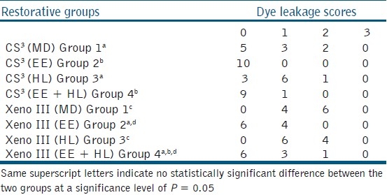
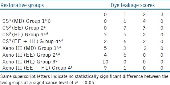
At enamel margins, Clearfil S3 showed significantly lesser leakage than Xeno III when applied according to manufacturer's directions. Additional acid etching of enamel significantly reduced the microleakage for both the one-step self-etch adhesives at the enamel margin [Table 3]. At dentin margins, Xeno III, when applied according to manufacturer's directions, depicted significantly lesser leakage than Clearfil S3. With the application of an additional hydrophobic, solvent-free resin layer, a decrease in leakage scores at dentin margins was observed for both the adhesives tested, but the effect was significant only for Xeno III [Table 4].
Table 3.
Microleakage scores at enamel margins (n=10)

Table 4.
Microleakage scores at dentin margins (n=10)

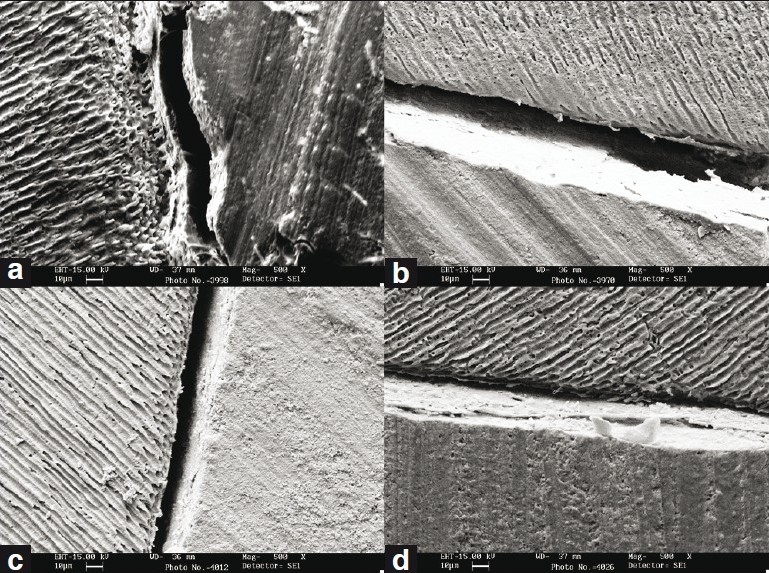
SEM observations of the resin–dentin interface are summarized in Figures 1a–d. Both Clearfil S3 and Xeno III, when applied according to the manufacturer's directions, showed the presence of interfacial gap at the dentinal surface with little or no resin tag formation. After additional application of hydrophobic resin layer, only Xeno III showed improved interfacial adaptation with absence of gap.
Figure 1.

(a) Cross-section of the interface obtained in vitro between CS3 and dentin. Generalized gap can be observed with very few and short resin tags, (b) Cross-section of the interface obtained in vitro between CS3 and dentin with additional hydrophobic layer application showing interfacial gap and absence of resin tags, (c) Cross-section of the interface obtained in vitro between Xeno III and dentin. Generalized gap can be observed with no resin tags, (d) Cross-section of the interface obtained in vitro between Xeno III and dentin with additional hydrophobic resin layer application showing better interfacial adaptation
DISCUSSION
When applied according to manufacturer's directions, Xeno III showed significantly lesser leakage at the dentin margins as compared to Clearfil S3. At enamel margins, however, Clearfil S3 performed significantly better than Xeno III, when applied according to manufacturer's directions. This could be due to the fact that Clearfil S3 contains 10-methacryloxydecyl dihydrogen phosphate (MDP), which has been reported to have a high chemical bonding potential to hydroxyapatite.[23] Furthermore, the calcium salt of MDP is highly insoluble. According to the adhesion–decalcification concept, the less soluble the calcium salt of an acidic molecule, the more intense and stable the molecular adhesion to a hydroxyapatite-based substrate.[24] A chemical interaction between hydroxyapatite and functional monomers in an adhesive leads to higher bond strengths compared with those that rely on micro-mechanical retention to the enamel substrate alone.
With prior phosphoric acid treatment of the enamel margins, a significant decrease in leakage was observed for both Xeno III and Clearfil S3 [Table 3]. Various studies have also indicated the potential benefit of additional enamel etching with phosphoric acid prior to the use of self-etch adhesives.[25–29] Brackett et al.[25] reported a significant decrease in leakage of self-etch adhesives at enamel margins of class V cavities when prior enamel etching with phosphoric acid was done. Luhrs et al.[29] showed that additional phosphoric acid etching significantly increased the shear bond strength to enamel of all the examined self-etch adhesives. Van Meerbeek et al.[28] also reported more marginal defects at the enamel margins of class V composite restorations when prior phosphoric acid etching was not done with mild two-step self-etch adhesives, although they recorded no difference in clinical performance. Ermis et al.[30] evaluated over a 3-year period, the clinical performance of class III composite restorations bonded with a mild two-step self-etch adhesive with and without additional enamel etching. Although a retention rate of 100% was reported for both the non-etch and the etch groups, the non-etched restorations revealed more marginal defects and superficial marginal discoloration than the etched restorations. Contrary to our study, Watanabe et al.[5] demonstrated that two of the five single-step self-etch adhesives tested showed no significant increase in bond strength due to prior acid etching. They suggested that not only the depth of enamel etching, but also the composition and mechanical properties of the adhesives might play important roles in bonding.
The most plausible explanation for decreased leakage at the enamel margin after additional phosphoric acid etching of enamel is the increase in enamel porosity, resulting in an increased resin interlocking and micro-mechanical retention. In spite of the weak correlation between enamel-etching depth/pattern and bond strength found in literature, the aggressiveness of the enamel treatment may play an important role.[31] Khosravi et al.[32] also concluded that simplified all-in-one adhesive systems need pre-etching of the enamel margins with phosphoric acid for an effective seal. Therefore, the null hypothesis that prior acid etching did not affect the microleakage was rejected.
In this study, additional application of hydrophobic, solvent-free resin layer improved the performance of both one-step self-etch adhesives in dentin. However, the effect was significant only for Xeno III [Table 4]. Our results are supported by various studies[9,20,33,34] which reported improvement in resin–dentin bonding of one-step self-etch adhesives after hydrophobic resin layer application. At the enamel margins, a slight increase in leakage was observed after hydrophobic resin layer application though the effect was not statistically significant.
In the SEM examination, both Clearfil S3 and Xeno III, when applied according to the manufacturer's directions, showed the presence of interfacial gap at the dentinal surface with little or no resin tag formation. After additional application of hydrophobic resin layer, only Xeno III showed improved interfacial adaptation with absence of gap. Minimal resin tag formation has been reported in one-step self-etch adhesives and resin tags are usually scant and thinner, indicating that most of the dentinal tubules remain obstructed by smear plugs, which is in contrast to etch and rinse adhesives.[35,36] With the use of SEM, an excellent characterization of the ultramorphological interface is possible due to an enhanced resolution. However, polishing the restored disks followed by demineralization and deproteinization can induce artifacts. Also, the dehydration techniques and the high vacuum may also have provoked changes to the interfaces analyzed. Yet, the challenging conditions during SEM examinations are an excellent test to reveal the weakest link of the evaluated interfaces.
Favorable results obtained after hydrophobic layer application may be attributed to the relative increase in the concentration of hydrophobic monomers within the adhesive interface, decreasing water diffusion which could have otherwise occurred rapidly.[9,37] This could in turn inhibit polymerization and thereby weaken the adhesive–resin composite interface. The additional layer of hydrophobic adhesive also increased the thickness of the adhesive layer that is known to reduce polymerization stresses.[38] Lodovici et al.[39] demonstrated that the application of an extra coat of hydrophobic, solvent-free bonding resin was not able to minimize the damage caused by thermal/mechanical load cycling to the adhesive interfaces obtained with flat dentin surfaces. However, their study was done using a three-step etch and rinse and two-step self-etch system, while most of the current literature regarding the application of an additional hydrophobic resin layer has demonstrated the effect to be beneficial for simplified one-step self-etch adhesives bonded to dentin. It is therefore suggested that further understanding of the factors that contribute to the quality of restorations and their bonding characteristics is required.
CONCLUSIONS
Phosphoric acid etching of enamel margins significantly improved the sealing effectiveness of both one-step self-etch adhesives. Application of hydrophobic resin coat with Xeno III decreased microleakage at dentin margins and showed better interfacial adaptation under SEM.
Footnotes
Source of Support: Nil
Conflict of Interest: None declared.
REFERENCES
- 1.Van Meerbeek B, De Munck J, Yoshida Y, Inoue S, Vargas M, Vijay P, et al. Buonocore Memorial Lecture. Adhesion to enamel and dentin: Current status and future challenges. Oper Dent. 2003;28:215–35. [PubMed] [Google Scholar]
- 2.Van Meerbeek B, Vargas M, Inoue S, Yoshida Y, Peumans M, Lambrechts P, et al. Adhesives and cements to promote preservation dentistry. Oper Dent. 2001;1:119–44. [Google Scholar]
- 3.Hannig M, Bock H, Bott B, Hoth-Hannig W. Intercrystallite nanoretention of self-etching adhesives at enamel imaged by transmission electron microscopy. Eur j Oral Sci. 2002;110:464–70. doi: 10.1034/j.1600-0722.2002.21326.x. [DOI] [PubMed] [Google Scholar]
- 4.Brackett WW, Ito S, Nishitani Y, Haisch LD, Pashley DH. The microtensile bond strength of self-etching adhesives to ground enamel. Oper Dent. 2006;31:332–7. doi: 10.2341/05-38. [DOI] [PubMed] [Google Scholar]
- 5.Watanabe T, Tsubota K, Takamizawa T, Kurokawa H, Rikuta A, Ando S, et al. Effect of prior acid etching on bonding durability of single-step adhesives. Oper Dent. 2008;33:426–33. doi: 10.2341/07-110. [DOI] [PubMed] [Google Scholar]
- 6.Barkmeier WW, Erickson RL, Kimmes NS, Latta MA, Wilwerding TM. Effect of enamel etching time on roughness and bond strength. Oper Dent. 2009;34:217–22. doi: 10.2341/08-72. [DOI] [PubMed] [Google Scholar]
- 7.Celiberti P, Lussi Use of self-etching adhesive on previously etched intact enamel and its effect on sealant microleakage and tag formation. J Dent. 2005;33:163–71. doi: 10.1016/j.jdent.2004.08.010. [DOI] [PubMed] [Google Scholar]
- 8.Hegde MN, Bhandary S. An evaluation and comparison of shear bond strength of composite resin to dentin, using newer dentin bonding agents. J Conserv Dent. 2008;11:71–5. doi: 10.4103/0972-0707.44054. [DOI] [PMC free article] [PubMed] [Google Scholar]
- 9.Albuquerque M, Pegoraro M, Mattei G, Reis A, Loguercio AD. Effect of double-application or the application of a hydrophobic layer for improved efficacy of one-step self-etch systems in enamel and dentin. Oper Dent. 2008;33:564–70. doi: 10.2341/07-145. [DOI] [PubMed] [Google Scholar]
- 10.Miyazaki M, Sato M, Onose H. Durability of enamel bond strength of simplified bonding systems. Oper Dent. 2000;25:75–80. [PubMed] [Google Scholar]
- 11.Bastioli C, Romano G, Migliaresi C. Water sorption and mechanical properties of dental composites. Biomater. 1990;11:219–23. doi: 10.1016/0142-9612(90)90159-n. [DOI] [PubMed] [Google Scholar]
- 12.Eliades G, Vougiouklakis G, Palaghias G. Heterogeneous distribution of single-bottle adhesive monomers in the resin-dentin interdiffusion zone. Dent Mater. 2001;17:277–83. doi: 10.1016/s0109-5641(00)00082-8. [DOI] [PubMed] [Google Scholar]
- 13.Spencer P, Wang Y. Adhesive phase separation at the dentin interface under wet bonding conditions. J of Biomed Mater Res. 2002;62:447–56. doi: 10.1002/jbm.10364. [DOI] [PubMed] [Google Scholar]
- 14.Tay FR, Pashley DH, Yoshiyama M. Two modes of nanoleakage expression in single-step adhesives. J Dent Res. 2002;81:472–76. doi: 10.1177/154405910208100708. [DOI] [PubMed] [Google Scholar]
- 15.Ferrari M, Tay FR. Technique sensitivity in bonding to vital acid-etched dentin. Oper Dent. 2003;28:3–8. [PubMed] [Google Scholar]
- 16.Tay FR, Pashley DH, Yiu CK, Sanares AM, Wei SH. Factors contributing to the incompatibility between simplified-step adhesives and chemically-cured or dual-cured composites. Part1. Single-step self-etching adhesive. J Adhes Dent. 2003;5:27–40. [PubMed] [Google Scholar]
- 17.Nyunt MM, Imai Y. Adhesion to dentin with resin using sulfinic acid initiator system. Dent Mater J. 1996;15:175–82. doi: 10.4012/dmj.15.175. [DOI] [PubMed] [Google Scholar]
- 18.Tay FR, Pashley DH. Have dentin adhesives become too hydrophilic? J Can Dent Assoc. 2003;69:726–31. [PubMed] [Google Scholar]
- 19.Tay FR, Lai CN, Chersoni S, Pashley DH, Mak YF, Suppa P, et al. Osmotic blistering in enamel bonded with one-step self-etch adhesives. J Dent Res. 2004;83:290–5. doi: 10.1177/154405910408300404. [DOI] [PubMed] [Google Scholar]
- 20.Brackett WW, Ito S, Tay FR, Haisch LD, Pashley DH. Microtensile dentin bond strength of self-etching resins: Effect of a hydrophobic layer. Oper Dent. 2005;30:733–8. [PubMed] [Google Scholar]
- 21.Van Landuyt KL, Peumans M, De Munck J, Lambrechts P, Van Meerbeek B. Extention of one-step self-etch adhesive into a multi–step adhesive. Dent Mater. 2006;22:533–4. doi: 10.1016/j.dental.2005.05.010. [DOI] [PubMed] [Google Scholar]
- 22.Munro GA, Hilton TJ, Hermesch CB. In vitro microleakage of etched and rebonded class V composite resin restorations. Oper Dent. 1996;21:203–8. [PubMed] [Google Scholar]
- 23.Yoshioka M, Yoshida Y, Inoue S, Lambrechts P, Vanherle G, Nomura Y, et al. Adhesion/decalcification mechanisms of acid interactions with human hard tissues. J Biomed Mater Res. 2002;59:56–62. doi: 10.1002/jbm.1216. [DOI] [PubMed] [Google Scholar]
- 24.Yoshida Y, Nagakane K, Fukuda R, Nakayama Y, Okazaki M, Shintani H, et al. Comparative study on adhesive performance of functional monomers. J Dent Res. 2004;83:454–8. doi: 10.1177/154405910408300604. [DOI] [PubMed] [Google Scholar]
- 25.Brackett MG, Brackett WW, Haisch LD. Microleakage of class V resin composites placed using self-etching resins: Effect of prior enamel etching. Quint Inter. 2006;37:109–13. [PubMed] [Google Scholar]
- 26.Peumans M, De Munck J, Van Landuyt KL, Lambrechts P, Van Meerbeek B. Three-year clinical effectiveness of a two-step self-etch adhesive in cervical lesions. Eur J Oral Sci. 2005;113:512–8. doi: 10.1111/j.1600-0722.2005.00256.x. [DOI] [PubMed] [Google Scholar]
- 27.Van Landuyt KL, Kanumilli P, De Munck J, Peumans M, Lambrechts P, Van Meerbeek B. Bond strength of a mild self-etch adhesive with and without prior acid-etching. J Dent. 2006;34:77–85. doi: 10.1016/j.jdent.2005.04.001. [DOI] [PubMed] [Google Scholar]
- 28.Van Meerbeek B, Kanumilli P, De Munck J, Van Landuyt KL, Lambrechts P, Peumans M. A randomized controlled study evaluating the effectiveness of a two-step self-etch adhesive with and without selective phosphoric-acid etching of enamel. Dent Mater. 2005;21:375–83. doi: 10.1016/j.dental.2004.05.008. [DOI] [PubMed] [Google Scholar]
- 29.Luhrs AK, Guhr S, Schilke R, Borchers L, Geurtsen W, Gunay H. Shear bond strength of self- etch adhesives to enamel with additional phosphoric acid etching. Oper Dent. 2008;33:155–62. doi: 10.2341/07-63. [DOI] [PubMed] [Google Scholar]
- 30.Ermis RB, Temel UB, Celik EU, Kam O. Clinical performance of a two-step self-etch adhesive with additional enamel etching in class III cavities. Oper Dent. 2010;35:147–55. doi: 10.2341/09-089-C. [DOI] [PubMed] [Google Scholar]
- 31.Perdigao J, Lopes L, Lambrechts P, Leitão J, Van Meerbeek B, Vanherle G. Effects of a self-etching primer on enamel shear bond strengths and SEM morphology. Am J Dent. 1997;10:141–6. [PubMed] [Google Scholar]
- 32.Khosravi K, Ataei E, Mousavi M, Khodaeian N. Effect of phosphoric acid etching of enamel margins on the microleakage of a simplified all-in-one and a self-etch adhesive system. Oper Dent. 2009;34:531–36. doi: 10.2341/08-026-L. [DOI] [PubMed] [Google Scholar]
- 33.Reis A, Albuquerque M, Pegoraro M, Mattei G, Bauer JR, Grande RH, et al. Can the durability of one-step self-etch adhesives be improved by double application or by an extra layer of hydrophobic resin? J Dent. 2008;33:309–15. doi: 10.1016/j.jdent.2008.01.018. [DOI] [PubMed] [Google Scholar]
- 34.Pushpa R, Suresh BS, Arunagiri D, Manuja N. Influence of hydrophobic layer and delayed placement of composite on the marginal adaptation of two self-etch adhesives. J Conserv Dent. 2009;12:60–64. doi: 10.4103/0972-0707.55619. [DOI] [PMC free article] [PubMed] [Google Scholar]
- 35.Margvelashvili M, Goracci C, Beloica M, Papacchini F, Ferrari M. In vitro evaluation of bonding effectiveness to dentin of all-in-one adhesives. J Dent. 2010;38:106–12. doi: 10.1016/j.jdent.2009.09.008. [DOI] [PubMed] [Google Scholar]
- 36.Radovic I, Vulicevic ZR, Garcia-Godoy F. Morphological evaluation of 2- and 1- step self-etching system interfaces with dentin. Oper Dent. 2006;31:710–8. doi: 10.2341/05-145. [DOI] [PubMed] [Google Scholar]
- 37.Tay FR, Pashley DH, Suh BI, Carvalho RM, Itthagarum Single-step adhesives are permeable membranes. J Dent. 2002;30:371–82. doi: 10.1016/s0300-5712(02)00064-7. [DOI] [PubMed] [Google Scholar]
- 38.Choi KK, Condon JR, Ferracane JL. The effects of adhesive thickness on polymerization contraction stress of composite. J Dent Res. 2000;79:812–7. doi: 10.1177/00220345000790030501. [DOI] [PubMed] [Google Scholar]
- 39.Lodovici E, Reis A, Geraldeli S, Ferracane JL, Ballester RY, Filho LE. Does adhesive thickness affect resin-dentin bond strength after thermal/load cycling? Oper Dent. 2009;34:58–64. doi: 10.2341/08-37. [DOI] [PubMed] [Google Scholar]


