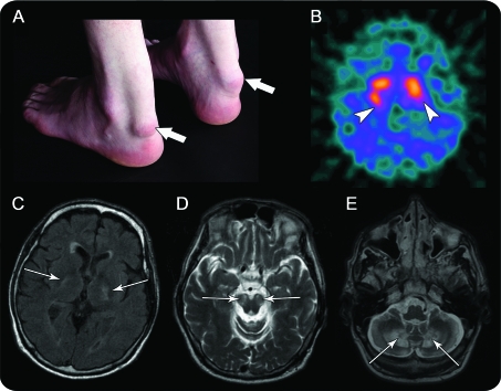
A 45-year-old man presented with rapidly progressive gait and balance deterioration, bradykinesia, and speech and swallowing difficulties. He had longstanding cognitive impairment and bilateral cataract surgery in childhood. On examination he had asymmetric parkinsonism with pyramidal signs and bilateral Achilles tendon xanthomas (figure) . Imaging at age 43 (figure) and increased urinary bile alcohols confirmed cerebrotendinous xanthomatosis (CTX).1,2 Treatment was started with chenodeoxycholic acid but clinical deterioration continued. Cocareldopa was added at age 45 with transient benefit.
Figure. Clinical and radiologic signs.
(A) Bilateral tendon xanthomas (arrows). (B) I-123-Ioflupane (I-123 FP-CIT) SPECT: reduced bilateral, asymmetric putaminal uptake (arrowheads). MRI (arrows): diffuse volume loss with signal abnormality, (C) in the globus pallidus, internal capsules on axial fluid-attenuated inversion recovery, (D) cerebral peduncles, substantia nigra, and (E) extensive white matter involvement of the cerebellar hemispheres including dentate nuclei on axial T2-weighted imaging.
CTX is an autosomal recessive disorder caused by reduced mitochondrial sterol-27-hydroxylase activity (CYP27A1 gene), leading to accumulation of cholestanols. Features include infantile diarrhea, childhood cataracts, xanthomas, and psychiatric and neurologic abnormalities. While clinical improvement has been described with chenodeoxycholic acid, statins, and levodopa for patients with parkinsonian features, other patients continue to progress.1
AUTHOR CONTRIBUTIONS
Olga Kirmi: primary author, organization, image collection. Elaine Murphy: author, clinical input, clinical photograph. Miryam Carecchio: author, clinical input. Tom Sulkin: figure author and interpretation of DAT scan and MRI. Julia Rankin: interpretation of clinical and biochemical findings, diagnosis. Fergus Robertson: author, neuroradiology input, MR interpretation, supervisor.
DISCLOSURE
Dr. Kirmi reports no disclosures. Dr. Murphy has received a speaker honorarium from Vitaflo International and receives research support from Shire plc and Genzyme Corporation. Dr. Carecchio has received research support from IRCSS Fondazione Maggiore Policlinico, University of Milan, Italy. Dr. Sulkin serves on the medical advisory committee of e-locum, UK. Dr. Rankin and Dr. Robertson report no disclosures.
REFERENCES
- 1. Su CS, Chang WN, Huang SH, et al. Cerebrotendinous xanthomatosis patients with and without parkinsonism: clinical characteristics and neuroimaging findings. Mov Disord 2010;25:452–458 [DOI] [PubMed] [Google Scholar]
- 2. Barkhof F, Verrips A, Wesseling P, et al. Cerebrotendinous xanthomatosis: the spectrum of imaging findings and the correlation with neuropathologic findings. Radiology 2000;217:869–876 [DOI] [PubMed] [Google Scholar]


