Abstract
Accumulating evidence has revealed that the tyrosine kinases play a major role in glioma proliferation and invasion. The largest family of tyrosine kinases, the Eph family, and its ligands, the ephrins, are frequently overexpressed in glioma, suggesting important roles for their bidirectional signals in glioma pathobiology. Ephs bind to cell surface–associated ephrin ligands on neighboring cells and have many biological functions during embryonic development of the central nervous system, including axon mapping, cell migration, and angiogenesis. Recent findings suggest that Eph/ephrin signaling affects glioma cell growth, migration, and invasion in vitro and in vivo. However, their roles in glioma seem complex, because both tumor growth promoter and suppressor potentials have been ascribed to Ephs and ephrins. Here, we review recent advances in research on the role of Eph/ephrin signaling in glioma and suggest that the Eph/ephrin system could be a potential target of glioma therapy.
Keywords: Eph, ephrin, glioma, tyrosine kinase
Protein tyrosine kinases are enzymes that are capable of adding a phosphate group to certain tyrosines on target proteins. A receptor tyrosine kinase (RTK) is a tyrosine kinase located at the cellular membrane that is activated by the binding of a ligand to its extracellular domain. Protein phosphorylation by kinases is an important intracellular communication mechanism and cellular activity regulator that functions as an “on” or “off” switch for many cellular functions. Ninety unique tyrosine kinase genes, including 58 RTKs, have been identified in the human genome, products of which regulate cellular proliferation, survival, differentiation, function, and motility. Tyrosine kinases have been shown not only to be the key regulators of normal cellular processes but also to play a critical role in the development and progression of many cancers. Thus, tyrosine kinases are now considered excellent targets for cancer chemotherapy.
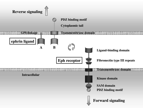
Erythropoietin-producing human hepatocellular carcinoma (Eph) receptors, which contain 14 distinct members, constitute the largest family of RTKs in the human genome.1–3 Eph receptors are separated into EphA (A1–A8) and EphB (B1–B6) subgroups on the basis of sequence homology and ligand-binding specificity. These receptors are located on cell surfaces and transduce signals upon binding to the ligands that are typically located on the surfaces of neighboring cells (Fig. 1). EphA receptors bind preferentially to glycosylphosphatidylinositol (GPI)–linked ephrin-A ligands (ephrins A1–A6), whereas EphB receptors bind to ephrin-B ligands (ephrins B1–B3) that contain transmembrane and intracellular domains. Ephs, like other RTKs, initiate signal transduction through autophosphorylation after ligand-receptor engagement, a phenomenon referred to as “forward signaling.” In contrast to other soluble RTK ligands, the ephrins possess unique features in that they are membrane bound and are capable of receptor-like active signaling (“reverse signaling”), which results in bidirectional cell-to-cell communication (Fig. 1). In general, ephrin-A and ephrin-B ligands interact with EphA and EphB receptors, respectively, but there are some exceptions in which ephrin-A5 functionally interacts with EphB2 and in which EphA4 binds to both ephrin-A and ephrin-B family members.2,4 The ensuing bidirectional signals have emerged as a major form of contact-dependent communication between cells. The mutual activation of Eph/ephrin generates repulsive signals and causes dispersive effects upon cell–cell contact. The signal is regulated at several levels, for example, by varying degrees of forward and reverse signaling, as well as multimerization of ligand-receptor complexes, which are involved with kinase-dependent and kinase-independent signaling.5 Eph/ephrin molecules are typically most highly expressed in neural and endothelial cells, and their signaling mainly regulates a number of cellular events during embryonic development, such as neural crest cell migration, axon guidance, boundary formation, hindbrain segmentation, and vasculogenesis.1,6 The different Eph/ephrin molecules are conceivably linked to different intracellular signaling pathways in a cell type–specific manner, which allows this system to perform a variety of functions.
Fig. 1.
Eph/ephrin structure and signaling. The figure shows a general diagram of the A and B ephrin ligands and their Eph receptor, including membrane orientation. Eph/ephrin activation can lead both to forward and reverse signaling.
Extensive evidence indicates that the Eph/ephrin signaling pathways are the key determinants for both physiological and pathological conditions.3,7 The expression of Ephs and ephrins is frequently altered in tumor tissue, compared with the tissue of origin. Emerging evidence suggests their strong involvement in tumor biology, including in metastasis, invasion, and angiogenesis.8–10 However, how these receptors affect cancer progression remains incompletely understood. The result of the Eph/ephrin interaction is remarkably divergent in different contexts. The same molecules can promote cell proliferation in one tissue but inhibit it in another and can even act as a tumor promoter and suppressor in the same tissue.11 It appears that the Eph/ephrin system may support the onco-phenotype, rather than that the activation of the system is the primary event in tumorigenesis. The challenge now is to better understand the complex and seemingly paradoxical signaling mechanisms of Eph receptors and ephrins, which will allow the development of effective strategies to target these proteins in the treatment of diseases such as cancer. In this article, we focus on Eph/ephrin function in the brain, especially in malignant glioma, the most common malignant primary brain tumor in adults.
Eph/ephrin in the normal brain
Eph receptors and ephrin ligands were initially characterized as important regulators in central nervous system development.12 The significance of Eph/ephrin signaling in the development of embryonic vasculature was initially proven by gene knockout studies in mice. Disruption of either ephrin-B2 or EphB4 resulted in similar defects in blood vessel remodeling,13,14 suggesting that ephrin-B2– and EphB4-mediated signaling between developing vessels may be required for proper vascular system morphogenesis and patterning. In addition, other Eph/ephrins may also be involved in vascular development, because embryonic arteries express ephrin-B1 whereas embryonic veins express ephrin-B1, EphB3, and EphB4.15 Recent progress in the field has shown that Eph receptors and ephrins affect multiple aspects of adult brain function, including regulation of synaptic structure and electrophysiological properties and excitatory synapse formation.16 In fact, certain Eph/ephrin molecules are expressed in the normal brain, as shown in a mouse system17 and in Homo sapiens.18–20 Specifically, ephrin-A3 expressed on astrocytes in the adult mouse hippocampus interacts with EphA4 on dendritic spines of hippocampal neurons, causing spine shortening and collapse. A similar phenomenon involves neuronal growth cone collapse mediated by the ephrin-A/EphA interaction during development.21
Eph/ephrin and neural stem cells
The discovery that pluripotent, self-renewing neural stem cells exist throughout life in the adult mammalian brain has only reemerged in the past decade,22,23 reflecting the discovery of evidence in the 1960s of a possible occurrence of neurogenesis in the adult brain.24 Neural stem cells are influenced by Eph/ephrin signaling during both development and adulthood. There is some evidence that Eph/ephrin function is required to limit the proliferative potential of neural stem cells, suggesting that Eph and ephrin are the key molecules maintaining the neural stem cell niche.25–27 Neural stem cells in the subventricular zone express ephrin-A2, whereas quiescent ependymal cells express EphA7. EphA7 induces ephrin-A2 reverse signaling, negatively regulating adult neural stem cell proliferation.26 Furthermore, blocking of the interaction between EphB receptors and ephrin-B ligands leads to an increase in the number of dividing cells in the subventricular zone.25 Therefore, both the A and B classes negatively regulate neural stem cell proliferation in the adult subventricular zone. In addition to influencing neural stem cells in the adult brain, EphB2 signaling also regulates niche cell plasticity and plays a critical role in stem cell niche maintenance and self-renewal.28 Cancer stem cells have been isolated from human brain tumors,29 resulting in a hierarchy of clonally derived populations in brain cancer. Malignant cancer stem cells exhibit extensive proliferative potential, whereas differentiated cancer cells have limited proliferative capabilities. Glioma stem cells become self-sufficient, undergoing uncontrolled proliferation, due to either internal mutations that allow for uncontrolled proliferation or changes in the niche signals in which the environment becomes overwhelmed with cell proliferation–favoring signals. Thus, specific Eph/ephrin activity might negatively regulate brain tumor development. To our knowledge, no data have yet been reported concerning the expression of Eph/ephrin in glioma stem cells, although various kinds of cancer stem cells have been shown to express Eph/ephrin.30 Elucidation of the signaling pathways that Ephs and ephrins employ to regulate the generation of new cells from neural stem cells in adults and to influence glioma pathobiology may contribute to the development of new therapeutic strategies for glioblastoma (GBM).
Eph/ephrin and glioma
To date, various studies have investigated the involvement of Eph/ephrin in several pathogenetic processes in the nervous system. Several studies in recent years have clearly indicated that altered Eph receptor and ephrin ligand expression is associated with histological grade in glioma and increased potential for GBM growth, angiogenesis, invasion, and adverse outcome. Certain Eph/ephrin members have already been recognized as potential molecular markers and targets in GBM for the development of novel biological therapeutic agents. The glioma cell invasion process appears to be similar to that of cell movement during neural development. The highly infiltrative nature of human gliomas recapitulates the migratory behavior of glial progenitors, suggesting that the activators, receptors, and signaling proteins that contribute to neural crest cell migration, such as the Eph/ephrin system, may be associated with glioma invasion.31 In fact, Eph/ephrin family members were identified as being overexpressed by the invading GBM cells by DNA microarray analysis comparing invasive and stationary tumor cells collected from GBM biopsy specimens using laser-capture microdissection.18,19,32 Glioma cells tend to migrate along certain preferred paths, particularly along blood vessels and fiber tracts,33 suggesting that invading glioma cells extensively interact with their respective microenvironment. Because Eph/ephrin is expressed in the normal human brain, it is reasonable to speculate that glioma cells possibly interact with normal cells via Eph/ephrin during both progression and invasion processes. Taken together, the Eph/ephrin system seems to play a role in the pathobiology of human glioma. The function, expression, and clinical relevance of Eph/ephrin in GBM reported to date are summarized in Table 1.
Table 1.
The function and expression of Eph/ephrin members in glioma
| Eph/ephrin | Function in GBM | Target molecules | Expression in GBM | Clinical relevance |
|---|---|---|---|---|
| EphA | ||||
| EphA2 | Ligand dependent; anti-proliferation, anti-invasion5 | MAPK37, Akt5 | Overexpression34,40,41 | Unfavorable prognostic factor37,38 |
| Ligand independent; invasion promoter | ||||
| Neovasculalization38 | ||||
| EphA4 | FGFR1, Akt/MAPK. Rac1, Cdc4245 | Overexpression45 | ||
| EphA5 | No effect on proliferation48 | Positive47 | Unfavorable prognostic factor46 | |
| EphA7 | Neovasculalization46 | Overexpression46 | ||
| ephrin-A | ||||
| ephrin-A1 | low-level34,40 | |||
| ephrin-A2 | Favorable prognostic factora | |||
| ephrin-A3 | Down regulateda | |||
| ephrin-A5 | Tumor suppressor49 | Negative regulation of EGFR49 | Low-level49 | |
| EphB | ||||
| EphB1 | Favorable prognostic factora | |||
| EphB2 | Invasion promoter18 | R-Ras50 | Overexpression18 | |
| EphB4 | Angiogenesis54 | |||
| ephrin-B | ||||
| ephrin-B1 | Overexpression20 | |||
| ephrin-B2 | Invasion promoter20, angiogenesis54 | VEGFR254 | Overexpression20 | Unfavorable prognostic factor20 |
| ephrin-B3 | Invasion promoter19,32 | Rac119 | Overexpression19 | |
Abbreviations: EGFR, epidermal growth factor receptor; FGFR, fibroblast growth factor receptor; GBM, glioblastoma; MAPK, mitogen-activated protein kinase; VEGFR2, vascular endothelial growth factor receptor-2.
aM. Nakada; unpublished data.
EphA/ephrin-A signaling in glioma
Among the EphA/ephrin-A proteins, most research to date has described the relationship between EphA2 and ephrin-A1 with respect to glioma biology.34–36 EphA2 is also highly expressed in different carcinomas, including breast, ovarian, gastric, pancreatic, and prostate cancers, suggesting that EphA2 might be a common oncoprotein in many cancers.34 A novel genome-wide screen combining patient outcome analyses with array comparative genomic hybridization and mRNA expression profiling found that EphA2 mRNA overexpression was inversely correlated with patient survival in a 21-GBM panel.37 Another report mentioned that EphA2 was strongly overexpressed in 60% of tumors in patients with GBM and was associated with poor prognosis.38 Increased EphA2 expression was found not only in tumor cells but also in highly vascular GBM areas, as well as in endothelial cells surrounding tumor areas, suggesting that EphA2 is involved in neovascularization.38 Regulation of EphA2 expression is not known in detail. However, a recent article revealed that microRNA-26b, which is down-regulated in GBM, is an important negative EphA2 regulator.39
On the contrary, the expression pattern of ephrin-A1 in tumors does not seem to be the same as what has been documented for EphA2. Ephrin-A1 is expressed at low levels in GBM when EphA2 is overexpressed.34 Another report demonstrated that ephrin-A1 and EphA2 are co-localized in normal brain tissues. In contrast, EphA2 is dominantly expressed in glioma regions, whereas almost no ephrin-A1 expression is observed.40,41 EphA2, although overexpressed, may be present in its biologically inactive state because of the low level of ephrin-A1 ligand in GBM.34 A similar pattern of differential ephrin-A1/EphA2 expression was found in breast cancer.42
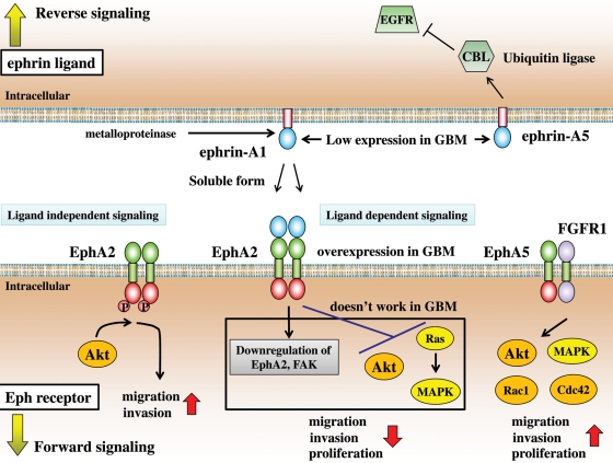
In several studies, EphA2 overexpression has been linked to malignant progression.43 Paradoxically, activation of EphA2 kinase by ephrin-A1 on glioma cells can trigger signaling events, such as anti-proliferation and anti-invasion, which are more consistent with a tumor suppressor.34 Ephrin-A1 possesses tumor-suppressing properties in this manner. Interestingly, prolonged exposure to ephrin-A1 leads to down-regulation of EphA2 receptor and focal adhesion kinase, resulting in reduced proliferation and migration activity.37,40 Down-regulation of EphA2 by ephrin-A1 is ascribed to the endocytosis of EphA2/ephrin-A1 complex.6 The low ephrin-A1 levels in GBM cells explain, at least in part, the lack of EphA2 receptor activation and the resultant persistent overexpression. These findings in vitro and the conflicting expression of EphA2 and ephrin-A1 in vivo suggest the possible existence of a feedback loop mechanism in which ephrin-A1 suppresses EphA2 expression and vice versa. Miao et al.5 recently revealed an interesting mechanism that converts the EphA2 receptor from a tumor suppressor (when activated by ephrin-A1 ligand; ligand-dependent signaling) to a tumor promoter (when phosphorylated by Akt; ligand-independent signaling). EphA2 inhibits Akt and Ras/mitogen-activated protein kinase (MAPK) when activated by ephrin-A1, resulting in inhibited cell migration and proliferation.5,44 However, ligand-dependent signaling may not work in GBM because of a lack of ephrin-A1 in GBM tissue. Instead, ligand-dependent signaling possibly works via autophosphorylation of EphA1 overexpressed in GBM without ephrin-A1 stimulation (Fig. 2).
Fig. 2.
Putative model of EphA/ephrin-A signaling in glioma. EphAs are enhancers of the malignant phenotype, whereas ephrin-As are tumor suppressors. In ligand-independent signaling of EphA2, Akt phosphorylates S897 in the carboxy-terminal tail of EphA2, leading to increased EphA2-dependent cell migration and invasion. In turn, ephrin-A1–induced EphA2 signaling inactivates Akt by causing its dephosphorylation at T308 and S473, thus decreasing EphA2 phosphorylation at S897 and, consequently, cell migration and invasion. Ligand-dependent signaling is ineffective in glioblastoma due to the lack of ephrin-A1. P, tyrosine phosphorylation.
Membrane-anchored ephrin-A1 is widely considered the ligand's endogenous functional form. Thus, the important physiological functions of ephrin-A1 appear to depend largely on cell-cell contact. However, Wykosky et al.36 reported an interesting finding: ephrin-A1 can be released from the GBM cell membrane and function as a soluble monomer in a paracrine manner without cell-cell contact. Similar to membrane-bound ephrin-A1, soluble monomeric ephrin-A1 also induces EphA2 down-regulation and decreases MAPK phosphorylation, resulting in inhibited cell migration and proliferation (Fig. 2). This finding may facilitate the design and enable a wider application of ephrinA1-based therapeutics targeting the EphA2 receptor.
In addition to the EphA2 receptor, several kinds of EphA receptors are reported as enhancers for the malignant glioma phenotype and might be potential therapeutic targets for malignant glioma. EphA4 is a specific Eph receptor for the ephrin-A and ephrin-B ligands. EphA4 receptor mRNA levels were significantly higher in glioma tissues than in normal brain tissues. Furthermore, EphA4 expression correlated with increasing tumor grade. Fukai et al.45 demonstrated that EphA4 forms a heteroreceptor complex with fibroblast growth factor receptor 1 (FGFR1) in glioma cells and that the EphA4-FGFR1 complex potentiated FGFR-mediated downstream signaling, such as Akt/MAPK, Rac1, and Cdc42, resulting in the promotion of proliferation and invasion (Fig. 2). Wang et al.46 found that overexpression of EphA7 protein determined in immunohistochemical analysis of the tissue was predictive of adverse outcomes in patients with GBM. In contrast, EphA5 was expressed in human glioma47 but did not affect proliferation in the U118 glioma cell line that expresses EphA5.48
Contrary to the EphA receptor, the majority of ephrin-A ligands have been reported as tumor suppressors in glioma as ephrin-A1. To date, little information has been available regarding the function of reverse signaling by ephrin-A1 in GBM. Interestingly, ephrin-A5 reverse signaling can negatively regulate epidermal growth factor receptor (EGFR), which is frequently aberrant in glioma. However, ephrin-A5 was expressed at a lower level in primary GBMs than in normal brain tissues, suggesting that glioma cells possibly silence ephrin-A5 expression to avoid its suppressive effect on EGFR (Fig. 2).49 Similarly, we found that ephrin-A2 functions as a tumor suppressor and that its expression is a favorable prognostic factor in GBM, although its expression level was similar in GBM and normal brain tissues (M. Nakada; unpublished data).
EphB/ephrin-B signaling in glioma
The function of EphB/ephrin-B signaling in GBM is less well known than that of EphA/ephrin-A. However, it has become clear that one of the major functions of EphB/ephrin-B in GBM is promoting migration and invasion. Interestingly, pathway enrichment analysis comparing whole human genome expression profiling between 2 distinct GBM cell phenotypes (invading cells and tumor core cells) from GBM specimens highlighted EphB/ephrin-B as the signaling system most tightly linked to the invading cell phenotype.20
EphB2 is overexpressed in several cancers, including GBM and gastrointestinal and liver cancers.18 A prior study showed that EphB2 plays a functional role in promoting GBM cell invasion by eliciting signaling through R-Ras and affecting integrin activity.50 Paradoxically, a tumor-suppressing role for EphB2 has been reported in both colorectal and prostate cancers.51,52 The dichotomy of EphB2 functioning as both an oncoprotein and a tumor suppressor may depend on cell type and the microenvironment in cancer.
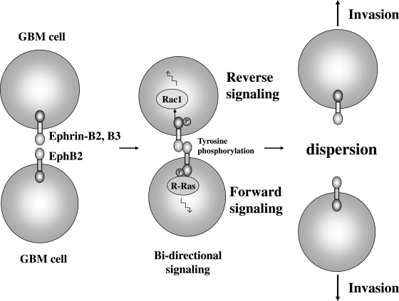
All ephrin-B ligand members are overexpressed in GBM, whereas all ephrin-A members reported thus far are down-regulated in GBM cells (Table 1). Co-expression of EphB2 and ephrin-B in GBM cells suggests the existence of an EphB/ephrin-B interaction through cell-cell contact in GBM; in contrast, EphA is speculated to interact with cleaved soluble ephrin-A but not with membrane-anchored ephrin-A in GBM. To date, ephrin-B2 and ephrin-B3 have been reported to be involved in GBM invasion as ligands for EphB2, suggesting a novel mechanism of promoting glioma invasion through direct interaction between individual tumor cells (Fig. 3).19,20 Ephrin-B2 and ephrin-B3 can activate EphB2 through cell-cell contact, inducing invasion via EphB2 forward signaling. Not much is yet known to date about reverse signaling through ephrin-A ligands, as mentioned above. However, reverse signaling of the ephrin-B2 and ephrin-B3 ligands was demonstrated to be an important factor regulating glioma cell invasion through Rac1 GTPase (Fig. 3).19 Moreover, ephrin-B2 expression levels were significantly associated with short-term survival in malignant astrocytomas.20
Fig. 3.
Putative model of EphB2/ephrin-B2, -B3 function in glioma. Cell–cell contact induces EphB2/ephrin-B3 phosphorylation, resulting in cell dispersion. This interaction contributes to glioma migration and invasion. P, tyrosine phosphorylation.
In addition, some EphB/ephrin-B members play an important role in vasculature organization, both in the central nervous system and in peripheral organs. EphB4 and ephrin-B2 have been extensively investigated from the standpoint of vascular biology. One report demonstrated that EphB4 and ephrin-B2 are overexpressed in endothelial cells of malignant brain tumors,53 but Eph/ephrin expression in adult normal brain vessels has not been studied. A recent study reported that ephrin-B2 reverse signaling induces vascular endothelial growth factor receptor (VEGFR)–2 internalization and activation and that it controls vessel sprouting during developmental angiogenesis.54 In addition, this system has been reported during tumor angiogenesis in a mouse GBM model.54 Thus, blocking of ephrin-B2 signaling in tumors might represent an intriguing strategy to simultaneously interfere with VEGFR2 function that could be used as an alternative or combinatorial anti-angiogenic treatment for tumor therapy.
From a different point of view, the finding that both glioma cells and endothelial cells express Eph/ephrin suggests interplay between Eph and ephrin at the glioma cell–endothelial cell interface. This may explain both GBM cell invasion along vessels and the difficulty of GBM cell invasion in vessels due to repulsion between tumor cells and endothelial cells.
Prospective
Eph/ephrin is a promising new therapeutic target in GBM. On the basis of basic research results, drug development is proceeding in fits and starts by several companies. EphA2 is a very attractive target for novel anticancer therapies. The highest degree of EphA2 expression in GBM and its low expression levels in the normal adult brain suggest that it is a striking target for GBM. Anti-EphA2 inhibitors are under development. Dasatinib, a multi-targeted kinase inhibitor already used in the treatment of chronic myelogenous leukemia and under clinical evaluation for treatment of solid tumors, potently inhibits EphA2 and other Eph receptors in addition to its primary targets, the Abl and Src family kinases.55 Other approaches that do not rely on the downstream consequences of EphA2 receptor activation or inhibition use EphA2 receptor-targeting molecules to selectively deliver drugs, toxins, and antigenic peptides to stimulate anti-tumor immune responses. Debinski et al.35,56 are developing an ephrin-A1 ligand that targets a Pseudomonas-derived toxin. Hatano et al.41,57 demonstrated that synthetic EphA2-derived peptide epitopes can elicit specific cytotoxic T-lymphocyte responses and anti-tumor responses in vivo and proposed a promising strategy against GBM by targeting EphA2 in peptide-based vaccine trials. In addition, it is notable that EphB4/ephrin-B2 signaling is a novel antiangiogenic target. A soluble EphB4 receptor and anti-EphB4 monoclonal antibodies have been developed to block EphB4 signaling.58
Conclusions
Emerging understanding of Eph/ephrin function provides new insight to glioma biology. The information stated here illustrates that we are only beginning to understand how this family of receptors and their ligands work in GBM. Because imbalance in the Eph/ephrin function apparently contributes in many aspects of glioma progression, the targeting of Eph/ephrin kinases to modulate aggressive glioma behavior might lead directly to the development of novel therapies that mandate expedient clinical exploration. The unique biological nature of Eph/ephrin, such as bidirectional signaling and the presence of a secreted form, may provide possible clues for manipulating the regulation of Eph/ephrin systems for GBM therapy.
Conflict of interest statement. None declared.
Funding
This work was supported by Grant-in-Aid for Young Scientists (A-21689038) from the Ministry of Education, Culture, Sports, Science and Technology and from the Japan Society for the Promotion of Science (M. N.), Foundation for Promotion of Cancer Research (M. N.), and Osaka Cancer Research Foundation (M. N.).
References
- 1.Wilkinson DG. Multiple roles of EPH receptors and ephrins in neural development. Nat Rev Neurosci. 2001;2:155–164. doi: 10.1038/35058515. doi:10.1038/35058515. [DOI] [PubMed] [Google Scholar]
- 2.Kullander K, Klein R. Mechanisms and functions of Eph and ephrin signalling. Nat Rev Mol Cell Biol. 2002;3:475–486. doi: 10.1038/nrm856. doi:10.1038/nrm856. [DOI] [PubMed] [Google Scholar]
- 3.Pasquale EB. Eph receptors and ephrins in cancer: bidirectional signalling and beyond. Nat Rev Cancer. 2010;10:165–180. doi: 10.1038/nrc2806. doi:10.1038/nrc2806. [DOI] [PMC free article] [PubMed] [Google Scholar]
- 4.Himanen JP, Chumley MJ, Lackmann M, et al. Repelling class discrimination: ephrin-A5 binds to and activates EphB2 receptor signaling. Nat Neurosci. 2004;7:501–509. doi: 10.1038/nn1237. doi:10.1038/nn1237. [DOI] [PubMed] [Google Scholar]
- 5.Miao H, Li DQ, Mukherjee A, et al. EphA2 mediates ligand-dependent inhibition and ligand-independent promotion of cell migration and invasion via a reciprocal regulatory loop with Akt. Cancer Cell. 2009;16:9–20. doi: 10.1016/j.ccr.2009.04.009. doi:10.1016/j.ccr.2009.04.009. [DOI] [PMC free article] [PubMed] [Google Scholar]
- 6.Pitulescu ME, Adams RH. Eph/ephrin molecules—a hub for signaling and endocytosis. Genes Dev. 2010;24:2480–2492. doi: 10.1101/gad.1973910. doi:10.1101/gad.1973910. [DOI] [PMC free article] [PubMed] [Google Scholar]
- 7.Pasquale EB. Eph-ephrin bidirectional signaling in physiology and disease. Cell. 2008;133:38–52. doi: 10.1016/j.cell.2008.03.011. doi:10.1016/j.cell.2008.03.011. [DOI] [PubMed] [Google Scholar]
- 8.Surawska H, Ma PC, Salgia R. The role of ephrins and Eph receptors in cancer. Cytokine Growth Factor Rev. 2004;15:419–433. doi: 10.1016/j.cytogfr.2004.09.002. doi:10.1016/j.cytogfr.2004.09.002. [DOI] [PubMed] [Google Scholar]
- 9.Pasquale EB. Eph receptor signalling casts a wide net on cell behaviour. Nat Rev Mol Cell Biol. 2005;6:462–475. doi: 10.1038/nrm1662. doi:10.1038/nrm1662. [DOI] [PubMed] [Google Scholar]
- 10.Merlos-Suarez A, Batlle E. Eph-ephrin signalling in adult tissues and cancer. Curr Opin Cell Biol. 2008;20:194–200. doi: 10.1016/j.ceb.2008.01.011. doi:10.1016/j.ceb.2008.01.011. [DOI] [PubMed] [Google Scholar]
- 11.Noberini R, Pasquale EB. Proliferation and tumor suppression: not mutually exclusive for Eph receptors. Cancer Cell. 2009;16:452–454. doi: 10.1016/j.ccr.2009.11.008. doi:10.1016/j.ccr.2009.11.008. [DOI] [PubMed] [Google Scholar]
- 12.Cheng N, Brantley DM, Chen J. The ephrins and Eph receptors in angiogenesis. Cytokine Growth Factor Rev. 2002;13:75–85. doi: 10.1016/s1359-6101(01)00031-4. doi:10.1016/S1359-6101(01)00031-4. [DOI] [PubMed] [Google Scholar]
- 13.Wang HU, Chen ZF, Anderson DJ. Molecular distinction and angiogenic interaction between embryonic arteries and veins revealed by ephrin-B2 and its receptor Eph-B4. Cell. 1998;93:741–753. doi: 10.1016/s0092-8674(00)81436-1. doi:10.1016/S0092-8674(00)81436-1. [DOI] [PubMed] [Google Scholar]
- 14.Gerety SS, Wang HU, Chen ZF, Anderson DJ. Symmetrical mutant phenotypes of the receptor EphB4 and its specific transmembrane ligand ephrin-B2 in cardiovascular development. Mol Cell. 1999;4:403–414. doi: 10.1016/s1097-2765(00)80342-1. doi:10.1016/S1097-2765(00)80342-1. [DOI] [PubMed] [Google Scholar]
- 15.Adams RH, Wilkinson GA, Weiss C, et al. Roles of ephrinB ligands and EphB receptors in cardiovascular development: demarcation of arterial/venous domains, vascular morphogenesis, and sprouting angiogenesis. Genes Dev. 1999;13:295–306. doi: 10.1101/gad.13.3.295. doi:10.1101/gad.13.3.295. [DOI] [PMC free article] [PubMed] [Google Scholar]
- 16.Yamaguchi Y, Pasquale EB. Eph receptors in the adult brain. Curr Opin Neurobiol. 2004;14:288–296. doi: 10.1016/j.conb.2004.04.003. doi:10.1016/j.conb.2004.04.003. [DOI] [PubMed] [Google Scholar]
- 17.Liebl DJ, Morris CJ, Henkemeyer M, Parada LF. mRNA expression of ephrins and Eph receptor tyrosine kinases in the neonatal and adult mouse central nervous system. J Neurosci Res. 2003;71:7–22. doi: 10.1002/jnr.10457. doi:10.1002/jnr.10457. [DOI] [PubMed] [Google Scholar]
- 18.Nakada M, Niska JA, Miyamori H, et al. The phosphorylation of EphB2 receptor regulates migration and invasion of human glioma cells. Cancer Res. 2004;64:3179–3185. doi: 10.1158/0008-5472.can-03-3667. doi:10.1158/0008-5472.CAN-03-3667. [DOI] [PubMed] [Google Scholar]
- 19.Nakada M, Drake KL, Nakada S, Niska JA, Berens ME. Ephrin-B3 ligand promotes glioma invasion through activation of Rac1. Cancer Res. 2006;66:8492–8500. doi: 10.1158/0008-5472.CAN-05-4211. doi:10.1158/0008-5472.CAN-05-4211. [DOI] [PubMed] [Google Scholar]
- 20.Nakada M, Anderson EM, Demuth T, et al. The phosphorylation of ephrin-B2 ligand promotes glioma cell migration and invasion. Int J Cancer. 2010;126:1155–1165. doi: 10.1002/ijc.24849. [DOI] [PMC free article] [PubMed] [Google Scholar]
- 21.Murai KK, Nguyen LN, Irie F, Yamaguchi Y, Pasquale EB. Control of hippocampal dendritic spine morphology through ephrin-A3/EphA4 signaling. Nat Neurosci. 2003;6:153–160. doi: 10.1038/nn994. doi:10.1038/nn994. [DOI] [PubMed] [Google Scholar]
- 22.Gage FH. Mammalian neural stem cells. Science. 2000;287:1433–1438. doi: 10.1126/science.287.5457.1433. doi:10.1126/science.287.5457.1433. [DOI] [PubMed] [Google Scholar]
- 23.Temple S. The development of neural stem cells. Nature. 2001;414:112–117. doi: 10.1038/35102174. doi:10.1038/35102174. [DOI] [PubMed] [Google Scholar]
- 24.Gross CG. Neurogenesis in the adult brain: death of a dogma. Nat Rev Neurosci. 2000;1:67–73. doi: 10.1038/35036235. doi:10.1038/35036235. [DOI] [PubMed] [Google Scholar]
- 25.Conover JC, Doetsch F, Garcia-Verdugo JM, Gale NW, Yancopoulos GD, Alvarez-Buylla A. Disruption of Eph/ephrin signaling affects migration and proliferation in the adult subventricular zone. Nat Neurosci. 2000;3:1091–1097. doi: 10.1038/80606. doi:10.1038/80606. [DOI] [PubMed] [Google Scholar]
- 26.Holmberg J, Armulik A, Senti KA, et al. Ephrin-A2 reverse signaling negatively regulates neural progenitor proliferation and neurogenesis. Genes Dev. 2005;19:462–471. doi: 10.1101/gad.326905. doi:10.1101/gad.326905. [DOI] [PMC free article] [PubMed] [Google Scholar]
- 27.Genander M, Holmberg J, Frisen J. Ephrins negatively regulate cell proliferation in the epidermis and hair follicle. Stem Cells. 2010;28:1196–1205. doi: 10.1002/stem.442. [DOI] [PubMed] [Google Scholar]
- 28.Nomura T, Goritz C, Catchpole T, Henkemeyer M, Frisen J. EphB signaling controls lineage plasticity of adult neural stem cell niche cells. Cell Stem Cell. 2010;7:730–743. doi: 10.1016/j.stem.2010.11.009. doi:10.1016/j.stem.2010.11.009. [DOI] [PMC free article] [PubMed] [Google Scholar]
- 29.Singh SK, Hawkins C, Clarke ID, et al. Identification of human brain tumour initiating cells. Nature. 2004;432:396–401. doi: 10.1038/nature03128. doi:10.1038/nature03128. [DOI] [PubMed] [Google Scholar]
- 30.Lazarova P, Wu Q, Kvalheim G, et al. Growth factor receptors in hematopoietic stem cells: EPH family expression in CD34+ and CD133+ cell populations from mobilized peripheral blood. Int J Immunopathol Pharmacol. 2006;19:49–56. [PubMed] [Google Scholar]
- 31.Nakada M, Nakada S, Demuth T, Tran NL, Hoelzinger DB, Berens ME. Molecular targets of glioma invasion. Cell Mol Life Sci. 2007;64:458–478. doi: 10.1007/s00018-007-6342-5. doi:10.1007/s00018-007-6342-5. [DOI] [PMC free article] [PubMed] [Google Scholar]
- 32.Hoelzinger DB, Mariani L, Weis J, et al. Gene expression profile of glioblastoma multiforme invasive phenotype points to new therapeutic targets. Neoplasia. 2005;7:7–16. doi: 10.1593/neo.04535. doi:10.1593/neo.04535. [DOI] [PMC free article] [PubMed] [Google Scholar]
- 33.Bellail AC, Hunter SB, Brat DJ, Tan C, Van Meir EG. Microregional extracellular matrix heterogeneity in brain modulates glioma cell invasion. Int J Biochem Cell Biol. 2004;36:1046–1069. doi: 10.1016/j.biocel.2004.01.013. doi:10.1016/j.biocel.2004.01.013. [DOI] [PubMed] [Google Scholar]
- 34.Wykosky J, Gibo DM, Stanton C, Debinski W. EphA2 as a novel molecular marker and target in glioblastoma multiforme. Mol Cancer Res. 2005;3:541–551. doi: 10.1158/1541-7786.MCR-05-0056. doi:10.1158/1541-7786.MCR-05-0056. [DOI] [PubMed] [Google Scholar]
- 35.Wykosky J, Gibo DM, Debinski W. A novel, potent, and specific ephrinA1-based cytotoxin against EphA2 receptor expressing tumor cells. Mol Cancer Ther. 2007;6:3208–3218. doi: 10.1158/1535-7163.MCT-07-0200. doi:10.1158/1535-7163.MCT-07-0200. [DOI] [PubMed] [Google Scholar]
- 36.Wykosky J, Debinski W. The EphA2 receptor and ephrinA1 ligand in solid tumors: function and therapeutic targeting. Mol Cancer Res. 2008;6:1795–1806. doi: 10.1158/1541-7786.MCR-08-0244. doi:10.1158/1541-7786.MCR-08-0244. [DOI] [PMC free article] [PubMed] [Google Scholar]
- 37.Liu F, Park PJ, Lai W, et al. A genome-wide screen reveals functional gene clusters in the cancer genome and identifies EphA2 as a mitogen in glioblastoma. Cancer Res. 2006;66:10815–10823. doi: 10.1158/0008-5472.CAN-06-1408. doi:10.1158/0008-5472.CAN-06-1408. [DOI] [PubMed] [Google Scholar]
- 38.Wang LF, Fokas E, Bieker M, et al. Increased expression of EphA2 correlates with adverse outcome in primary and recurrent glioblastoma multiforme patients. Oncol Rep. 2008;19:151–156. [PubMed] [Google Scholar]
- 39.Wu N, Zhao X, Liu M, et al. Role of MicroRNA-26b in glioma development and its mediated regulation on EphA2. PLoS One. 2011;6:e16264. doi: 10.1371/journal.pone.0016264. doi:10.1371/journal.pone.0016264. [DOI] [PMC free article] [PubMed] [Google Scholar]
- 40.Liu DP, Wang Y, Koeffler HP, Xie D. Ephrin-A1 is a negative regulator in glioma through down-regulation of EphA2 and FAK. Int J Oncol. 2007;30:865–871. [PubMed] [Google Scholar]
- 41.Hatano M, Eguchi J, Tatsumi T, et al. EphA2 as a glioma-associated antigen: a novel target for glioma vaccines. Neoplasia. 2005;7:717–722. doi: 10.1593/neo.05277. doi:10.1593/neo.05277. [DOI] [PMC free article] [PubMed] [Google Scholar]
- 42.Macrae M, Neve RM, Rodriguez-Viciana P, et al. A conditional feedback loop regulates Ras activity through EphA2. Cancer Cell. 2005;8:111–118. doi: 10.1016/j.ccr.2005.07.005. doi:10.1016/j.ccr.2005.07.005. [DOI] [PubMed] [Google Scholar]
- 43.Zelinski DP, Zantek ND, Stewart JC, Irizarry AR, Kinch MS. EphA2 overexpression causes tumorigenesis of mammary epithelial cells. Cancer Res. 2001;61:2301–2306. [PubMed] [Google Scholar]
- 44.Chen J, Zhuang G, Frieden L, Debinski W. Eph receptors and ephrins in cancer: common themes and controversies. Cancer Res. 2008;68:10031–10033. doi: 10.1158/0008-5472.CAN-08-3010. doi:10.1158/0008-5472.CAN-08-3010. [DOI] [PMC free article] [PubMed] [Google Scholar]
- 45.Fukai J, Yokote H, Yamanaka R, Arao T, Nishio K, Itakura T. EphA4 promotes cell proliferation and migration through a novel EphA4-FGFR1 signaling pathway in the human glioma U251 cell line. Mol Cancer Ther. 2008;7:2768–2778. doi: 10.1158/1535-7163.MCT-07-2263. doi:10.1158/1535-7163.MCT-07-2263. [DOI] [PubMed] [Google Scholar]
- 46.Wang LF, Fokas E, Juricko J, et al. Increased expression of EphA7 correlates with adverse outcome in primary and recurrent glioblastoma multiforme patients. BMC Cancer. 2008;8:79. doi: 10.1186/1471-2407-8-79. doi:10.1186/1471-2407-8-79. [DOI] [PMC free article] [PubMed] [Google Scholar]
- 47.Miescher GC, Taylor V, Olivieri G, Mindermann T, Schrock E, Steck AJ. Extensive splice variation and localization of the EHK-1 receptor tyrosine kinase in adult human brain and glial tumors. Brain Res Mol Brain Res. 1997;46:17–24. doi: 10.1016/s0169-328x(96)00268-9. doi:10.1016/S0169-328X(96)00268-9. [DOI] [PubMed] [Google Scholar]
- 48.Bruce V, Olivieri G, Eickelberg O, Miescher GC. Functional activation of EphA5 receptor does not promote cell proliferation in the aberrant EphA5 expressing human glioblastoma U-118 MG cell line. Brain Res. 1999;821:169–176. doi: 10.1016/s0006-8993(99)01112-9. doi:10.1016/S0006-8993(99)01112-9. [DOI] [PubMed] [Google Scholar]
- 49.Li JJ, Liu DP, Liu GT, Xie D. EphrinA5 acts as a tumor suppressor in glioma by negative regulation of epidermal growth factor receptor. Oncogene. 2009;28:1759–1768. doi: 10.1038/onc.2009.15. doi:10.1038/onc.2009.15. [DOI] [PubMed] [Google Scholar]
- 50.Nakada M, Niska JA, Tran NL, McDonough WS, Berens ME. EphB2/R-Ras signaling regulates glioma cell adhesion, growth, and invasion. Am J Pathol. 2005;167:565–576. doi: 10.1016/S0002-9440(10)62998-7. doi:10.1016/S0002-9440(10)62998-7. [DOI] [PMC free article] [PubMed] [Google Scholar]
- 51.Cortina C, Palomo-Ponce S, Iglesias M, et al. EphB-ephrin-B interactions suppress colorectal cancer progression by compartmentalizing tumor cells. Nat Genet. 2007;39:1376–1383. doi: 10.1038/ng.2007.11. doi:10.1038/ng.2007.11. [DOI] [PubMed] [Google Scholar]
- 52.Huusko P, Ponciano-Jackson D, Wolf M, et al. Nonsense-mediated decay microarray analysis identifies mutations of EPHB2 in human prostate cancer. Nat Genet. 2004;36:979–983. doi: 10.1038/ng1408. doi:10.1038/ng1408. [DOI] [PubMed] [Google Scholar]
- 53.Erber R, Eichelsbacher U, Powajbo V, et al. EphB4 controls blood vascular morphogenesis during postnatal angiogenesis. EMBO J. 2006;25:628–641. doi: 10.1038/sj.emboj.7600949. doi:10.1038/sj.emboj.7600949. [DOI] [PMC free article] [PubMed] [Google Scholar]
- 54.Sawamiphak S, Seidel S, Essmann CL, et al. Ephrin-B2 regulates VEGFR2 function in developmental and tumour angiogenesis. Nature. 2010;465:487–491. doi: 10.1038/nature08995. doi:10.1038/nature08995. [DOI] [PubMed] [Google Scholar]
- 55.Karaman MW, Herrgard S, Treiber DK, et al. A quantitative analysis of kinase inhibitor selectivity. Nat Biotechnol. 2008;26:127–132. doi: 10.1038/nbt1358. doi:10.1038/nbt1358. [DOI] [PubMed] [Google Scholar]
- 56.Garber K. Of Ephs and ephrins: companies target guidance molecules in cancer. J Natl Cancer Inst. 2010;102:1692–1694. doi: 10.1093/jnci/djq479. doi:10.1093/jnci/djq479. [DOI] [PubMed] [Google Scholar]
- 57.Hatano M, Kuwashima N, Tatsumi T, et al. Vaccination with EphA2-derived T cell-epitopes promotes immunity against both EphA2-expressing and EphA2-negative tumors. J Transl Med. 2004;2:40. doi: 10.1186/1479-5876-2-40. doi:10.1186/1479-5876-2-40. [DOI] [PMC free article] [PubMed] [Google Scholar]
- 58.Kuijper S, Turner CJ, Adams RH. Regulation of angiogenesis by Eph-ephrin interactions. Trends Cardiovasc Med. 2007;17:145–151. doi: 10.1016/j.tcm.2007.03.003. doi:10.1016/j.tcm.2007.03.003. [DOI] [PubMed] [Google Scholar]


