Abstract
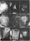
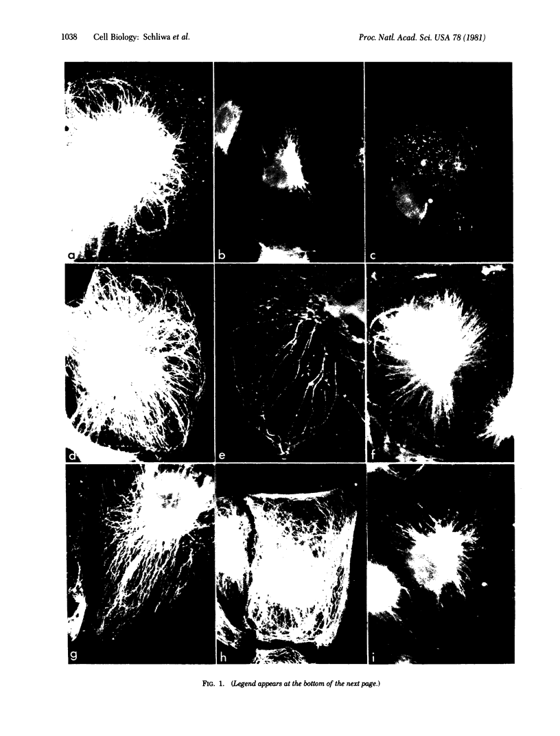
Detergent-extracted BSC-1 monkey cells have been used as a model system to study the Ca2+ sensitivity of in vivo polymerized microtubules under in vitro conditions. The effects of various experimental treatments were observed by immunofluorescence microscopy. Whereas microtubules are completely stable at Ca2+ concentrations below 1 μM, Ca2+ at greater than 1-4 μM induces microtubule disassembly that begins in the cell periphery and proceeds towards the cell center. At concentrations of up to 500 μM, both the pattern and time course of disassembly are not markedly altered, suggesting that, within this concentration range, Ca2+ effects are catalytic rather than stoichiometric. Higher (millimolar) Ca2+ concentration results in rapid destruction of microtubules. Of other divalent cations, only Sr2+ has a slight depolymerizing effect, whereas millimolar Ba2+, Mg2+, or Mn2+ is ineffective. Disassembly induced by micromolar Ca2+ is inhibited by pharmacological agents known to bind to calmodulin and inhibit its function, suggesting that calmodulin mediates Ca2+ effects. Both the addition of exogenous brain microtubule-associated proteins (MAPs) after lysis and the retention of endogenous cellular MAPs normally extracted during the lysis step stabilize microtubules against the depolymerizing effect of micromolar Ca2+. The results indicate that, in this model system, microtubules are sensitive to physiological Ca2+ concentrations and that this sensitivity may be conferred by calmodulin associated with the microtubules. MAPs appear to have a modulating effect on microtubular Ca2+ sensitivity and thus may function as a discriminating factor in cellular functions performed by calmodulin. It is hypothesized that Ca2+-stimulated microtubule disassembly depends on the relative amount of MAPs.
Keywords: detergent extraction, calmodulin, calmodulin inhibitors, immunofluorescence microscopy
Full text
PDF
Images in this article
Selected References
These references are in PubMed. This may not be the complete list of references from this article.
- Andersen B., Osborn M., Weber K. Specific visualization of the distribution of the calcium dependent regulatory protein of cyclic nucleotide phosphodiesterase (modulator protein) in tissue culture cells by immunofluorescence microscopy: mitosis and intercellular bridge. Cytobiologie. 1978 Aug;17(2):354–364. [PubMed] [Google Scholar]
- Bulinski J. C., Borisy G. G. Immunofluorescence localization of HeLa cell microtubule-associated proteins on microtubules in vitro and in vivo. J Cell Biol. 1980 Dec;87(3 Pt 1):792–801. doi: 10.1083/jcb.87.3.792. [DOI] [PMC free article] [PubMed] [Google Scholar]
- Bulinski J. C., Borisy G. G. Widespread distribution of a 210,000 mol wt microtubule-associated protein in cells and tissues of primates. J Cell Biol. 1980 Dec;87(3 Pt 1):802–808. doi: 10.1083/jcb.87.3.802. [DOI] [PMC free article] [PubMed] [Google Scholar]
- Cheung W. Y. Calmodulin plays a pivotal role in cellular regulation. Science. 1980 Jan 4;207(4426):19–27. doi: 10.1126/science.6243188. [DOI] [PubMed] [Google Scholar]
- Fujiwara K., Pollard T. D. Simultaneous localization of myosin and tubulin in human tissue culture cells by double antibody staining. J Cell Biol. 1978 Apr;77(1):182–195. doi: 10.1083/jcb.77.1.182. [DOI] [PMC free article] [PubMed] [Google Scholar]
- Fuller G. M., Brinkley B. R. Structure and control of assembly of cytoplasmic microtubules in normal and transformed cells. J Supramol Struct. 1976;5(4):497(349)–514(366). doi: 10.1002/jss.400050407. [DOI] [PubMed] [Google Scholar]
- Haga T., Abe T., Kurokawa M. Polymerization and depolymerization of microtubules in vitro as studied by flow birefringence. FEBS Lett. 1974 Mar 1;39(3):291–295. doi: 10.1016/0014-5793(74)80133-x. [DOI] [PubMed] [Google Scholar]
- Izant J. G., McIntosh J. R. Microtubule-associated proteins: a monoclonal antibody to MAP2 binds to differentiated neurons. Proc Natl Acad Sci U S A. 1980 Aug;77(8):4741–4745. doi: 10.1073/pnas.77.8.4741. [DOI] [PMC free article] [PubMed] [Google Scholar]
- Kinoshita S., Yazaki I. The behaviour and localization of intracellular relaxing system during cleavage in the sea urchin egg. Exp Cell Res. 1967 Sep;47(3):449–458. doi: 10.1016/0014-4827(67)90003-1. [DOI] [PubMed] [Google Scholar]
- Levin R. M., Weiss B. Mechanism by which psychotropic drugs inhibit adenosine cyclic 3',5'-monophosphate phosphodiesterase of brain. Mol Pharmacol. 1976 Jul;12(4):581–589. [PubMed] [Google Scholar]
- Marcum J. M., Dedman J. R., Brinkley B. R., Means A. R. Control of microtubule assembly-disassembly by calcium-dependent regulator protein. Proc Natl Acad Sci U S A. 1978 Aug;75(8):3771–3775. doi: 10.1073/pnas.75.8.3771. [DOI] [PMC free article] [PubMed] [Google Scholar]
- Means A. R., Dedman J. R. Calmodulin--an intracellular calcium receptor. Nature. 1980 May 8;285(5760):73–77. doi: 10.1038/285073a0. [DOI] [PubMed] [Google Scholar]
- Nishida E., Sakai H. Calcium-sensitivity of the microtubule reassembly system. Difference between crude brain extract and purified microtubular proteins. J Biochem. 1977 Jul;82(1):303–306. doi: 10.1093/oxfordjournals.jbchem.a131685. [DOI] [PubMed] [Google Scholar]
- Olmsted J. B., Borisy G. G. Ionic and nucleotide requirements for microtubule polymerization in vitro. Biochemistry. 1975 Jul;14(13):2996–3005. doi: 10.1021/bi00684a032. [DOI] [PubMed] [Google Scholar]
- Rosenfeld A. C., Zackroff R. V., Weisenberg R. C. Magnesium stimulation of calcium binding to tubulin and calcium induced depolymerization of microtubules. FEBS Lett. 1976 Jun 1;65(2):144–147. doi: 10.1016/0014-5793(76)80466-8. [DOI] [PubMed] [Google Scholar]
- Schliwa M. The role of divalent cations in the regulation of microtubule assembly. In vivo studies on microtubules of the heliozoan axopodium using the ionophore A23187. J Cell Biol. 1976 Sep;70(3):527–540. doi: 10.1083/jcb.70.3.527. [DOI] [PMC free article] [PubMed] [Google Scholar]
- Solomon F. Binding sites for calcium on tubulin. Biochemistry. 1977 Feb 8;16(3):358–363. doi: 10.1021/bi00622a003. [DOI] [PubMed] [Google Scholar]
- Solomon F., Magendantz M., Salzman A. Identification with cellular microtubules of one of the co-assemlbing microtubule-associated proteins. Cell. 1979 Oct;18(2):431–438. doi: 10.1016/0092-8674(79)90062-x. [DOI] [PubMed] [Google Scholar]
- Summers K., Kirschner M. W. Characteristics of the polar assembly and disassembly of microtubules observed in vitro by darkfield light microscopy. J Cell Biol. 1979 Oct;83(1):205–217. doi: 10.1083/jcb.83.1.205. [DOI] [PMC free article] [PubMed] [Google Scholar]
- Vallee R. B., Borisy G. G. The non-tubulin component of microtubule protein oligomers. Effect on self-association and hydrodynamic properties. J Biol Chem. 1978 Apr 25;253(8):2834–2845. [PubMed] [Google Scholar]
- Weber K., Rathke P. C., Osborn M. Cytoplasmic microtubular images in glutaraldehyde-fixed tissue culture cells by electron microscopy and by immunofluorescence microscopy. Proc Natl Acad Sci U S A. 1978 Apr;75(4):1820–1824. doi: 10.1073/pnas.75.4.1820. [DOI] [PMC free article] [PubMed] [Google Scholar]
- Weisenberg R. C. Microtubule formation in vitro in solutions containing low calcium concentrations. Science. 1972 Sep 22;177(4054):1104–1105. doi: 10.1126/science.177.4054.1104. [DOI] [PubMed] [Google Scholar]
- Welsh M. J., Dedman J. R., Brinkley B. R., Means A. R. Calcium-dependent regulator protein: localization in mitotic apparatus of eukaryotic cells. Proc Natl Acad Sci U S A. 1978 Apr;75(4):1867–1871. doi: 10.1073/pnas.75.4.1867. [DOI] [PMC free article] [PubMed] [Google Scholar]


