Abstract
Epidermal cysts are the most common cysts of the skin. Aconventional epidermal cyst rarely reaches a size of more than 5 cm in diameter. We report on two cases of giant epidermal cyst occurring in the neck. One patient had a cyst measuring 12×9×9 cm and the other patient had a non-pulsatile, dome-shaped lesion in the neck, which measured 6×5×3 cm. The lesions were totally excised. Histopathologically, both were confirmed as giant epidermal cysts.
Keywords: Epidermal cyst, Neck
INTRODUCTION
Epidermal cyst is a common cutaneous tumor. It usually involves the scalp, face, neck, back, and trunk1. A conventional epidermal cyst is generally small, and an epidermal cyst with a diameter of 5 cm or more is rare2. Epidermal cysts are commonly located on the head and neck. However, giant epidermal cysts occurring in the neck have rarely been reported in the literature. Herein, we present two cases of giant epidermal cysts occurring in the neck.
CASE REPORT
Case 1
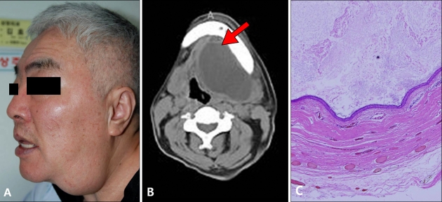
A 61-year-old man with a history of a painless, soft neck mass for 2 years was assessed in the outpatient clinic (Fig. 1A). The mass lesion had shown rapid growth in the last 2 months. He had no history of trauma. The patient complained of minimal respiratory discomfort. Neck computed tomography (CT) revealed a 12×9×9 cm hypodense multilocular mass extending to the tracheal area (Fig. 1B). During surgery, the entire mass was completely removed. No connections were observed between the mass and adjacent tissues, such as the sublingual gland or hyoid bone. Histologic sections showed a cyst having a fibrovascular wall with lining of the cyst cavity by squamous epithelium (Fig. 1C). The cyst did not contain any hair or hair follicles, but was filled with keratinaceous debris. The histopathological diagnosis was reported as an epidermal cyst. No postoperative complications were observed.
Fig. 1.
(A) A 61-year-old man with a neck mass. (B) Computed tomographic scan showed a hypodense multilocular mass arrow), which measured 12×9×9 cm, extending to the tracheal area. (C) The cyst was lined by flattened, keratinized stratified squamous epithelium with typical epidermal differentiation (H&E, ×40).
Case 2
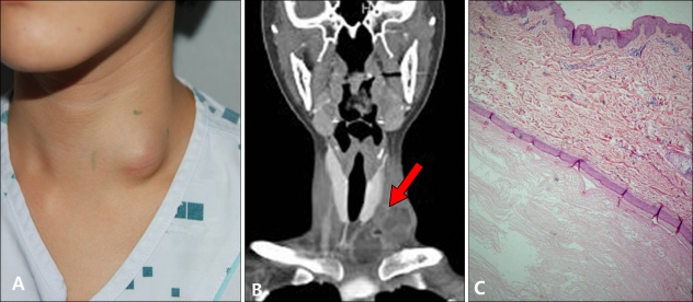
A 25-year-old woman presented to our department with a mass in the neck (Fig. 2A). She first became aware of the mass 1 year earlier, and it had enlarged slowly over that time. There was no history of trauma. Three months earlier, the lesion was excised at a local clinic. After two months, the lesion recurred and showed gradual enlargement. She presented with minimal respiratory discomfort. CT of the neck revealed a multilocular cystic mass localized within the left neck region, which measured approximately 6×5×3 cm. The mass showed expansion toward the trachea and compression of the tracheal air column (Fig. 2B). During surgery, the mass was excised. A macroscopic view of the specimen revealed no mesodermal extensions, such as hair or hair follicles. Histopathologic examination revealed a keratinized squamous epithelial lining with the inner surface lined with keratin lamellas (Fig. 2C). There were no histological findings of malignancy. Based on these findings, a histologic diagnosis of an epidermal cyst was made. There were no signs of recurrence during the 12 month follow-up period.
Fig. 2.
(A) A 25-year-old woman with a dome-shaped subcutaneous tumor located on the left lower neck. (B) Computed tomographic scan showed a multilocular cystic mass measuring 6×5×3 cm (arrow). The mass showed expansion toward the trachea. (C) The mass showed a keratinized squamous epithelial lining with the inner surface lined with keratin lamellas (H&E, ×40).
DISCUSSION
A conventional epidermal cyst is a common benign tumor of the skin that is most frequently found in young and middle-aged adults1. It is usually a slow-growing mass ranging in size from a few millimeters to a few centimeters2. However, a giant epidermal cyst more than 5 cm in diameter is rare1. In our case series, the dimensions of each of the epidermal cysts were 12×9×9 cm and 6×5×3 cm. Giant epidermal cyst was first described by Juhász and Szócska3 and Shah4. Giant epidermal cysts occurring in the neck, such as in our case, have rarely been reported.
A conventional epidermal cyst can be clinically diagnosed upon observance of the characteristic central, black punctum on the epidermis2,5. However, a central punctum was not observed in our patients. This made clinical diagnosis of the giant epidermal cyst more difficult.
A conventional epidermal cyst is usually a unilocular lesion and many giant epidermal cysts have also been reported as unilocular masses6-8. However, the 2 cases we have described were multilocular lesions. Multilocular giant epidermal cysts have been reported as having unique clinical features that differ considerably from those of a conventional epidermal cyst. Multilocular epidermal cysts are more common in elderly men, and affect areas with thick skin, such as the trunk and soles of the feet, and the lesions were unattended for long periods6. However, one of our patients was a 25-year old woman, and both patients had cervical lesions that became enlarged over a relatively short period.
An epidermal cyst is usually asymptomatic until it becomes infected or has become enlarged enough to cause damage to adjacent anatomic structures1. Hence, a giant epidermal cyst may produce adverse pressure effects on surrounding structures. In our patients, the lesions were found to be localized more deeply into the subcutaneous level. Fortunately, our patients presented with minimal discomforts, despite partial compression of the trachea by the giant epidermal cyst.
The pathophysiology of an epidermal cyst is thought of as either duct obstruction of a sebaceous gland, developmental defect of the sebaceous duct, or deep implantation of epidermal cells resulting from a blunt penetrating injury or previous surgery7. Trauma is considered to be the main cause; however, our patients had no trauma history. However, it is possible that the trauma occurred several years before development of the lesion and the patient could not recall the event. According to one report, a multilocular giant epidermal cyst was not produced by fusion of smaller epidermal cysts because the lobules were not completely independent and communicated with each other8. Tampieri et al.9 reported that lobules are formed because the cyst tends to spread in the direction of least resistance from the surrounding harder structures as it becomes larger, causing the cyst walls to extend like partitions.
Our cases of giant epidermal cyst presented as multilocular lesions. A large soft tissue mass with multilocular lesions may be confused with soft tissue cancer6. It should be remembered that a multilocular lesion can also occur in giant epidermal cysts. Among multilocular cysts lined with stratified squamous epithelium, proliferative epidermal cysts and dermoid cysts are lesions that can become large and multilocular lesions. These should be distinguished from multilocular giant epidermal cysts10. The cyst wall of a multilocular giant epidermal cyst is composed of non-atypical keratinocytes with normal cellularity and flattening, rather than proliferation. A proliferative epidermal cyst is locally aggressive and is a potentially malignant tumor. In a proliferative epidermal cyst, epithelial proliferation from the cyst wall projecting into the lumen or peripherally into the surrounding dermis can be seen10. In contrast to a multilocular giant epidermal cyst, dermoid cysts have hair follicles with sebaceous and sweat glands in the epithelium2.
In summary, two patients with a giant epidermal cyst in the neck area, which differs from a conventional epidermal cyst, are described in this article. These are rare cases, considering the unusual location. It must be recognized that a giant epidermal cyst may present as a multilocular lesion. A giant epidermal cyst necessitates a histopathologic examination in order to confirm the benign nature of the lesion.
References
- 1.Basterzi Y, Sari A, Ayhan S. Giant epidermoid cyst on the forefoot. Dermatol Surg. 2002;28:639–640. doi: 10.1046/j.1524-4725.2002.01314.x. [DOI] [PubMed] [Google Scholar]
- 2.Elder DE, Elenitsas R, Johnson B, Jr, Loffreda M, Miller J, Miller OF., III . Atlas and synopsis of Lever's histopathology of the skin. 10th ed. Philadelphia: Lippincott Williams & Wilkins; 2008. [Google Scholar]
- 3.Juhász G, Szócska J. Giant epidermoid cyst in the floor of the mouth. Fogorv Sz. 1979;72:23–25. [PubMed] [Google Scholar]
- 4.Shah SS, Varea EG, Farsaii A, Fernandez R, Richardson C, Schutte H. Giant epidermoid cyst of penis. Urology. 1979;14:389–391. doi: 10.1016/0090-4295(79)90089-x. [DOI] [PubMed] [Google Scholar]
- 5.Yamamoto T, Nishikawa T, Fujii T, Mizuno K. A giant epidermoid cyst demonstrated by magnetic resonance imaging. Br J Dermatol. 2001;144:217–218. doi: 10.1046/j.1365-2133.2001.03997.x. [DOI] [PubMed] [Google Scholar]
- 6.Ito R, Fujiwara M, Kaneko S, Takagaki K, Nagasako R. Multilocular giant epidermal cysts. J Am Acad Dermatol. 2008;58(5 Suppl 1):S120–S122. doi: 10.1016/j.jaad.2007.05.028. [DOI] [PubMed] [Google Scholar]
- 7.Polychronidis A, Perente S, Botaitis S, Sivridis E, Simopoulos C. Giant multilocular epidermoid cyst on the left buttock. Dermatol Surg. 2005;31:1323–1324. doi: 10.1111/j.1524-4725.2005.31211. [DOI] [PubMed] [Google Scholar]
- 8.Fujiwara M, Nakamura Y, Ozawa T, Kitoh A, Tanaka T, Wada A, et al. Multilocular giant epidermal cyst. Br J Dermatol. 2004;151:943–945. doi: 10.1111/j.1365-2133.2004.06227.x. [DOI] [PubMed] [Google Scholar]
- 9.Tampieri D, Melanson D, Ethier R. MR imaging of epidermoid cysts. AJNR Am J Neuroradiol. 1989;10:351–356. [PMC free article] [PubMed] [Google Scholar]
- 10.Sau P, Graham JH, Helwig EB. Proliferating epithelial cysts. Clinicopathological analysis of 96 cases. J Cutan Pathol. 1995;22:394–406. doi: 10.1111/j.1600-0560.1995.tb00754.x. [DOI] [PubMed] [Google Scholar]


