Abstract
The development of medical tissue adhesives has a long history without finding an all-purpose tissue adhesive for clinical daily routine. This is caused by the specific demands which are made on a tissue adhesive, and the different areas of application. In otorhinolaryngology, on the one hand, this is the mucosal environment as well as the application on bones, cartilage and periphery nerves. On the other hand, there are stressed regions (skin, oral cavity, pharynx, oesophagus, trachea) and unstressed regions (middle ear, nose and paranasal sinuses, cranial bones). But due to the facts that adhesives can have considerable advantages in assuring surgery results, prevention of complications and so reduction of medical costs/treatment expenses, the search for new adhesives for use in otorhinolaryngology will be continued intensively. In parallel, appropriate application systems have to be developed for microscopic and endoscopic use.
Keywords: tissue adhesives, cyanoacrylates, fibrin glue, GRG/GRF glue, albumin glutaraldehyde glue, applicators
1 Introduction
The idea to use adhesives for wound closure or to stabilize and fix tissues can be traced back to many centuries. After the use of different adhesives and glutinous substances (pitch, bee wax, natural rubber) for wound cover with more or less good results, the development of fibrin glues (1940) and later the cyanocrylates (1960) offered new ways in tissue adhesion.
The gold standard of wound closure, the suture, becomes less and less possible because of continuous miniaturisation and the development of minimally invasive surgery methods, particularly in mucosal areas. But a sufficient wound closure, a secured fixation of skin grafts, transplants and implants can be of vital importance for the success of a surgical therapy. In these areas, tissue adhesives virtually present themselves as method of choice.
The technical definition of adhesives can be conveyed offhand on medicine:
“An adhesive is a non-metallic substance that adheres or bonds compounds together by adhesion or cohesion without nonessential substantial changing the texture of the compounds.” (Definition of adhesives, DIN standard 16920)
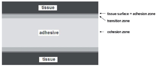
Gluing ensures a constant laminar force spreading. (Figure 1 (Fig. 1)) Unevenness of the material can be compensated by the adhesive [1]. Additionally to mechanical and chemical basics of adhesion, the characteristics of a living system must be considered for medical application.
Figure 1. cross-section of glued surfaces (tissue).
This results in following requirements to a medical tissue adhesive:
Biocompatibility:
biodegradation and resorbability in a defined period
no local or systemic toxicity, carcinogenity or teratogenity of the adhesive or its degradation products
marginal heat development during hardening
Compound strength:
high bond strength in wet environment with immediate functional stress
adequate elasticity
Application:
easy preparation
adequate flow characteristics and curing times
application systems for different areas of application
miniaturisation (microscopic and endoscopic surgical methods)
Other:
sterilisability
stable for storage
Up to now, there is no ideal tissue adhesive that fulfils all demands. Different biologic and synthetic adhesives for sealing wounds were used in the last years with partly good results. Below, the current and most important used commercial adhesives are introduced.
2 Biologic tissue adhesives
2.1 Fibrin glues: Tissucol®, Tisseel®, Crosseal®, Beriplast®
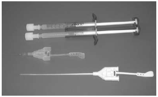
Fibrin glues (Figure 2 (Fig. 2)) are used since 1940 for sealing in surgery and are the mostly common tissue adhesives.
Figure 2. Fibrin glue with application system (Tissucol®).
Mechanism of adhesion
The components thrombin and fibrinogen cause, analogue to the last phase of blood coagulation, the formation of cross-linked fibrin. Here, the concentration of fibrinogen is 15 to 25 times higher than in circulating blood plasma. Therefore, fibrin is formed much faster. The other key factor of fibrin glue is factor XIII, which causes an indissoluble fibrin matrix.
Besides, most fibrin glues contain anti-fibrinolytic substances (tranexamic acid, aprotinin), which are responsible for stabilisation of the adhesion by inhibition of the fibrinolysis [2].
Advantages:
good adhesion in wet environments
no heat development during sealing
advantageous curing time (30 sec)
degradation without major tissue irritation in a few days
Disadvantages:
little adhesive strength
adhesion is not stress-stable
aprotinin with bovine origin with (even if low) risk of transmission of pathogens/prions [3]
blood product with (even if low) risk of transmission of blood-borne pathogens [4]
Indications:
tissue adhesion, haemostasis, support of wound healing
Contraindications:
arterial or heavy venous bleeding, allergic heparin-induced thrombocytopenia, intolerance to bovine products
All in all, fibrin glues are well-suited for fixation and wound closure as well as for haemostasis of moderate bleedings in non-stressed regions. But the adhesion strength is not sufficient for a reliable adhesion in mechanically stressed regions.
The time of resorption depends on the applicated quantity and on the location. A fast resorption can be found in tissues with good blood supply (mucosa, nasal, oral, gastrointestinal, parenchymatic organs), in the bradytrophic implant sites (middle ear, nasal septum, bones) the resorption is noticeable extended [5]. Cell culture studies report about convenient influences of fibrin glue on wound healing processes. Here, positive effects of cellular wound healing (fibroblasts) play a major role [6], [7].
3 Synthetic adhesives
3.1 Cyanoacrylates
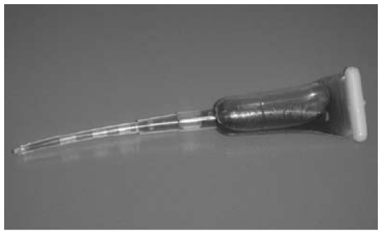
Since the adhesive attributes of cyanoacrylates (Figure 3 (Fig. 3)) have been described for the first time in 1959, there are interests for their medical use [8].
Figure 3. Cyanoacrylate glue (Histoacryl®).
But the first short-chain cyanoacrylates (methyl cyanoacrylates) turned out to be histotoxic and caused distinct foreign-body reactions. The long-chain cyanoacrylates of the second generation are more biocompatible [9], [10].
With rising chain-length, toxicity and adhesion strength decrease, elasticity and polymerisation time increase.
first generation:
methyl-cyanoacrylate
second generation:
ethyl-2-cyanoacrylate: Epiglu®
n-butyl-2-cyanoacrylate: Histoacryl®, Liquiband®
2-octyl-cyanoacrylate: Dermabond®
isobutyl-cyanoacrylate: Indermil®
ethyl-2-cyanoacrylate + butyl acrylate + methacryloxysulpholane: Glubran®
n-butyl-2-cyanoacrylate + methacryloxysulpholane: Glubran 2®
Mechanism of adhesion
In contact with hydroxide ions (liquids like blood or water, air humidity), the cyanoacrylates form long, strong and waterproof chains in an exothermic reaction. The resulting polymer leads to a stable adhesive bond. The polymerisation time is within 20 sec and 2 min. With too much moisture, the reaction runs too fast for a tissue adhesion.
Advantages:
good adhesion in moderate wet environments
high adhesion strength
Disadvantages:
toxic degradation products
heat development during polymerisation
Administration for:
topical skin adhesive
3.2 Gelatine resorcinol formaldehyde/glutaraldehyde glues (GRF/GRG): Gluetiss®
These adhesives were introduced 1966, particularly to seal parenchymatic organs and blood vessels. Because of the exchange of formaldehyde with less toxic dialdehydes the biocompatibility was highly improved.
Mechanism of adhesion
Resorcinol and dialdehyde react to a 3-dimensional network. Gelatine serves as filler. Polymerisation time is 2 min, the degradation products are much less histotoxic as those of the cyanoacrylates.
Advantages:
better adhesive strength than fibrin glues
moderate adhesion in wet environments
Disadvantages:
use in otorhinolaryngology difficult due to application (direct application of both components)
Administration for:
aortic dissection, reconstruction of insufficient aortic valves
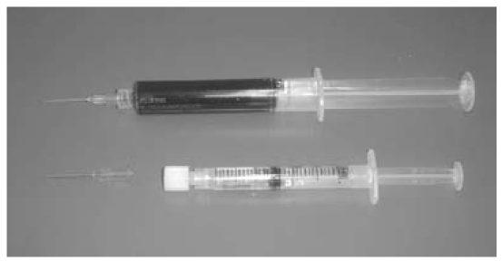
GRG adhesives (Figure 4 (Fig. 4)) are used in vascular surgery with good results. Due to their adhesion attributes, they seem to be appropriate for otorhinolaryngology. Sporadic animal experiments show a good suitability to fix cartilage and mucosa [11], [12].
Figure 4. Gelatine resorcinol glutaraldehyde glue (Gluetiss®).
3.3 Albumin glutaraldehyde glue: BioGlue®
Mechanism of adhesion
The glutaraldehyde molecules band together by covalent bond with the added albumine as well as with the proteins of the tissue. The polymerisation time starts immediately after mixture of the components; the entire adhesion strength is achieved after 2 min.
Advantages:
good adhesion strength in wet environments
Disadvantages:
albumin is of bovine origin (risk of allergic reaction)
Administration for:
aortic dissection, skin
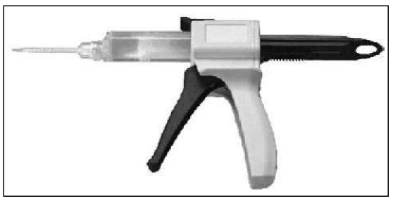
Due its adhesion attributes in wet environments, this adhesive (Figure 5 (Fig. 5)) seems to be appropriate for otorhinolaryngology, too. In vascular surgery, it is used with good results [13].
Figure 5. Albumin glutaraldehyde glue (BioGlue®), www.cryolife.com.
4 Areas of application
Most of the above-named adhesives were and are used experimentally and clinically in otorhinolaryngology, but a particularly suitable tissue adhesive for all applications does not exist yet.
In the following, the experiences which were made until now with adhesives in various areas of otorhinolaryngology are described and the special requirements to an adhesive in the particular regions are summarised.
4.1 Auricle/middle ear
Potential application areas:
otoplasty for protruding ears, skin closure
closure of tympanic membrane – readaption of the edges after traumatic rupture, fixation of fascia or cartilage during reconstruction
reconstruction of the ossicles chain – gluing of the ossicles among each other, fixation of transplants (cartilage, ossicles), fixation of implants (TORP, PORP)
fixation of implantable hearing systems – cochlea implant electrode, vibration elements of partly implantable hearing devices
Cyanoacrylates were tested on rabbits to fixate auricle cartilage – with good fixation and uncomplicated handling. There were no increased inflammation reactions during an adhesion between cartilage and bone. In contrast, the application in tissues with good blood supply showed a significant higher histotoxicity [14], [15].
Samuel et al. used isobutyl-cyanoarylate (Indermil®) to fixate transplants during closure of tympanic membrane perforations. In 10 patients, they found delayed implant healing with granulations and dislocation of the transplant [16]. Case reports describe good results in the adhesion of ossicles among each other [17]. There are no systematic studies to the use of cyanoacrylates in ear surgery. The reported failures with creation of granulations and overshooting inflammation reaction are based on the one hand on early cyanoacrylates (methyl cyanoacrylates) [18]. On the other hand, the main problem in the use of long-chain cyanoacrylates (Histoacryl®, Dermabond®, Indermil®) is the application of the adhesives in minimal dose at a microscopic small place. If too much adhesive is unsighted applied, the polymerisation runs too fast with higher heat development and can lead to tissue damages. Also, the controverse description of ototoxic effects has its origins in the mostly uncontrollable application and so in the varying concentrations of degradation products [19], [20], [21].
Fibrin glues were used to fix transplants in myringoplasty. Sakagami et al. report about a success rate of 78% in underlay myringoplasty with fascia or connective tissue in 391 ear surgeries [22]. These patients solely suffered from a tympanic membrane perforation in a mesotympanal form of chronic otitis media, after trauma or after tympanum drainage. The success rate of tympanic membrane closure without fibrin glue is lower; and the cost problems and the (even if low) transmission risk of pathogens have to be discussed here.
For the reconstruction of the ossicles chain in defects of the long arm of the incus, fibrin glue is due to its insufficient stiffness not recommendable [23]. Punctual adhesion should be a further problem.
All in all, appropriate adhesives could solve some of the problems of ear surgery: certain fixation of transplants (fascia, cartilage) and implants (ossicle replacement, cochlea-implant electrodes, floating mass transducer), and certain fixation of cartilage in otoplasty for protruding ears. Systematic studies to commercial and adequate adhesives are lacking, and cannot be replaced by singular case reports.
Another main problem is the development of suitable application systems for microscopic ear surgery with punctual application of very small doses of glue.
4.2 Nose/paranasal sinuses
Potential application areas:
sealing of mucosa in septal plasty, closure of septum perforations, turbinoplasty, epistaxis
fixation of transplants: cartilage, bone, composite grafts
fixation of implants: bone and cartilage replacement, stents and place holder
dural plasty
Cyanoacrylates (here: n-butyl-2-cyanoacrylate) were used in animal experiments to fix the septum to anterior nasal spine. A stable fixation was achieved in rabbits. A temporary inflammatory reaction appeared in the glued area, that abated after 8 weeks [24]. Dabb et al. reported about the use of 2-octyl-cyanoacrylate to fix bone transplants in the range of the nose tip and bridge. In the follow-up examination (up to 18 months), no complications appeared [25]. In the range of the paranasal sinuses, reports about closure of maxillary sinus mucosa in animal experiments with n-butyl-cyanoacrylate with good results in sinus lift surgeries can be found [26].
Fibrin glues are used to fix transplants in endonasal dural plasty with good results for many years [27], [28], [29]. For clinical purposes, the insufficient adhesive strength in larger areas is problematic (dislocation of transplants into the inner nose). Also the application systems are not adapted to endoscopic technologies – so a selective gluing of small patches is not possible, or only with difficulties.
The use of fibrin glues to prevent septal haematomas and the fixation of mucosa is reported in septal surgery. The clinical use showed uncomplicated healing processes (in 100 patients [30], 57 patients [31] and 11 patients [32]), whilst animal tests showed suboptimal results. Erkan et al. tested the effect of fibrin glue on nasal mucosa tissues in rabbits. Histologically, a distinct inflammatory reaction was found with increased mucosal thickness, reduction of perichondrium and cartilage thickness as well as cartilage damage [33].
The form of application and the dose of glue seem to play significant roles. If a thin film of glue is applicated with a spray applicator, the glue dose can be reduced with equal functionality – the consequences of degradation of the glue can be reduced [34].
Albumin glutaraldehyde glue (BioGlue®) is used to fix the middle turbinate in endonasal paranasal sinus surgery in a field report with good results [12]. After a lesion of mucosa at middle turbinate and septum, the middle turbinate was fixed to septum with BioGlue®. In the follow-up (six months postoperatively), only six of 212 patients showed a lateralisation of the turbinate. Complications were not observed.
Current domain of tissue adhesives in the range of the nose and paranasal sinuses is the use of fibrin glues to fix transplants (fascia, turbinate mucosa) and implants (allogenic bioimplants, alloplastic materials) for dural plasty [35]. Other published uses of different tissue adhesives have been tested only little systematic in vivo and in vitro and describe mostly clinical monitoring.
At this time, a use of tissue adhesives in the range of cartilaginous and bony nose has to be questioned critically. Systematic long term studies and the development of better suited application systems (similar to the ear) are necessary.
4.3 Pharynx/oesophagus
Potential application areas:
fixation of skin grafts, closure of mucosa
supply of bleedings
closure of fistulae
There are high requirements to a tissue adhesive for the closure of mucosa in the upper gastrointestinal tract concerning bonding in wet environments and adhesion strength. A suture is often difficult or not feasible, so only a suitable tissue adhesive permits a wound closure.
Until now, there are only few publications about the use of adhesives for mucosa closure in the upper gastrointestinal tract. Perez et al. tried an adhesion of the mucosa with cyanoacrylates (n-butyl-2-cyanoacrylat) after tooth extraction in 30 patients – with good results [36]. Weerda et al. described the use of fibrin glue to seal a mucosal lesion after transaction of the wall of Zenker's diverticel [37]. Fibrin glues were applicated in small iatrogenic oesophagus lesions with partly good results [38], [39], [40]. Cyanoacrylates have been used for haemostasis in oesophagus varices and bleeding gastric ulcers for a long time [41].
For the upper gastrointestinal tract as domain of flexible and inflexible endoscopy, a tissue adhesive for wound closure in mucosal environments is absolutely desirable. The gold standard, the suture, is no alternative due to the access and technical borders. A biocompatible tissue adhesive in these stressed implant sites could prevent disturbed wound healing, and complications (bleedings, fistulae) can be controlled. The current studies are only a decent beginning to solve this complex problem. Also, the construction of application system for endoscopy in parallel to the development of the adhesive must take place.
4.4 Larynx/trachea
Potential application areas:
fixation of transplants: mucosa, cartilage
fixation of implants: stents and place holder
closue of fistulae
In the range of the respiratory tract, a fixation of inserted materials is essential to avoid a dislocation with dyspnoea. There are only sporadic experiences fixing transplants or implants. Naumann und Lang describe the use of fibrin glue to fix the mucosa in laryngeal widening [42]. Adhesions in the deep trachea and the bronchial system, especially in lung emphysema (GRG adhesive), bronchopleural fistulae (BioGlue®) and to prevent leakages during wedge resection of the lung showed good results [43], [44], [45]. Takagi et al. tested the effect of wound healing of fibrin glues in tracheal anastomoses in rats and found decreased development of collagen and hydroxyproline in the group with fibrin glue additionally to suture [46]. Good results regarding the stability of the sutures and the appearance of leakages in tracheal anastomoses report Takahashi et al. by using GRG adhesive in comparison with sutures alone [47].
In summary, the adhesives can additionally secure sutures in tracheal range, because an insufficient suture can be associated here always with life-threatening complications. But to secure the use on human beings, systematic in vitro and in vivo studies are necessary.
4.5 Skin
The adhesion of skin is the best validated and in vivo and in vitro tested application of tissue adhesives. The most used adhesives here are the cyanoacrylates. Many animal studies showed results that are equivalent to the conventional skin suture [48], [49], [50], [51], [52], [9]. Precondition for functional and aesthetic results is, on the one hand, a subcutaneous suture that intercepts the skin tension to converge the skin edges optimally. On the other hand, all major bleedings must have stopped, because the polymerisation would run too fast for adequate adhesion with to much heat development.
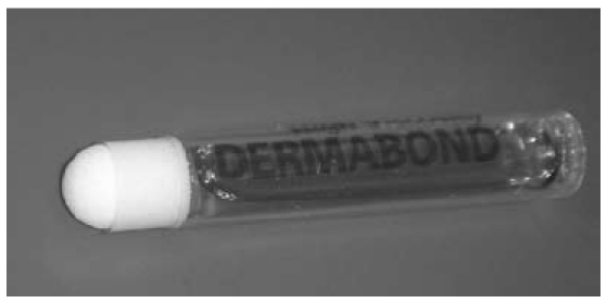
Contraindications for the use of cyanoacrylates in skin closure are: infection of the wound, substantial defects, wounds in the range of the eyes and mucous membranes, bites, and very hairy skin. Particularly noteworthy is the development of optimally adapted applicators, in contrary to the other reported uses for skin adhesion. This spectrum ranges from a brush (LiquiBand®) to canule-like applicators (Histoacryl®) or absorbing round applicator tips (Dermabond®) (Figure 6 (Fig. 6)).
Figure 6. Cyanoacrylate glue (Dermabond®).
Fibrin glues has been tested separately in animal experiments for skin closure. The test showed insufficient adhesive strength, but good haemostatic properties [53]. Marchac et al. used sprayed fibrin glue subcutaneously in face-lift surgeries and report about a decrease of swelling and oedemas [54].
From clinical purpose, the adhesion of skin can – considering the costs compared to suture materials – be kept in mind by
children and uncooperative patients to avoid removal of suture rests
on skin under the risk of impact of saliva and tracheal secretions (perioral, tracheostoma), because cyanoacrylates ensure a waterproof closure of the wound
4.6 Nerves
In suturing nerves, as a primary suture after injuries or interponates, adhesives have been also tested to ensure the suture or to seal anastomoses. Cyanoacrylates alone were used to seal anastomoses in animal experiments [55]. Octyl-2-cyanoacrylate did not show any differences concerning toxicity or inflammatory reaction compared with nerve sutures. The quantitative functional tests in the group with adhesives showed a faster return of the neural functions compared with the group with sutures. As a reason, the possible effect of the outer splint of the nerves due to the adhesive is discussed, which is known from other studies [56]. Other authors report of foreign-body reactions and fibrosis of the glued nerves due to cyanoacrylate (Glubran®) [57]. The reason may be the problematic application and dosage of the adhesives as well as another composition.
Fibrin glues had also been tested to glue nerves, with advantages over nerve sutures alone, regarding to the return of neural functions [58], [59], [60]. A clinical study on 36 patients after gluing the ends of the facial nerve (directly: 11 patients; or with interponate: 25 patients) showed no differences to neural sutures [61].
All in all, adhesives have advantages in surgery of the peripheral nerves. A problem is the punctual application with as little doses of glue as possible between the ends of the nerve with use of the effects of outer splinting. A complete replacement of the nerve sutures is currently not in sight, but a combination of both methods could make sense.
4.7 Bones
The adhesion of bone and fragments has a manifold spectrum in traumatology and reconstructive surgery. Besides, an ensured fixation of implants by adhesives is worthwhile.
The actual use of bone adhesives is currently limited to a few special indications [62]. Cyanoacrylates (butyl-2-cyanoacrylate) were tested in animal experiments to fix cranial bones compared with plate osteosynthesis with same good results [63], [64], [65], [66].
Clinical uses comprised only a few patients. Kim (10 patients, [67]) showed good results in the fixation of multiple fractures in the facial maxillary sinus, orbital floor and the anterior wall of the frontal sinus without inflammatory complications. Mehta et al. (10 patients, [68]) glued fractures of the mandibula and could reduce the duration of intermaxillary fixation.
Fibrin glues generally show only little adhesion and cause increased development of connective tissue and thus to a decelerated osteogenesis [5]. Other authors describe high osteoinductive effects in animal experiments, whereas the different time for osteogenesis in different species has to be considered [69]. Fast resorption of the glue (few days) leads to instability, because osteogenesis is not effective in this time [70]. Fibrin glues seem to be not suitable for fixation in stressed implant sites with low osteogenesis rate (cranial bone) [71]. A replacement with bone adhesives is not expected in the next time, but the use combined with conventional methods seems to be realizable.
5 Application systems
Compared to the adhesives, the development of special application systems has been intended, except these for skin sealing. But not only is the adhesive important for a successful adhesion, it has to be carried easy and with clear view to its destination. Current developments are limited to moderate-adapted systems for skin closure or adhesion of larger areas.
But otorhinolaryngology with microscopic and endoscopic minimal invasive surgery has special demands on an application system – and again on adhesive characteristics like texture, flow characteristics, or hardening time.
In the middle ear, a punctual gluing of smallest structures should be possible without disturbance of the view through the microscope. Texture, flow characteristics, and hardening time have to allow the adhesion in a range of millimetres.
Endonasal adhesion requires applicators with navigable tip to applicate adhesives punctual, for example at the skull base.
In endotracheal or oesophagus adhesion, an endoscope is used to get to the destination. So the application system has to be compatible to the instruments’ channel of a flexible endoscope. An adaptation of the flow characteristics and hardening time to the long distances between external and internal destination is also necessary. Adhesives with two components need a mixing chamber at the end of the applicator. Navigable tips of the application system would be very helpful in flexible and inflexible endoscopy to reach angular structures.
6 Current and future developments
6.1 Own studies
Since 2006, in a cooperative project that is supported by the Thüringer Aufbaubank, tissue adhesives for application in mucosal environment were studied. Cooperation partners are INNOVENT e.V. Technologieentwicklung Jena, Department for Biomaterials (development of new tissue adhesives), Occlutech GmbH (development of applicators, up to 2008), and the Laboratory for Biomaterials of the Department for Otorhinolaryngology, Friedrich Schiller University Jena (in vitro tests of adhesives and applicators).
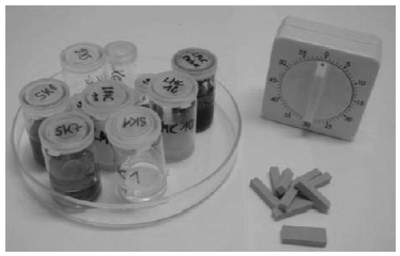
For the in vitro tests of the adhesives, a two-stage test was developed to simulate the conditions of wet environments. In the first step, defined test specimen (2.5x1.0x0.5 cm), stored in physiological saline for 24 hours, were glued (Figure 7 (Fig. 7)). Application characteristics, handling, fixation and hardening time were evaluated. Adhesion strength was tested a. during dry storage after 10 and 30 min, and b. during wet storage (physiological saline) after 10 min and 12 days. This procedure was used for 45 developed one- and two-component adhesives. 20 adhesives were regarded as qualified.
Figure 7. Testing the adhesives on specimens.
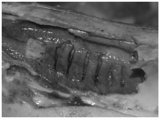
In the second step, wet environments of nearly biologic conditions were simulated. The cavum nasii with septal cartilage and the buccal musculature of a fresh pig’s head (ca. 2 hours after slaughtering) were preparated.
At the septal cartilage, defined mucosal flaps (0.5x2cm) were prepared to fix them at the cartilage (Figure 8 (Fig. 8)). The buccal musculature was cut into stripes to fix them with each other. The adhesion sites were moistened with physiological saline, the adhesion strength was tested after 10, 30 and 60 minutes. After this second step, eight suitable adhesives were selected.
Figure 8. Gluing of mucosal flaps to septal cartilage (pig’s head).
Additionally, adhesive strength tests were made with a texture analyzer on buccal mucosal flaps (Figure 9 (Fig. 9)). For comparison, fibrin glue (Tissucol®), cyanoacrylates (Histoacryl®, Dermabond®) and GRG glue (Gluetiss®) were tested. The new developed adhesives (based on GRG and methacrylates) showed better results than fibrin glue and GRG glue, but weaker than the tested cyanoacrylates [72].
Figure 9. Tensile strength measurements with texture analyzer.

In a new project, supported by the Bundesministerium für Wirtschaft und Technologie, the adhesives have to be modified, adapted to different developed application systems, and tested in animal experiments. The development of the applicators is made by the new partner bess pro gmbh.
6.2 Protein-based adhesives
In the last years, there were many approaches to develop tissue adhesives analogue to the adhesives of different mussel species. These adhesives tend to form very stable, multifunctional and resolvable connections. The adhesive proteins from Mytilus edulis (= Mytilus edulis foot proteins – Mefp 1-3) were identified, but their exact adhesion mechanism could not be analysed yet. First adhesive approaches run, e.g., at the Fraunhofer Institute for Manufacturing Technology and Applied Materials Research in Bremen [73].
6.3 Polylactide- and methacrylate-based tissue adhesives
Polylactides are used as bioresorbable plate systems and as carrier for pharmaceuticals in medicine in medicine. Free terminal hydroxyl groups are able to import cross-linking groups and so to polymerise. First polylactide-based adhesives were used to seal lung parenchyma [74].
Methacrylate-based adhesives consist of low-molecular components, which polymerise to complex hydrolysable networks. In animal experiments, a high biocompatibility was shown as well as adhesive strength in bode adhesions [75].
7 Conclusions
The use of tissue adhesives is very interesting for all fields of otorhinolaryngology and can ensure surgery results, prevent complications, and so minimize medical costs. Currently, the usage of adhesives is only established in dural plasty, haemostasis and skin adhesion. In all the other fields, adhesives are applied and studied in animal experiments and clinical case reports. The synthetic adhesives lack of systematic in vivo and in vitro tests for the wide use. In future, the fields of research will be located here. Additional to the tissue adhesives, particularly in otorhinolaryngology the development of application systems for microscopic and endoscopic use must take place.
References
- 1.Heiss C, Schnettler R. Bioresorbierbare Knochenklebstoffe - Historischer Rückblick und heutiger Stand. Unfallchirurg. 2005;108:348–355. doi: 10.1007/s00113-005-0951-y. [DOI] [PubMed] [Google Scholar]
- 2.Pursifull NF, Morey AF. Tissue glues and nonsuturing techniques. Curr Opin Urol. 2007;17:396–401. doi: 10.1097/MOU.0b013e3282f0d683. Available from: http://dx.doi.org/10.1097/MOU.0b013e3282f0d683. [DOI] [PubMed] [Google Scholar]
- 3.Petersen B, Barkun A, Carpenter S, et al. Tissue adhesives and fibrin glues. Gastrointest Endosc. 2004;60:327–333. doi: 10.1016/S0016-5107(04)01564-0. Available from: http://dx.doi.org/10.1016/S0016-5107(04)01564-0. [DOI] [PubMed] [Google Scholar]
- 4.Panis R, Scheele J. Hepatitisrisiko bei der Fibrinklebung in der HNO-Chirurgie. Laryngol Rhinol Otol (Stuttg) 1981;60:367–368. doi: 10.1055/s-2007-997150. Available from: http://dx.doi.org/10.1055/s-2007-997150. [DOI] [PubMed] [Google Scholar]
- 5.Reck R, Bernal-Sprekelsen M. Fibrinkleber und Tricalciumphosphat-Implantate in der Mittelohrchirurgie. Eine tierexperimentelle Studie. Laryngorhinootologie. 1989;68:152–156. doi: 10.1055/s-2007-998307. Available from: http://dx.doi.org/10.1055/s-2007-998307. [DOI] [PubMed] [Google Scholar]
- 6.Gosain AK, Lyon VB. The current status of tissue glues: part II. For adhesion of soft tissues. Plast Reconstr Surg. 2002;110:1581–1584. doi: 10.1097/01.PRS.0000033993.30838.3E. Available from: http://dx.doi.org/10.1097/01.PRS.0000033993.30838.3E. [DOI] [PubMed] [Google Scholar]
- 7.Wolf G, Plinkert PK, Schick B. Zelltransplantationen zum Liquorfistelverschluss: Erfahrungen mit Fibrinkleber und Fibroblasten. HNO. 2005;53:439–445. doi: 10.1007/s00106-004-1156-3. Available from: http://dx.doi.org/10.1007/s00106-004-1156-3. [DOI] [PubMed] [Google Scholar]
- 8.Coover H, Joyner F, Shearer N, Wicker T. Chemistry and performance of cyanoacrylate adhesives. Special Technical Papers. 1959;5:413–417. [Google Scholar]
- 9.Alamouti D, von Kobyletzki G, Allard P, Hoffmann K. Ein prospektiver Vergleich von Octylcyanoacrylat-Gewebekleber und konventionellen Wundverschlüssen. Hautarzt. 1999;50:58–59. doi: 10.1007/s001050050867. Available from: http://dx.doi.org/10.1007/s001050050867. [DOI] [PubMed] [Google Scholar]
- 10.Leggat PA, Smith DR, Kedjarune U. Surgical applications of cyanoacrylate adhesives: a review of toxicity. ANZ J Surg. 2007;77:209–213. doi: 10.1080/19338240903241291. Available from: http://dx.doi.org/10.1080/19338240903241291. [DOI] [PubMed] [Google Scholar]
- 11.Costa HJ, Pereira CS, Costa MP, et al. Comparison of butyl-2-cyanoacrylate, gelatin-resorcin-formaldehyde (GRF) compound and suture in stabilization of cartilage grafts in rabbits. Rev Bras Otorrinolaringol (Engl Ed) 2006;72:61–71. doi: 10.1016/S1808-8694(15)30036-7. [DOI] [PMC free article] [PubMed] [Google Scholar]
- 12.Friedman M, Schalch P. Middle turbinate medialization with bovine serum albumin tissue adhesive (BioGlue) Laryngoscope. 2008;118:335–338. doi: 10.1097/MLG.0b013e318158198f. Available from: http://dx.doi.org/10.1097/MLG.0b013e318158198f. [DOI] [PubMed] [Google Scholar]
- 13.Chao HH, Torchiana DF. BioGlue: albumin/glutaraldehyde sealant in cardiac surgery. J Card Surg. 2003;18:500–503. doi: 10.1046/j.0886-0440.2003.00304.x. Available from: http://dx.doi.org/10.1046/j.0886-0440.2003.00304.x. [DOI] [PubMed] [Google Scholar]
- 14.Toriumi DM, Raslan WF, Friedman M, Tardy ME., Jr Variable histotoxicity of histoacryl when used in a subcutaneous site: an experimental study. Laryngoscope. 1991;101:339–343. doi: 10.1288/00005537-199104000-00001. Available from: http://dx.doi.org/10.1288/00005537-199104000-00001. [DOI] [PubMed] [Google Scholar]
- 15.Brown PN, McGuff HS, Noorily AD. Comparison of N-octyl-cyanoacrylate vs suture in the stabilization of cartilage grafts. Arch Otolaryngol Head Neck Surg. 1996;122:873–877. doi: 10.1001/archotol.1996.01890200063014. [DOI] [PubMed] [Google Scholar]
- 16.Samuel PR, Roberts AC, Nigam A. The use of Indermil (n-butyl cyanoacrylate) in otorhinolaryngology and head and neck surgery. A preliminary report on the first 33 patients. J Laryngol Otol. 1997;111:536–540. doi: 10.1017/S0022215100137855. Available from: http://dx.doi.org/10.1017/S0022215100137855. [DOI] [PubMed] [Google Scholar]
- 17.Adamson RM, Jeannon JP, Stafford F. A traumatic ossicular disruption successfully repaired with n-butyl cyanoacrylate tissue adhesive. J Laryngol Otol. 2000;114:130–131. doi: 10.1258/0022215001904860. Available from: http://dx.doi.org/10.1258/0022215001904860. [DOI] [PubMed] [Google Scholar]
- 18.Koltai PJ, Eden AR. Evaluation of three cyanoacrylate glues for ossicular reconstruction. Ann Otol Rhinol Laryngol. 1983;92:29–32. doi: 10.1177/000348948309200107. [DOI] [PubMed] [Google Scholar]
- 19.Wells JR, Gernon WH. Bony ossicular fixation using 2-cyano-butyl-acrylate adhesive. Arch Otolaryngol Head Neck Surg. 1987;113:644–646. doi: 10.1001/archotol.1987.01860060070017. [DOI] [PubMed] [Google Scholar]
- 20.Chen E, Harner SG. The effect of butyl 2-cyanoacrylate on the middle and inner ear of the chinchilla. Otolaryngol Head Neck Surg. 1986;95:187–192. doi: 10.1177/019459988609500210. [DOI] [PubMed] [Google Scholar]
- 21.Maw JL, Kartush JM. Ossicular chain reconstruction using a new tissue adhesive. Am J Otol. 2000;21:301–305. doi: 10.1016/S0196-0709(00)80035-6. Available from: http://dx.doi.org/10.1016/S0196-0709(00)80035-6. [DOI] [PubMed] [Google Scholar]
- 22.Sakagami M, Yuasa R, Yuasa Y. Simple underlay myringoplasty. J Laryngol Otol. 2007;121:840–844. doi: 10.1017/S0022215106005561. Available from: http://dx.doi.org/10.1017/S0022215106005561. [DOI] [PubMed] [Google Scholar]
- 23.Zahnert T, Huttenbrink KB. Fehlermöglichkeiten bei der Ossikelkettenrekonstruktion. HNO. 2005;53:89–102. doi: 10.1007/s00106-004-1168-z. Available from: http://dx.doi.org/10.1007/s00106-004-1168-z. [DOI] [PubMed] [Google Scholar]
- 24.Alkan S, Dadas B, Celik D, et al. The efficacy of N-2-butyl cyanoacrylate in the fixation of nasal septum to the anterior nasal spine in rabbits: experimental study. Eur Arch Otorhinolaryngol. 2007;264:1425–1430. doi: 10.1007/s00405-007-0407-9. Available from: http://dx.doi.org/10.1007/s00405-007-0407-9. [DOI] [PubMed] [Google Scholar]
- 25.Dabb R, Gaffield J, Camp L. Use of Cyanoacrylate (Super Glue) for the Fixation and Prefabrication of Nasal Cartilage Grafts. Aesthetic Surg J. 2001;21:328–333. doi: 10.1067/maj.2001.117709. Available from: http://dx.doi.org/10.1067/maj.2001.117709. [DOI] [PubMed] [Google Scholar]
- 26.Choi BH, Kim BY, Huh JY, et al. Cyanoacrylate adhesive for closing sinus membrane perforations during sinus lifts. J Craniomaxillofac Surg. 2006;34:505–509. doi: 10.1016/j.jcms.2006.07.859. Available from: http://dx.doi.org/10.1016/j.jcms.2006.07.859. [DOI] [PubMed] [Google Scholar]
- 27.Draf W. Erfahrungen mit der Technik der Fibrinklebung in der Hals-Nasen-Ohren-Chirurgie. Laryngol Rhinol Otol (Stuttg) 1980;59:99–107. doi: 10.1055/s-2007-1008826. Available from: http://dx.doi.org/10.1055/s-2007-1008826. [DOI] [PubMed] [Google Scholar]
- 28.Hosemann W, Nitsche N, Rettinger G, Wigand ME. Die endonasale, endoskopisch kontrollierte Versorgung von Duradefekten der Rhinobasis. Laryngorhinootologie. 1991;70:115–119. doi: 10.1055/s-2007-998001. Available from: http://dx.doi.org/10.1055/s-2007-998001. [DOI] [PubMed] [Google Scholar]
- 29.Aletsee C, Konopik V, Dazert S, Dieler R. Operative Versorgung von Verletzungen der Frontorhinobasis. Laryngorhinootologie. 2003;82:626–631. doi: 10.1055/s-2003-42688. Available from: http://dx.doi.org/10.1055/s-2003-42688. [DOI] [PubMed] [Google Scholar]
- 30.Daneshrad P, Chin GY, Rice DH. Fibrin glue prevents complications of septal surgery: findings in a series of 100 patients. Ear Nose Throat J. 2003;82:196–197. [PubMed] [Google Scholar]
- 31.Boenisch M, Nolst Trenite GJ. Fibrinkleber bei Septumplastik. [Fibrin glue for operative correction of septal deviations]. HNO. 2004;52:963–967. doi: 10.1007/s00106-004-1130-0. (Ger). Available from: http://dx.doi.org/10.1007/s00106-004-1130-0. [DOI] [PubMed] [Google Scholar]
- 32.Lee JY, Lee SH, Kim SC, Koh YW, Lee SW. Usefulness of autologous cartilage and fibrin glue for the prevention of septal perforation during septal surgery: a preliminary report. Laryngoscope. 2006;116:934–937. doi: 10.1097/01.MLG.0000215174.47864.9A. Available from: http://dx.doi.org/10.1097/01.MLG.0000215174.47864.9A. [DOI] [PubMed] [Google Scholar]
- 33.Erkan AN, Cakmak O, Kocer NE, Yilmaz I. Effects of fibrin glue on nasal septal tissues. Laryngoscope. 2007;117:491–496. doi: 10.1097/MLG.0b013e31802dc5bc. Available from: http://dx.doi.org/10.1097/MLG.0b013e31802dc5bc. [DOI] [PubMed] [Google Scholar]
- 34.Vaiman M, Eviatar E, Segal S. The use of fibrin glue as hemostatic in endonasal operations: a prospective, randomized study. Rhinology. 2002;40:185–188. [PubMed] [Google Scholar]
- 35.Schneider G. Bioimplantate - Eigenschaften und Anwendungshinweise. Laryngorhinootologie. 2003;82:839–852. doi: 10.1055/s-2004-814134. [DOI] [PubMed] [Google Scholar]
- 36.Perez M, Fernandez I, Marquez D, Bretana RM. Use of N-butyl-2-cyanoacrylate in oral surgery: biological and clinical evaluation. Artif Organs. 2000;24:241–243. doi: 10.1046/j.1525-1594.2000.06519.x. Available from: http://dx.doi.org/10.1046/j.1525-1594.2000.06519.x. [DOI] [PubMed] [Google Scholar]
- 37.Weerda H, Ahrens KH, Schlenter WW. Maßnahmen zur Verringerung der Komplikationsrate bei der endoskopischen Operation des Zenkerschen Divertikels. Laryngorhinootologie. 1989;68:675–677. doi: 10.1055/s-2007-998428. Available from: http://dx.doi.org/10.1055/s-2007-998428. [DOI] [PubMed] [Google Scholar]
- 38.Agha-Mir-Salim P, Beck R, Bloching M, Berghaus A. Endoskopische Behandlung einer iatrogenen Ösophagusperforation. Laryngorhinootologie. 2000;79:39–42. doi: 10.1055/s-2000-8780. Available from: http://dx.doi.org/10.1055/s-2000-8780. [DOI] [PubMed] [Google Scholar]
- 39.Truong S, Bohm G, Klinge U, Stumpf M, Schumpelick V. Results after endoscopic treatment of postoperative upper gastrointestinal fistulas and leaks using combined Vicryl plug and fibrin glue. Surg Endosc. 2004;18:1105–1108. doi: 10.1007/s00464-003-8286-7. Available from: http://dx.doi.org/10.1007/s00464-003-8286-7. [DOI] [PubMed] [Google Scholar]
- 40.Lautermann J, Radecke K, Sudhoff H, et al. Management of iatrogenic esophageal perforations. HNO. 2007;55:723–728. doi: 10.1007/s00106-006-1456-x. Available from: http://dx.doi.org/10.1007/s00106-006-1456-x. [DOI] [PubMed] [Google Scholar]
- 41.Seewald S, Sriram PV, Naga M, et al. Cyanoacrylate glue in gastric variceal bleeding. Endoscopy. 2002;34:926–932. doi: 10.1055/s-2002-35312. Available from: http://dx.doi.org/10.1055/s-2002-35312. [DOI] [PubMed] [Google Scholar]
- 42.Naumann C, Lang G. Fibrinkleber in der Larynxchirurgie. Laryngol Rhinol Otol (Stuttg) 1981;60:364–366. doi: 10.1055/s-2007-1008740. Available from: http://dx.doi.org/10.1055/s-2007-1008740. [DOI] [PubMed] [Google Scholar]
- 43.Olearchyk AS. Diffuse bullous emphysema of the lung: conservative resection with a local application of a biological glue. J Card Surg. 2004;19:542–543. doi: 10.1111/j.0886-0440.2004.200378.x. Available from: http://dx.doi.org/10.1111/j.0886-0440.2004.200378.x. [DOI] [PubMed] [Google Scholar]
- 44.Passage J, Tam R, Windsor M, O'Brien M. Bioglue: a review of the use of this new surgical adhesive in thoracic surgery. ANZ J Surg. 2005;75:315–318. doi: 10.1111/j.1445-2197.2005.03350.x. Available from: http://dx.doi.org/10.1111/j.1445-2197.2005.03350.x. [DOI] [PubMed] [Google Scholar]
- 45.Nomori H, Horio H, Suemasu K. The efficacy and side effects of gelatin-resorcinol formaldehyde-glutaraldehyde (GRFG) glue for preventing and sealing pulmonary air leakage. Surg Today. 2000;30:244–248. doi: 10.1007/s005950050053. Available from: http://dx.doi.org/10.1007/s005950050053. [DOI] [PubMed] [Google Scholar]
- 46.Takagi M, Akiba T, Yamazaki Y, Nariai K, Iwaki T. The wound-healing effect of fibrin glue for tracheal anastomosis in experimental pulmonary surgery. Surg Today. 2001;31:845–847. doi: 10.1007/s005950170063. Available from: http://dx.doi.org/10.1007/s005950170063. [DOI] [PubMed] [Google Scholar]
- 47.Takahashi N, Ichimiya Y, Mawatari T, Kusajima K, Komatsu S. The reinforcement of tracheoplasty with a self-fascia lata and Gelatin-Resorcin-Formal (GRF) glue. Surg Today. 1997;27:1046–1050. doi: 10.1007/BF02385786. Available from: http://dx.doi.org/10.1007/BF02385786. [DOI] [PubMed] [Google Scholar]
- 48.Graefe H, Wollenberg B, Brocks C. Einsatz von Hautklebern in der Kopf-Hals-Chirurgie - Analyse von kosmetischen Ergebnissen, Anwendbarkeit und Wirtschaftlichkeit cyanoacrylatbasierter Klebstoffe. HNO. 2008;56:947–954. doi: 10.1007/s00106-008-1819-6. Available from: http://dx.doi.org/10.1007/s00106-008-1819-6. [DOI] [PubMed] [Google Scholar]
- 49.Blondeel PNV, Murphy JW, Debrosse D, et al. Closure of long surgical incisions with a new formulation of 2-octylcyanoacrylate tissue adhesive versus commercially available methods. American Journal of Surgery. 2004;188:307–313. doi: 10.1016/j.amjsurg.2004.04.006. Available from: http://dx.doi.org/10.1016/j.amjsurg.2004.04.006. [DOI] [PubMed] [Google Scholar]
- 50.Toriumi DM, O'Grady K, Desai D, Bagal A. Use of octyl-2-cyanoacrylate for skin closure in facial plastic surgery. Plast Reconstr Surg. 1998;102:2209–2219. doi: 10.1097/00006534-199811000-00062. Available from: http://dx.doi.org/10.1097/00006534-199811000-00062. [DOI] [PubMed] [Google Scholar]
- 51.Pelissier P, Casoli V, Le Bail B, Martin D, Baudet J. Internal use of n-butyl 2-cyanoacrylate (Indermil) for wound closure: an experimental study. Plast Reconstr Surg. 2001;108:1661–1666. doi: 10.1097/00006534-200111000-00034. Available from: http://dx.doi.org/10.1097/00006534-200111000-00034. [DOI] [PubMed] [Google Scholar]
- 52.Bhalla RK, Lesser TH. Simple, painless, cosmetic closure of endaural incisions. J Laryngol Otol. 2003;117:67–68. doi: 10.1258/002221503321046685. Available from: http://dx.doi.org/10.1258/002221503321046685. [DOI] [PubMed] [Google Scholar]
- 53.Park W, Kim WH, Lee CH, et al. Comparison of two fibrin glues in anastomoses and skin closure. J Vet Med A Physiol Pathol Clin Med. 2002;49:385–389. doi: 10.1046/j.1439-0442.2002.00468.x. Available from: http://dx.doi.org/10.1046/j.1439-0442.2002.00468.x. [DOI] [PubMed] [Google Scholar]
- 54.Marchac D, Pugash E, Gault D. The use of sprayed fibrin glue for face lifts. Eur J Plast Surg. 1987;10:139–143. doi: 10.1007/BF00539254. Available from: http://dx.doi.org/10.1007/BF00539254. [DOI] [Google Scholar]
- 55.Pineros-Fernandez A, Rodeheaver PF, Rodeheaver GT. Octyl 2-cyanoacrylate for repair of peripheral nerve. Ann Plast Surg. 2005;55:188–195. doi: 10.1097/01.sap.0000164386.72523.3c. Available from: http://dx.doi.org/10.1097/01.sap.0000164386.72523.3c. [DOI] [PubMed] [Google Scholar]
- 56.Guntinas-Lichius O, Angelov DN. Experimentelle Untersuchungen zur Verbesserung der Fazialisregeneration. HNO. 2008;56:122–130. doi: 10.1007/s00106-007-1610-0. Available from: http://dx.doi.org/10.1007/s00106-007-1610-0. [DOI] [PubMed] [Google Scholar]
- 57.Wieken K, Angioi-Duprez K, Lim A, Marchal L, Merle M. Nerve anastomosis with glue: comparative histologic study of fibrin and cyanoacrylate glue. J Reconstr Microsurg. 2003;19:17–20. doi: 10.1055/s-2003-37186. Available from: http://dx.doi.org/10.1055/s-2003-37186. [DOI] [PubMed] [Google Scholar]
- 58.Maragh H, Meyer BS, Davenport D, Gould JD, Terzis JK. Morphofunctional evaluation of fibrin glue versus microsuture nerve repairs. J Reconstr Microsurg. 1990;6:331–337. doi: 10.1055/s-2007-1006838. Available from: http://dx.doi.org/10.1055/s-2007-1006838. [DOI] [PubMed] [Google Scholar]
- 59.Ornelas L, Padilla L, Di Silvio M, et al. Fibrin glue: an alternative technique for nerve coaptation--Part I. Wave amplitude, conduction velocity, and plantar-length factors. J Reconstr Microsurg. 2006;22:119–122. doi: 10.1055/s-2006-932506. Available from: http://dx.doi.org/10.1055/s-2006-932506. [DOI] [PubMed] [Google Scholar]
- 60.Ornelas L, Padilla L, Di Silvio M, et al. Fibrin glue: an alternative technique for nerve coaptation--Part II. Nerve regeneration and histomorphometric assessment. J Reconstr Microsurg. 2006;22:123–128. doi: 10.1055/s-2006-932507. Available from: http://dx.doi.org/10.1055/s-2006-932507. [DOI] [PubMed] [Google Scholar]
- 61.Grayeli AB, Mosnier I, Julien N, et al. Long-term functional outcome in facial nerve graft by fibrin glue in the temporal bone and cerebellopontine angle. European Archives of Oto-Rhino-Laryngology. 2005;262:404–407. doi: 10.1007/s00405-004-0829-6. Available from: http://dx.doi.org/10.1007/s00405-004-0829-6. [DOI] [PubMed] [Google Scholar]
- 62.Tittel K. Zytotoxizität bioresorbierbarer Hartgewebekleber in vitro (Dissertation) München: 2006. [Google Scholar]
- 63.Shermak MA, Wong L, Inoue N, et al. Fixation of the craniofacial skeleton with butyl-2-cyanoacrylate and its effects on histotoxicity and healing. Plast Reconstr Surg. 1998;102:309–318. doi: 10.1097/00006534-199808000-00003. Available from: http://dx.doi.org/10.1097/00006534-199808000-00003. [DOI] [PubMed] [Google Scholar]
- 64.Ahn DK, Sims CD, Randolph MA, et al. Craniofacial skeletal fixation using biodegradable plates and cyanoacrylate glue. Plast Reconstr Surg. 1997;99:1508–1515. [PubMed] [Google Scholar]
- 65.Gosain AK. The current status of tissue glues: I. For bone fixation. Plast Reconstr Surg. 2002;109:2581–2583. doi: 10.1097/00006534-200206000-00066. Available from: http://dx.doi.org/10.1097/00006534-200206000-00066. [DOI] [PubMed] [Google Scholar]
- 66.Dadas B, Alkan S, Cifci M, Basak T. Treatment of tripod fracture of zygomatic bone by N-2-butyl cyanoacrylate glue fixation, and its effects on the tissues. Eur Arch Otorhinolaryngol. 2007;264(5):539–544. doi: 10.1007/s00405-006-0227-3. Available from: http://dx.doi.org/10.1007/s00405-006-0227-3. [DOI] [PubMed] [Google Scholar]
- 67.Kim YO. Use of cyanoacrylate in facial bone fractures. J Craniofac Surg. 1997;8:229–234. doi: 10.1097/00001665-199705000-00017. Available from: http://dx.doi.org/10.1097/00001665-199705000-00017. [DOI] [PubMed] [Google Scholar]
- 68.Mehta MJ, Shah KH, Bhatt RG. Osteosynthesis of Mandibular Fractures with N-Butyl Cyanoacrylate - a Pilot-Study. J Oral Maxillofacial Surg. 1987;45:393–396. doi: 10.1016/0278-2391(87)90006-1. Available from: http://dx.doi.org/10.1016/0278-2391(87)90006-1. [DOI] [PubMed] [Google Scholar]
- 69.Le Nihouannen D, Saffarzadeh A, Aguado E, et al. Osteogenic properties of calcium phosphate ceramics and fibrin glue based composites. J Mater Sci Mater Med. 2007;18:225–235. doi: 10.1007/s10856-006-0684-7. Available from: http://dx.doi.org/10.1007/s10856-006-0684-7. [DOI] [PubMed] [Google Scholar]
- 70.Donkerwolcke M, Burny F, Muster D. Tissues and bone adhesives--historical aspects. Biomaterials. 1998;19:1461–1466. doi: 10.1016/S0142-9612(98)00059-3. Available from: http://dx.doi.org/10.1016/S0142-9612(98)00059-3. [DOI] [PubMed] [Google Scholar]
- 71.Sawamura Y, Terasaka S, Ishii N, Tada M, Abe H. Osteoregenerative lateral suboccipital craniectomy using fibrin glue. Acta Neurochir (Wien) 1997;139:446–451. doi: 10.1007/BF01808882. [DOI] [PubMed] [Google Scholar]
- 72.Voigt S, Schneider G. Zweistufiges in-vitro-Testmodell zur Validierung von Gewebeklebstoffen im Schleimhautbereich. Biomaterialien. 2007;8:218. [Google Scholar]
- 73.Groß A. Medizintechnik: Miesmuscheln liefern den Bioklebstoff der Zukunft. Bremen: Fraunhofer Instituts für Fertigungstechnik und Materialforschung; 2010. Available from: www.ifam.fraunhofer.de. [Google Scholar]
- 74.Heiss C, Schnettler R. Bioresorbable adhesives in trauma and othopaedic surgery. Biomaterialien. 2003;4:298–304. [Google Scholar]
- 75.Heiss C, Hahn N, Wenisch S, et al. The tissue response to an alkylene bis(dilactoyl)-methacrylate bone adhesive. Biomaterials. 2005;26:1389–1396. doi: 10.1016/j.biomaterials.2004.04.048. Available from: http://dx.doi.org/10.1016/j.biomaterials.2004.04.048. [DOI] [PubMed] [Google Scholar]


