

Abstract
Nanoparticle assemblies interconnected with DNA triple helixes can be used to colorimetrically screen for triplex DNA binding molecules and simultaneously determine their relative binding affinities based on melting temperatures. Nanoparticles assemble only when DNA triple helixes form between DNA from two different particles and a third strand of free DNA. In addition, the triple helix structure is unstable at room temperature and only forms in the presence of triplex DNA binding molecules which stabilize the triple helix. The resulting melting transition of the nanoparticle assembly is much sharper and at a significantly higher Tm than the analogous triplex structure without nanoparticles. Upon nanoparticle assembly, a concomitant red-to-blue color change occurs. The assembly process and color change does not occur in the presence of duplex DNA binders and therefore provides a significantly better screening process for triplex DNA binding molecules compared to standard methods.
Regulating gene expression by controlling nucleic acid transcription is a potential strategy for the treatment of genetic-based diseases. A promising approach involves the use of triplex forming oligonucleotides (TFOs).1 Triple helix nucleic acids, or triplex structures, are formed through sequence specific Hoogsteen, or reverse Hoogsteen, hydrogen bond formation between a single-stranded TFO and purine bases in the major groove of a target duplex.2 Because TFOs can achieve sequence-specific recognition of genomic DNA, they can, in principle, be used to modulate gene expression by interfering with transcription factors that bind to DNA. However, at present, only purine-rich sequences can be targeted and the resultant triplex structure is less stable than the analogous duplex. This inherent instability has prompted research efforts to develop molecules that selectively bind to such triplex structures to stabilize the TFO-duplex complex. Potentially, triplex specific binding molecules could be used in conjunction with TFOs to achieve control of gene expression.3 Molecules identified as triplex binders include benzoindoloquinoline, benzopyridoquinoxaline, naphthyquinoline, acridine, and anthraquinone derivatives.4 In the past, typical screening processes for identifying triplex binders have included competitive dialysis, mass spectroscopy, electrophoresis and UV/Vis melting experiments, most of which are not applicable to high-throughput screening processes.5 However, with the development of combinatorial libraries which can produce large numbers of potential drug candidates, high-throughput screening strategies have become a necessary part of drug development.6
Herein, we describe a colorimetric assay which can screen for potential triple helix specific DNA binders and simultaneously determine their relative binding affinities using DNA functionalized Au nanoparticles.7 Au nanoparticle probes are ideal for this purpose due to their intense optical properties, enhanced binding properties, and sharp melting transitions.8, 9a In the past, DNA functionalized Au nanoparticles have been used to detect DNA, proteins, and metal ions.9
The assay consists of two sets of gold nanoparticles, NP-1 and NP-2, and a free strand of DNA, DNA-3. NP-1 and NP-2 are functionalized with either 3′ or 5′ pyrimidine rich thiol-modified oligonucleotide strands which are noncomplementary and do not interact. DNA-3 is complementary to NP-1 with a two base dangling end to prevent noncross-linked NP-1 aggregation.10 When NP-1 and DNA-3 are combined, they form nonaggregate-linking duplexes on the nanoparticle surface. NP-2 DNA has the proper sequence to form a triplex with the initial NP-1/DNA-3 duplex, but due to the low stability of the triplex structure, aggregation does not form at room temperature. However, introduction of a triplex binding agent, either benzo[e]pyridoindole (BePI)11 or coralyne (CORA)12, stabilizes triplex formation through Hoogsteen-type Py·PuPy triplet base hydrogen bonds and induces reversible nanoparticle aggregation resulting in a concomitant red-to-blue color change due to a red-shifting and dampening of the nanoparticle plasmon resonance, Scheme 1. Introduction of a duplex binder does not stabilize the triplex structure and no aggregation is seen.
Scheme 1.

Representation of structure and color change of nanoassembly in the presence of triplex binder at room temperature.
This assay is prepared by combining NP-1 and NP-2 (1.5 nM each) in a 1:1 molar ratio in 10 mM PBS buffer (pH = 7.0, NaCl 0.5 M) with DNA-3 (150 nM) and triplex binder (5 μM). Kinetic analysis indicates that triplex formation and subsequent aggregation is dependent on triplex binder and DNA-3 concentrations, Figure 1. In the absence of triplex binder, due to instability at room temperature, the triplex structure does not form. As a result, nanoparticle aggregation does not occur to a significant extent as evidenced by the minimal change in the maximum of the surface plasmon at 520 nm. However, in the presence of a triplex binder, BePI or CORA, the triplex structure is stabilized and nanoparticle aggregation occurs with a concomitant red to blue color change and decrease in absorbance at 520 nm. Note that in the absence of DNA-3, NP-1 and NP-2 cannot form aggregates even in the presence of a triplex binder, Figure 1B. These results demonstrate that nanoparticle aggregation is dependent on the presence of both DNA-3 and a triplex stabilizing binder. Increasing the temperature reverses the aggregation process exhibiting a sharp melting transition (Figure 2A) consistent with nanoparticle aggregate melting and a corresponding color change from blue-to-red.
Figure 1.
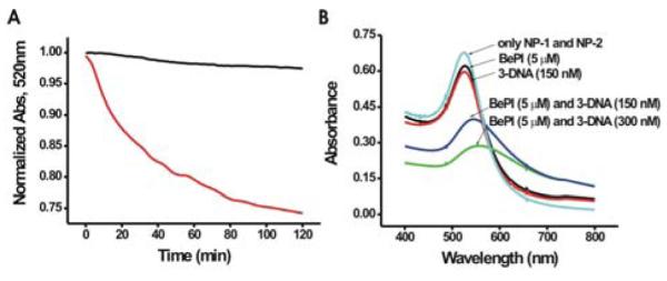
A) The hybridization kinetics monitored at 520 nm without stirring of NP-1 and NP-2 (1.5 nM each) in the presence DNA-3 (150 nM) (red) and DNA-3 + BePI (5 μM) (black). B) The UV spectrum of NP-1 and NP-2 after six hours incubation (1.5 nM each).
Figure 2.

Melting curves of A) NP-1, NP-2 and DNA-3 assemblies in the presence of DNA binders, B) DNA-1, DNA-2 and DNA-3 (no nanoparticles) in the presence of DNA binders.
To further investigate the importance of the triplex structure and rule out aggregation due to duplex formation between the nanoparticles, we performed the assay in the presence of seven duplex binders 4′,6-diamidino-2-phenylindole (DAPI), ellipticine (EIPT), amsacrine (AMSA), daunorubicin (DNR), anthraquinone-2-carboxylic acid (AQ2A), ethidium bromide (EB), and 9-aminoacridine (9-AA))(5 μM). Unlike with BePI and CORA, nanoparticle aggregation was not seen in the presence of the duplex binders. The absence of a melting transition in each sample containing a duplex DNA binder confirmed the absence of nanoparticle aggregates, Figure 2A. The results show that only BePI and CORA, triplex binders, can induce aggregation through triplex stabilization, thus leading to a screening process for triplex binding molecules. In addition, the sharp melting transition of the nanoparticle aggregates provides excellent differentiation between the melting temperatures, giving information about the relative binding strengths of the triplex binding molecules. The sample involving BePI, a strong triplex binding molecule, melts at a higher temperature than the sample with CORA which is a weaker triplex binding molecule. Incidentally, due to the similarities in the DNA sequence of NP1- and NP-2, similar results can be found in the absence of NP-2. This process does not detract from the results shown here (see Supporting Info).
To confirm the consistency of our results with more traditional screening techniques, we screened all nine DNA binders plus a control by measuring the melting properties (260 nm) of unmodified DNA-1, DNA-2, and DNA-3, with each DNA binder, Figure 2B (Supporting Info.). The melting experiments performed in the presence of the triplex binders, BePI and CORA, have two melting transitions (Figure 2B). The first extremely weak transitions (34.8 and 17.0 °C, respectively) are associated with the denaturation of the triplex structure and the second (61.4 and 60.1 °C, respectively) are representative of the corresponding duplex. None of the seven duplex binders or the control showed two melting transitions. This confirms that, of the DNA binders used here, only BePI and CORA are triplex binders. This is consistent with our results found using the gold nanoparticle colorimetric method described here. These results show dramatically improved differentiation between the triplex and duplex DNA binders as compared to the nanoparticle-free DNA measurements performed by monitoring the spectroscopic signature of DNA at 260 nm (compare Figure 2A and 2B, respectively). The enhanced differentiation is due to the intense optical properties of the nanoparticle probes as compared with the UV-Vis signature of DNA at 260 nm
In general, assay methods that can detect drug candidates by colorimetric changes are convenient, and for this reason, an assay that could screen for triplex binders would be of great interest. At present, there are no assays that provide this capability. The use of DNA functionalized Au nanoparticles for this purpose is demonstrated in Figure 3. The mixtures of NP-1, NP-2 and DNA-3 containing the control and duplex DNA binders remain red in color. Only the mixtures containing the triplex binders, BePI and CORA, turn blue/purple in color. This shows discrimination between triplex stabilizing and nonstabilizing binders by an easily identifiable color change. This result is consistent with the control experiments involving serial analysis of each DNA binder with nanoparticle-free triplex DNA.
Figure 3.

The color change of nanoassembly (NP-1 and NP-2, and DNA-3) in the absence and presence of DNA binders at room temperature.
In summary, we have developed a colorimetric assay which can screen for triplex binding molecules and simultaneously determine their relative binding capabilities by monitoring color change. This method allows dramatically improved discrimination between stabilizing and nonstabilizing triplex binders. The simplicity of this assay makes it more convenient than other methods such as competitive dialysis, mass spectroscopy, electrophoresis and UV/Vis melting experiments. In addition, this assay can be easily adapted to high-throughput screening methods, which can be used to determine potential triplex binders from large combinatorial libraries.
Supplementary Material
ACKNOWLEDGEMENTS
This work was supported by the NCI through a CCNE, the NSF, and the AFOSR. CAM is grateful for a Director’s Pioneer Award. MSH is grateful for a fellowship from the Korea Research Foundation. We thank Cole Krumbholz for graphical assistance.
Footnotes
SUPPORTING INFORMATION Assemblies formed using NP-1, nanoparticle-free DNA sequences.
REFERENCES
- 1 a).Buchini S, Leumann CJ. Curr. Opin. Chem. Biol. 2003;7:717. doi: 10.1016/j.cbpa.2003.10.007. [DOI] [PubMed] [Google Scholar]; b) Curcio LD, Bouffard DY, Scunlont KJ. Phamacol. Ther. 1997;74:317. doi: 10.1016/s0163-7258(97)00005-3. [DOI] [PubMed] [Google Scholar]; c) Giovannangeli C, Hélène C. Nature Biotech. 2000;18:1245. doi: 10.1038/82348. [DOI] [PubMed] [Google Scholar]; d) Karen M, Vasquez KM, Narayanan L, Glazer PM. Science. 2000;290:530. doi: 10.1126/science.290.5491.530. [DOI] [PubMed] [Google Scholar]
- 2.Fox KR. Curr. Med. Chem. 2000;7:17. doi: 10.2174/0929867003375506. [DOI] [PubMed] [Google Scholar]
- 3 a).Praseuth D, Guieysse AL, Hélène C. Biochim. Biophy. Acta. 1999;1489:181. doi: 10.1016/s0167-4781(99)00149-9. [DOI] [PubMed] [Google Scholar]; b) Haq I, Ladbury J. J. Mol. Recognit. 2000;13:188. doi: 10.1002/1099-1352(200007/08)13:4<188::AID-JMR503>3.0.CO;2-1. [DOI] [PubMed] [Google Scholar]
- 4 a).Nguyen CH, Marchand C, Delage S, Sun J-S, Garestier T, Hélène C, Bisagni E. J. Am. Chem. Soc. 1998;120:2501. [Google Scholar]; b) Escudé C, Nguyen CH, Mergny J-L, Sun J-S, Bisagni E, Garestier T, Hélène C. J. Am. Chem. Soc. 1995;117:10212. [Google Scholar]; c) Wilson WD, Tanious FA, Mizan S, Yao S, Kiselyov AS, Zon G, Strekowskit L. Biochemistry. 1993;32:10614. doi: 10.1021/bi00091a011. [DOI] [PubMed] [Google Scholar]; d) Heald RA, Modi C, Cookson JC, Hutchinson I, Laughton CA, Gowan SM, Kelland LR, Stevens MFG. J. Med. Chem. 2002;45:590. doi: 10.1021/jm011015q. [DOI] [PubMed] [Google Scholar]; e) Venitt S, Crofton-Sleigh C, Agbandje M, Jenkins TC, Neidle S. J. Med. Chem. 1998;41:3748. doi: 10.1021/jm980167r. [DOI] [PubMed] [Google Scholar]
- 5 a).Chaires JB, Ren J, Hamelberg D, Kumar A, Pandya V, Boykin DW, Wilson WD. J. Med. Chem. 2004;47:5729. doi: 10.1021/jm049491e. [DOI] [PubMed] [Google Scholar]; b) Rosu F, De Pauw E, Guittat L, Alberti P, Lacroix L, Mailliet P, Riou J-F, Mergny J-L. Biochemistry. 2003;42:10361. doi: 10.1021/bi034531m. [DOI] [PubMed] [Google Scholar]; c) Marchand C, Bailly C, Nguyen CH, Bisagni E, Garestier T, Hélène C, Waring MJ. Biochemistry. 1996;35:5022. doi: 10.1021/bi952908l. [DOI] [PubMed] [Google Scholar]; c) Silver GC, Sun J-S, Nguyen CH, Boutorine AS, Bisagni E, Hélène C. J. Am. Chem. Soc. 1997;119:263. doi: 10.1021/bc9600675. [DOI] [PubMed] [Google Scholar]
- 6 a).Thompson LA, Ellman JA. Chem. Rev. 1996;96:555. doi: 10.1021/cr9402081. [DOI] [PubMed] [Google Scholar]; b) Johnston PA, Johnston PA. Drug Discov. Today. 2002;6:353. doi: 10.1016/s1359-6446(01)02140-7. [DOI] [PubMed] [Google Scholar]; c) Boger DL, Desharnais J, Capps K. Angew. Chem. Int. Ed. Engl. 2003;42:4138. doi: 10.1002/anie.200300574. [DOI] [PubMed] [Google Scholar]
- 7.Mirkin CA, Letsinger RL, Mucic RC, Storhoff JJ. Nature. 1996;382:607. doi: 10.1038/382607a0. [DOI] [PubMed] [Google Scholar]
- 8.Lytton-Jean AKR, Mirkin CA. J. Am. Chem. Soc. 2005;127:12754. doi: 10.1021/ja052255o. [DOI] [PubMed] [Google Scholar]
- 9 a).Elghanian R, Storhoff JJ, Mucic RC, Letsinger RL, Mirkin CA. Science. 1997;277:1078. doi: 10.1126/science.277.5329.1078. [DOI] [PubMed] [Google Scholar]; b) He L, Musick MD, Nicewarner SR, Salinas FG, Benkovic SJ, Natan MJ, Keating CD. J. Am. Chem. Soc. 2000;122:9071. [Google Scholar]; c) Maxwell DJ, Taylor JR, Nie S. J. Am. Chem. Soc. 2002;124:9606. doi: 10.1021/ja025814p. [DOI] [PubMed] [Google Scholar]; d) Niazov T, Pavlov V, Xiao Y, Gill R, Willner I. Nano Lett. 2004;4:1683. [Google Scholar]; e) Pavlov V, Xiao Y, Shlyahovsky B, Willner I. J. Am. Chem. Soc. 2004;126:11768. doi: 10.1021/ja046970u. [DOI] [PubMed] [Google Scholar]; f) Liu J, Lu Y. J. Am. Chem. Soc. 2003;125:6642. doi: 10.1021/ja034775u. [DOI] [PubMed] [Google Scholar]
- 10.Sato K, Hosokawa K, Maeda M. J. Am. Chem. Soc. 2003;125:8102. doi: 10.1021/ja034876s. [DOI] [PubMed] [Google Scholar]
- 11.Mergny JL, Duval-Valentin G, Nguyen CH, Perrouault L, Faucon B, Rougée M, Montenay-Garestier T, Bisagni E, Hélène C. Science. 1992;256:1681. doi: 10.1126/science.256.5064.1681. [DOI] [PubMed] [Google Scholar]
- 12.Lee JS, Latimer LJP, Hampel KJ. Biochemistry. 1993;32:5591. doi: 10.1021/bi00072a014. [DOI] [PubMed] [Google Scholar]
Associated Data
This section collects any data citations, data availability statements, or supplementary materials included in this article.