Abstract
Purpose/Background:
Previous research studies by Bolga, Ayotte, and Distefano have examined the level of muscle recruitment of the gluteal muscles for various clinical exercises; however, there has been no cross comparison among the top exercises from each study. The purpose of this study is to compare top exercises from these studies as well as several other commonly performed clinical exercises to determine which exercises recruit the gluteal muscles, specifically the gluteus medius and maximus, most effectively.
Methods:
Twenty-six healthy subjects participated in this study. Surface EMG electrodes were placed on gluteus medius and maximus to measure muscle activity during 18 exercises. Maximal voluntary muscle contraction (MVIC) was established for each muscle group in order to express each exercise as a percentage of MVIC and allow standardized comparison across subjects. EMG data were analyzed using a root-mean-square algorithm and smoothed with a 50 millisecond time reference. Rank ordering of the exercises was performed utilizing the average percent MVIC peak activity for each exercise.
Results:
Twenty-four subjects satisfied all eligibility criteria and consented to participate in the research study. Five of the exercises produced greater than 70%MVIC of the gluteus medius muscle. In rank order from highest EMG value to lowest, these exercises were: side plank abduction with dominant leg on bottom (103%MVIC), side plank abduction with dominant leg on top (89%MVIC), single limb squat (82%MVIC), clamshell (hip clam) progression 4 (77%MVIC), and font plank with hip extension (75%MVIC). Five of the exercises recruited gluteus maximus with values greater than 70%MVIC. In rank order from highest EMG value to lowest, these exercises were: front plank with hip extension (106%MVIC), gluteal squeeze (81%MVIC), side plank abduction with dominant leg on top (73%MVIC), side plank abduction with dominant leg on bottom (71%MVIC), and single limb squat (71%MVIC). Four of the exercises produced greater than 70%MVIC for both gluteus maximus and medius muscles.
Conclusions:
Higher %MVIC values achieved during performance of exercises correlate to muscle hypertrophy.20,22 By knowing the %MVIC of the gluteal musculature that occurs during various exercises, potential for strengthening of the gluteal muscles can be inferred. Additionally, exercises may be rank ordered to appropriately challenge the gluteal musculature during rehabilitation.
Keywords: gluteus medius, gluteus maximus, muscle recruitment, rehabilitation exercise
INTRODUCTION
The lower extremity functions in a kinematic chain, leading many researchers in recent years to examine the mechanical effect of weak proximal musculature on more distal segments.1,2 Previous research by Distefano,3 Bolgla,4 and Ayotte5 has sought to determine the most appropriate exercises to strengthen the gluteal muscles due to their role in maintaining a level pelvis and preventing hip adduction and internal rotation during single limb support.1,6 Measurement of such femoral torsion and pelvic rotation in the transverse plane, along with measurement of pelvic tilt in the sagittal plane can indicate abnormal alignment of the hip joint.7 Numerous pathologies have been described which are related to the inability to maintain proper alignment of the pelvis and the femur, including: tibial stress fracture,8 low back pain,9,10 iliotibial band friction syndrome,1,11 anterior cruciate ligament injury,1,12 and patellofemoral pathology.2,13–17 While Distefano,3 Bolgla,4 and Ayotte5 have examined a wide range of exercises used to strengthen the hip musculature, to the knowledge of the authors, no cross comparison amongst the top exercises from each study has been performed.
Similar to Distefano,3 Ayotte,4 and Bolgla,5 exercises examined in the current study were rank ordered according to their recruitment of specific gluteal musculature and expressed as a percent of the subject's maximum volitional isometric contraction (MVIC). By knowing the approximate percentage of MVIC (%MVIC) recruitment of each of the gluteal muscles in a wide variety of exercises, the exercises may be ranked to appropriately challenge the gluteal musculature. MVIC was established in the standard manual muscle testing positions for gluteus medius and maximus, as described by Daniels and Worthingham.18 The use of the sidelying abduction position is supported by the results of Widler,19 where similarity in EMG activity for weight bearing and sidelying abduction (ICC's 0.880 and 0.902 for the respective positions) demonstrated that it is acceptable to use the MVIC value obtained during the standard manual muscle test position in order to establish a percentage MVIC for a weight bearing exercise.
Several previously published research articles helped to establish the parameters for determining a sufficient level of muscle activation for strength gains referenced in the current study. Anderson found that in order for strengthening adaptation to occur, muscle stimuli of at least 40-60% of a subject's MVIC must occur.20 When quantifying muscular strength, work by Visser correlates the use of a MVIC and a one-repetition maximum.21 In order to gain maximal muscular hypertrophy, Fry's work suggests an 80-95% of a subject's one repetition maximum must be achieved.22 Based on the work by Anderson,20 Visser,21 and Fry,22 for the purposes of this study, exercises producing greater than 70%MVIC were deemed acceptable for enhancement of strength.
Distefano examined electromyography (EMG) signal amplitude normalized values of gluteus medius and gluteus maximus muscles during exercises of varying difficulty in order to determine which exercises most effectively recruit these muscles.3 Rank order of exercises and %MVIC of Distefano's study can be viewed in Table 1. Of the top five exercises for the gluteus medius described by Distefano, the authors of the current study chose to reexamine sidelying hip abduction, single limb squat, and the single limb deadlift. Lateral band walk was not included in the current study as the researchers wished to only examine exercises that required no external resistance.
Table 1.
Findings of Distefano et al.3 Values are described as %MVIC, followed by rank in parentheses.
| Exercise Condition | Glut Max %MVIC (rank) | Glut Med %MVIC (rank) |
|---|---|---|
| Side-lying hip abd | 39 (6) | 81 (1) |
| Clam with 30 hip flex | 34 (10) | 40 (10) |
| Clam with 60 hip flex | 39 (6) | 38 (12) |
| Single-limb squat | 59 (1) | 64 (2) |
| Single-limb deadlift | 59 (1) | 58 (4) |
| Lateral band walk | 27 (12) | 61 (3) |
| Forward lunge | 44 (4) | 42 (9) |
| Sideways lunge | 41 (5) | 39 (11) |
| Transverse lunge | 49 (3) | 48 (6) |
| Forward hop | 35 (8) | 45 (8) |
| Sideways hop | 30 (11) | 57 (5) |
| Transverse hop | 35 (8) | 48 (6) |
Research by Bolgla and Uhl also examined the magnitude of hip abductor muscle activation during rehabilitative exercises.4 Their results may be viewed in Table 2. Of the exercises studied by Bolgla et al, the authors of the current study chose only to look at the pelvic drop and sidelying hip abduction. These two exercises were chosen since the primary intention of the current study was to compare an exercise's recruitment of the gluteal musculature, and not the activation effects of weight bearing versus non-weight bearing on the musculature.
Table 2.
Findings by Bolgla and Uhl,4 represented as %MVIC.
| Exercise condition | Glut Med % MVTC |
|---|---|
| Pelvic drop | 57 |
| WB with flexion left hip abd | 46 |
| WB left hip abd | 42 |
| NWB sidelying hip abd | 42 |
| NWB standing hip abd | 33 |
| NWB standing flexed hip abd | 28 |
*WB = Weight Bearing, NWB = Non-Weight Bearing, Abd = Abduction
Finally, Ayotte et al. used EMG to analyze lower extremity muscle activation of the pelvic stabilizers as well as the quadriceps complex during five unilateral weight bearing exercises,5 displayed in Table 3. The authors of the current study elected to forgo analyzing a single-limb wall squat and a single-limb mini-squat due to their similarity to the single-limb squat. Forward step-up and lateral step-up were included in the current analysis. The current study serves to compare top exercises from these previously published studies, as well as several other commonly performed clinical exercises in order to determine the exercises that are most effective at recruiting the gluteus maximus and medius.
Table 3.
Findings of Ayotte et al.5 Values are described as %MVIC, followed by rank in parentheses.
| Exercise condition | Glut Max %MVIC (rank) | Glut Med % MV1C (rank) |
|---|---|---|
| Wall squat | 86 (1) | 52 (1) |
| Mini-squat | 57 (4) | 36 (5) |
| Front step up | 74 (2) | 44 (2) |
| Lateral step up | 56 (5) | 38 (3) |
| Retro step up | 59 (3) | 37 (4) |
METHODS
Subjects
This study was approved by the Institutional Review Board of Belmont University. A total of 26 subjects were recruited from within the university and surrounding community through flyers and word of mouth. Healthy subjects who were able to perform exercise for approximately one hour were included in the study and reported to the laboratory for a single testing session. At this time they completed an informed consent form as well as a health history form and comprehensive lower quarter screen to identify exclusionary criteria. Pain when performing exercises, current symptoms of injury, history of ACL injury or any lower extremity surgery within past two years, and age of less than 21 years were criteriariteria for exclusion.
Testing Procedures
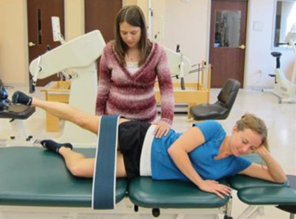
EMG data were collected and analyzed on the dominant leg, identified by which leg the subject used to kick a ball.3,5,23 Alcohol wipes were used to clean the skin over the gluteal region prior to electrode placement. Schiller Blue Surface electrodes (Schiller America Inc.; Doral, FL) were placed over the gluteus medius and gluteus maximus muscles of the subject's dominant side,4 per standard EMG protocol.24 In order to ensure consistent electrode placement throughout testing, electrodes were secured with surgical tape. Placement was confirmed by viewing EMG signals while separately activating each muscle. Subjects then performed a sub-maximal warm-up for five minutes on a stationary bicycle while watching a brief video of the exercises to be performed in order to familiarize subjects with exercise technique. A five-second MVIC was performed three times in the standard manual muscle testing protocol positions for each gluteal muscle18,19 with one minute of rest between each contraction. A strap was secured around the distal femur during muscle testing for both muscles to ensure standardization of resistance (Figure 1). Verbal encouragement was given with each trial.
Figure 1.
Maximum voluntary isometric contraction testing example set up.
Exercise order was randomized using a random pattern generator25 in order to avoid any order bias due to fatigue. Subjects were barefoot while performing exercises to prevent any potential variations that may have occurred due to footwear. Two minutes of rest was given between the performance of each exercise. Subjects performed eight repetitions of each exercise, three practice repetitions and five repetitions that were used for data collection. Exercises were performed to a metronome set at 60 beats per minute to standardize the rate of movement across subjects.
To replicate a clinical setting, researchers chose to use visual analysis of movement to ensure proper exercise technique rather than an electrogoniometer or movement analysis software since both of these procedures are unlikely to be available in a clinic. To ensure proper exercise technique, each subject was allowed three practice repetitions prior to data collection and any necessary verbal and tactile cues by the instructing researcher. A description of each exercise may be found in Appendix A. After completing all exercises, the subject's MVIC was reassessed to ensure electrodes had not been displaced during testing.
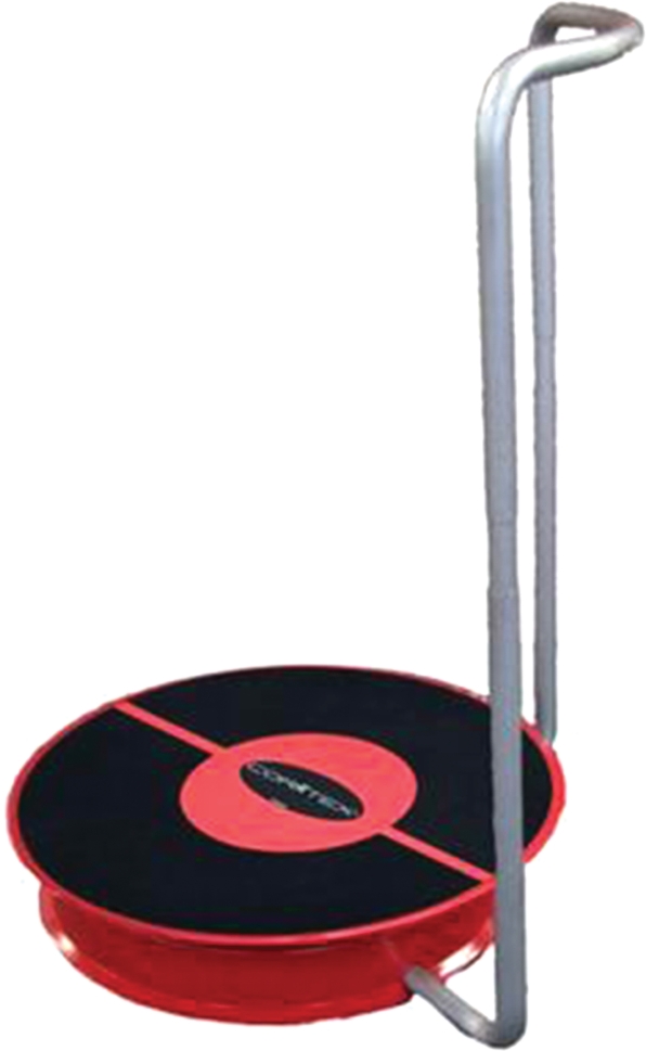
The equipment used for the conditions which required an unstable surface is the Core-Tex Balance Trainer™ (Performance Dynamics; San Diego, CA), a new piece of exercise equipment which is a platform mounted on a half-sphere atop a circular basin lined with ball bearings, creating an unstable and rapidly accelerating surface (Figure 2). The Core-Tex™ was developed to train a healthy fitness population; however, it may also be used to train individuals during rehabilitation in a clinical setting.
Figure 2.
CorTexTM equipment (Performance Dynamics, San Diego, CA).
Data Analysis
All data were rectified and smoothed using a root-mean-square algorithm, and smoothed with a 50 millisecond (msec) time reference. Peak amplitudes were averaged over a 100 msec window of time, 50 msec prior to peak and 50 msec after the peak.
To determine MVIC, the middle 3/5ths time for each manual muscle test trial was isolated and the peak value determined. The highest peak value out of the three trials was recorded and determined to be the MVIC.
In order to establish %MVIC for each exercise performed by an individual subject, data were collected for the last five repetitions of each exercise. If the EMG data were clearly cyclic, the middle three repetitions were analyzed. If it was difficult to determine when a repetition started and stopped on visual analysis of EMG data, then the middle 3/5ths of the total time to perform the five repetitions was analyzed. The highest peak out of the three repetitions was then divided by MVIC to yield %MVIC for that individual.
To determine %MVIC values for rank ordering of exercises, the %MVIC for each muscle was averaged between all subjects for each exercise.
RESULTS
Twenty-four subjects satisfied all eligibility criteria and consented to participate in the research study. Data from one subject were excluded due to faulty data from the EMG leads for both muscles, and data from another subject were excluded due to faulty data from the EMG lead for gluteus maximus only. There were a few other isolated instances of faulty data from EMG leads, in which case the subject's data were excluded from analysis for that specific exercise. The number of subjects included in data analysis for each exercise can be referenced in Tables 4 and 5. Due to the advanced level of some of the exercises included in the current study, such as single limb bridge on unstable surface and side plank, some subjects were unable to successfully complete all exercises. In these instances, subject data were not included in data analysis for that specific exercise. Peak amplitudes, expressed as %MVIC for gluteus medius and gluteus maximus, are rank ordered in Tables 4 and 5. Five of the exercises produced greater than 70%MVIC of the gluteus medius muscle. In rank order from highest EMG value to lowest, these exercises were: side plank abduction with dominant leg on bottom (103%MVIC), side plank abduction with dominant leg on top (89%MVIC), single limb squat (82%MVIC), clamshell (hip clam) progression 4 (77%MVIC), and font plank with hip extension (75%MVIC). Five of the exercises recruited gluteus maximus with values greater than 70%MVIC. In rank order from highest EMG value to lowest, these exercises were: front plank with hip extension (106%MVIC), gluteal squeeze (81%MVIC), side plank abduction with dominant leg on top (73%MVIC), side plank abduction with dominant leg on bottom (71%MVIC), and single limb squat (71%MVIC). Table 6 displays the exercises that produced greater than 70%MVIC for both gluteus medius and maximus muscles. These exercises included front plank with hip extension (75%MVIC, 106%MVIC), side plank abduction with dominant leg on top (89%MVIC, 73%MVIC), side plank abduction with dominant leg on bottom (103%MVIC, 71%MVIC), and single limb squat (82%MVIC, 71%MVIC) for gluteus medius and maximus respectively.
Table 4.
Results for Gluteus Medius recruitment, %MVIC and rank for all exercises.
| Exercise condition | # Subjects Included for analysis | %MVIC Gluteus Medius | Rank Gluteus Medius |
|---|---|---|---|
| Side plank abd, DL down | 21 | 103.11 | 1 |
| Side plank abd, DL up | 22 | 88.82 | 2 |
| Single limb squat | 22 | 82.26 | 3 |
| Clamshell (Hip Clam) 4 | 23 | 76.88 | 4 |
| Front plank with Hip Ext | 23 | 75.13 | 5 |
| Clamshell (Hip Clam) 3 | 22 | 67.63 | 6 |
| Side-lying abd | 23 | 62.91 | 7 |
| Clamshell (Hip Clam) 2 | 22 | 62.45 | 8 |
| Lateral step-up | 21 | 59.87 | 9 |
| Skater squat | 22 | 59.84 | 10 |
| Pelvic Drop | 23 | 58.43 | 11 |
| Hip circumduction, stable | 23 | 57.39 | 12 |
| Dynamic Leg Swing | 22 | 57.30 | 13 |
| Single limb deadlift | 22 | 56.08 | 14 |
| Single limb bridge, stable | 22 | 54.99 | 15 |
| Forward step-up | 22 | 54.62 | 16 |
| Single limb bridge, unstable | 20 | 47.29 | 17 |
| Clamshell (Hip Clam) 1 | 22 | 47.23 | 18 |
| Quadruped hip ext, DOM | 23 | 46.67 | 19 |
| Gluteal squeeze | 23 | 43.72 | 20 |
| Hip circumduction, unstable | 23 | 37.88 | 21 |
| Quadruped hip ext, non-DOM | 23 | 22.03 | 22 |
Table 5.
Results for Gluteus Medius recruitment, %MVIC and rank for all exercises.
| Exercise condition | # Subjects Included for analysis | %MVIC Gluteus Maximus | Rank Gluteus Maximus |
|---|---|---|---|
| Front plank with Hip Ext | 22 | 106.22 | 1 |
| Gluteal squeeze | 22 | 80.72 | 2 |
| Side plank abd, DL up | 22 | 72.87 | 3 |
| Side plank abd, DL down | 21 | 70.96 | 4 |
| Single limb squat | 22 | 70.74 | 5 |
| Skater squat | 21 | 66.18 | 6 |
| Lateral step-up | 20 | 63.83 | 7 |
| Quadruped hip ext, DOM | 22 | 59.70 | 8 |
| Single limb deadlift | 21 | 58.84 | 9 |
| Forward step-up | 22 | 54.67 | 10 |
| Single limb bridge, stable | 21 | 54.24 | 11 |
| Clamshell (Hip Clam) 1 | 22 | 53.10 | 12 |
| Side-lying abd | 22 | 51.13 | 13 |
| Single limb bridge, unstable | 18 | 49.35 | 14 |
| Hip circumduction, stable | 22 | 37.85 | 15 |
| Dynamic leg swing | 22 | 33.65 | 16 |
| Hip circumduction, unstable | 22 | 28.87 | 17 |
| Clamshell (Hip Clam) 3 | 22 | 26.63 | 18 |
| Clamshell (Hip Clam) 4 | 22 | 26.22 | 19 |
| Pelvic Drop | 22 | 25.10 | 20 |
| Quadruped hip ext, non-DOM | 22 | 21.04 | 21 |
| Clamshell (Hip Clam) 2 | 22 | 12.36 | 22 |
Table 6.
Top exercises for muscle activation of both gluteus medius and gluteus maximus (>70% MVIC).
| Exercise condition | %MVIC Gluteus Medius | %MVIC Gluteus Maximus |
|---|---|---|
| Front plank with Hip Ext | 75.13 | 106.22 |
| Side plank abd, DL up | 88.82 | 72.87 |
| Side plank abd, DL down | 103.11 | 70.96 |
| Single limb squat | 82.26 | 70.74 |
DISCUSSION
The main objective of this study was to examine muscle activity during common clinical exercises used to strengthen the gluteus medius and gluteus maximus muscles. This study sought to analyze and compare information reported in previous studies by Distefano, Bolga, and Ayotte regarding ranking of various therapeutic exercises using %MVIC. The secondary objective was to describe %MVIC for other commonly used therapeutic exercises not previously reported upon. The authors of this study chose to examine peak amplitude averaged over a 100 ms window, 50 ms prior to peak and 50 ms after the peak, during repetitions five, six and seven, the highest of which was converted to %MVIC. This methodology is similar to studies by both Distefano3 and Bolgla.4 Ayotte et al. averaged EMG activity over a 1.5 sec window during the concentric phase of each exercise.5 Due to slight differences in data collection and data analysis between the current study, and studies conducted by Distefano, Bolgla and Ayotte, interpretation of results and similarities across studies will predominantly address the sequence of rank order as opposed to absolute values for the %MVIC.3–5
There were two exercises where %MVIC was found to be higher than MVIC, side plank abduction with dominant leg down (103%MVIC) for gluteus medius and front plank with hip extension (106%MVIC) for gluteus maximus. There are several possibilities as to why these findings may have occurred. One possibility is that subjects lacked sufficient motivation to perform a true maximal contraction during MVIC testing, despite the fact that verbal encouragement was given to all subjects during max testing of both muscles. Another possibility is that subjects were not able to truly give a maximum effort during the manual muscle test. Authors of previous research have reported that in order to obtain a true maximum contraction, it is necessary to superimpose an interpolated twitch, which is an electrically stimulated contraction, on top of the maximum voluntary contraction.26 Current research in electrophysiology is further examining this phenomenon with mixed results regarding sensitivity of various interpolated twitch techniques, differences in methodology, and interpretation of their results.27–29 Future researchers using MVIC for standardization across subjects should follow this research closely in order to ensure the most accurate methodology is used for establishing maximal voluntary muscle contractions. A final possibility is that with these exercises there was substantial co-contraction of the core musculature, which may have led to higher values than could be obtained during isolated volitional contraction. In the MMT positions used to establish MVIC the pelvis is stabilized against the surface of the table with relatively isolated muscle recruitment. In both of the above mentioned exercises, the pelvis does not have external support and higher EMG values could reflect increased activity due to an increased need for stabilization resulting in synergistic co-contraction. Future research may need to examine differences in muscle recruitment and activation patterns in exercises that test isolated muscle function versus ones that require core stabilization resulting in co-contraction.
Gluteus Medius
Table 7 depicts the top gluteus medius exercises determined by the authors of the current study as referenced to the exercises examined in studies performed by Distefano,3 Bolgla,4 and Ayotte.5 The authors of the current study found highest %MVIC peak values for side plank abduction with dominant leg on bottom (103%MVIC), side plank abduction with dominant leg on top (89%MVIC), single limb squat (82%MVIC), clamshell progression 4 (77%MVIC), and front plank (75%MVIC) as outlined in Table 7. Four of the top five exercises were not previously examined by Distefano,3 Bolga,4 or Ayotte.5 All of these exercises exhibited greater than 70%MVIC, the peak amplitude necessary for enhancement of strength, suggesting they may have benefits for gluteus medius strengthening. However, these exercises are all very challenging and would not be appropriate for initial strengthening in patients with weak core musculature due to their high degree of difficulty and the amount of core stabilization required. The possible exception may be clamshell progression 4, due to the stabilization provided to the subject when lying on the floor to perform the exercise. While the top exercises in this study produced the greatest peak amplitude EMG values, it is also important to consider functional demands and dosage when selecting an exercise for muscle training and strengthening, especially in early stages of rehabilitation of a weak or under-recruited muscle.
Table 7.
Comparison of rank order of exercises for recruitment of gluteus medius between the current study and Distefano,3 Bolgla,4 and Ayotte,5 using %MVIC
| Exercise condition | Current Study | Distefano3 | Bolgla4 | Ayotte5 | |
|---|---|---|---|---|---|
| 1 | Side plank abd, DL down | 103 | |||
| 2 | Side plank abd, DL up | 89 | |||
| 3 | Single Limb Squat | 82 | 64 | 52* | |
| 4 | Clamshell (Hip Clam) 4 | 77 | |||
| 5 | Front plank with Hip Ext | 75 | |||
| 7 | Side-lying abd | 63 | 81 | 42 | |
| 9 | Lateral step-up | 60 | 38 | ||
| 11 | Pelvic Drop | 58 | 57 | ||
| 14 | Single limb deadlift | 56 | 58 | ||
| 16 | Forward step-up | 55 | 44 | ||
| 18 | Clamshell (Hip Clam) 1 | 47 | 40 |
Single-limb wall squat
The top gluteus medius exercises from Distefano's study were sidelying hip abduction (81%MVIC), single-limb squat (64%MVIC), and single limb dead lift (58%MVIC).3 With the exception of single limb squat, the current study found similar rank order with values of 63%MVIC, 82%MVIC, and 56%MVIC respectively. Of note, Distefano's subjects performed the single limb squat to a predetermined knee flexion angle of approximately 30 degrees,3 while the current study had the subjects perform the exercise to a predetermined chair height of 47 cm. This difference in methodology may account for the difference in findings across the two studies. The methodology used by Distefano may allow for greater normalization, as squatting to a predetermined knee flexion angle allows for equal challenge to all subjects, where as squatting to a predetermined height creates a greater challenge for taller subjects.
Bolga's top exercise for gluteus medius was the pelvic drop (57%MVIC).4 The current study found a similar value at 58%MVIC, although this exercise was ranked 11th out of the 22 exercises evaluated. This exercise should not be discounted; however, as it is a functional training exercise for pelvic stabilization in single limb stance, and many gait abnormalities and lower extremity pathologies are the result of the gluteus medius muscle's inability to properly and effectively stabilize the pelvis during single limb stance.
Bolga found sidelying abduction to have a value of 42%MVIC,4 which is significantly lower than the findings in either the Distefano3 or the current study. In general, qualitative movement analysis during performance of sidelying abduction reveals poor technique with frequent substitution using the tensor fascia lata muscle demonstrated through increased hip flexion during abduction, which may have accounted for the low value found in the Bolga study.6 Furthermore, subjects in both the Distefano and the Bolgla study maintained the bottom leg in neutral hip extension and knee extension,3,4 while subjects in the current study were allowed to flex the bottom hip and knee in order to provide greater support and stabilization during abduction of the top leg.
Ayotte's top exercise was the unilateral wall squat (52%MVIC),5 which is comparable to the single limb squat, ranking in the top three exercises in both the current study and in Distefano's study,3 although the external stabilization provided in the unilateral wall squat should be considered. Ayotte ranked forward step-up (44%MVIC) higher than lateral step-up (38%MVIC),5 whereas the authors of the current study ranked lateral step-up (60%MVIC) higher than forward step-up (55%MVIC). It should be noted that subjects were allowed upper extremity external support during the exercise in Ayotte's study which may account for these differences,5 along with differences in data analysis described previously.
Gluteus Maximus
Table 8 depicts the top exercises for gluteus maximus of the current study. These include front plank with hip extension (106%MVIC), gluteal squeeze (81%MVIC), side plank abduction with dominant leg on top (73%MVIC), side plank abduction with dominant leg on bottom (71%MVIC), and single limb squat (71%MVIC). The top four exercises from the current study were not performed in other studies. Bolgla's study did not include assessment of performance of the gluteus maximus so will not be included in the discussion below.4
Table 8.
Comparison of rank order of exercises for recruitment of gluteus maximus between the current study and Distefano,3 and Ayotte,5 using %MVIC.
| Exercise condition | Current Study | Distefano3 | Ayotte5 | |
|---|---|---|---|---|
| 1 | Front plank with Hip Ext | 106 | ||
| 2 | Gluteal squeeze | 81 | ||
| 3 | Side plank abd, DL up | 73 | ||
| 4 | Side plank abd, DL down | 71 | ||
| 5 | Single limb squat | 71 | 59 | 86* |
| 7 | Lateral step-up | 64 | 56 | |
| 9 | Single limb deadlift | 59 | 59 | |
| 10 | Forward step-up | 55 | 74 | |
| 12 | Clamshell (Hip Clam) 1 | 53 | 34 | |
| 13 | Side-lying abd | 51 | 39 |
Single-limb squat
Distefano's top exercises were single limb squat (59%MVIC), single limb dead lift (59%MVIC), and sidelying hip abduction (39%MVIC).3 Subjects performing these same exercises in the current study produced results of 71%MVIC, 59%MVIC, and 51%MVIC, respectively, demonstrating the same rank order of muscle activity as these exercises in the Distefano study.3 The only differences in rank ordering between the current study and Distefano's for gluteus maximus were between clamshell progression 1 and sidelying abduction;3 however, within each study there was less than 5%MVIC difference for each exercise when determining rank order (Table 8). As previously noted, differences in technique and substitution are common occurrences during the performance of sidelying abduction which may account for the differences found between the two studies.
Ayotte ranked forward step-up (74%MVIC) higher than lateral step-up (56%MVIC),5 whereas the current study ranked lateral step-up (64%MVIC) higher than forward step-up (55%MVIC). Again, differences could be attributed to variances in technique or the ability of subjects in Ayotte's study to use external upper extremity support5 as well as differences in data analysis.
The low ranking for stable single limb bridge (11th) and unstable single limb bridge (14th) was somewhat surprising as both are common exercises used clinically for gluteus maximus strengthening. There were several instances of subjects reporting hamstring cramping during bridging on the unstable surface, which led the researchers to suspect substitution with the hamstrings during this exercise. The same may hold true for bridging on the stable surface, however there were fewer complaints. Future studies should examine muscle recruitment and activation patterns of gluteus maximus and the hamstrings during various bridging activities.
The effect of a subject's attention to volitional contraction of a muscle during an exercise should also be considered. The gluteal squeeze was the only exercise where verbal cues were explicitly given to maximally contract the gluteal muscles while performing the exercise, which could possibly have contributed to its high ranking for performance by the gluteus maximus. Future research should examine the difference in amount, if any, noted in muscle recruitment when verbal instructions are given to concentrate on the muscle contraction while performing the exercise versus no verbal instructions during performance. The effects of tone of voice, volume of cues, and frequency of verbal cueing are unknown.
CONCLUSION
Anderson and Fry have previously reported that higher %MVIC values with exercises correlate to muscle hypertrophy.20,22 By knowing the %MVIC of the gluteus maximus and medius that occurs during various exercises, the potential for strengthening these muscles can be inferred. Subsequently, exercises may be ranked to appropriately challenge the gluteus maximus and medius during rehabilitation. The authors of the current study found patterns within their results consistent with previous research published by Distefano and Bolgla.3,4 The authors conclude that differences in data collection and analysis as well as the use of external upper extremity support may have accounted for the differences noted between the current study and the study by Ayotte.5 One of the purposes of the current study was to provide a rank ordered list of exercises for the recruitment of the gluteus maximus and medius. These rank ordered lists may help form the basis for a graded rehabilitation program. For patients early in the rehabilitation process, the clinician should systematically determine which muscle they are wishing to strengthen and use less difficult (lower %MVIC) exercises. In order to maximally challenge a patient's gluteus maximus and medius, the authors recommend using a front plank with hip extension, a single limb squat, and a side plank on either extremity with hip abduction.
APPENDIX A
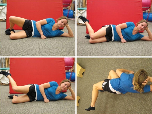
- Clamshell (hip clam) Progression: Each exercise is performed with the subject sidelying on the non-dominant side. (Figure 3)
- Progression 1 (upper left): Start position is sidelying with hips flexed to approximately 45 degrees, knees flexed, and feet together. Subject externally rotates the top hip to bring the knees apart for one metronome beat and returns to start position during the next beat.
- Progression 2 (upper right): Start position identical to progression 1; however, in this progression subject keeps the knees together while internally rotating the top hip to lift the top foot away from the bottom foot for one metronome beat, returning to the start position during the next beat
- Progression 3 (lower left): The subject is positioned identical to progressions 1 and 2, but with the top leg raised parallel to the ground. The subject maintains the height of the knee while internally rotating at the hip by bringing the foot toward the ceiling for one beat and then returns to the start position during the next beat.
- Progression 4 (lower right): The subject is positioned the same as progression 3, but with the hip fully extended. As in progression 3, the subject maintains the height of the knee and internally rotates at the hip by bringing the foot toward the ceiling for one beat and returns to the start position with knee and ankle in line during the next beat.
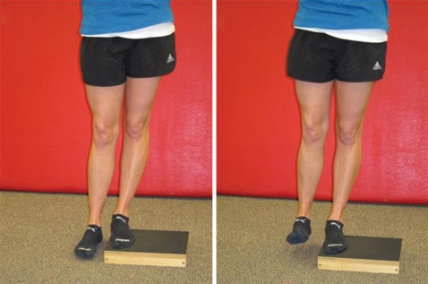
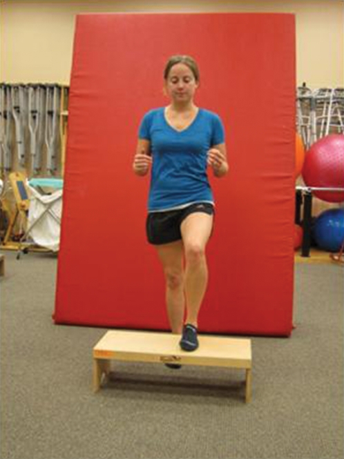
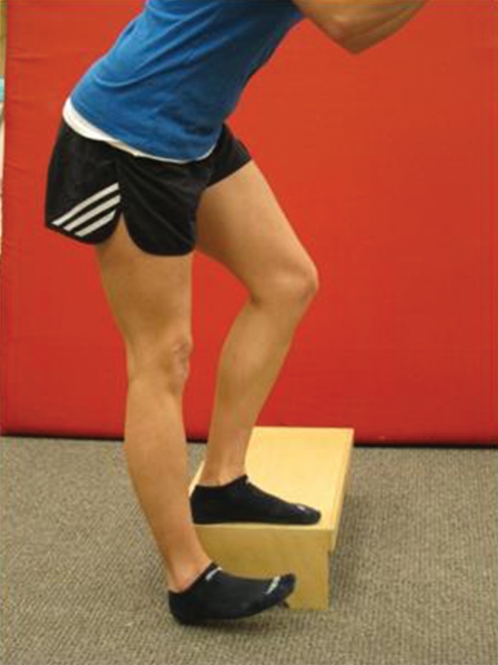
Pelvic drop: Subject stands with dominant leg on the edge of a 5 cm box (right), and then lowers the heel of the non-dominant leg to touch the ground without bearing weight, for one beat (left). Subject returns foot to the height of the box while keeping the hips and knees extended for one beat. (Figure 4)
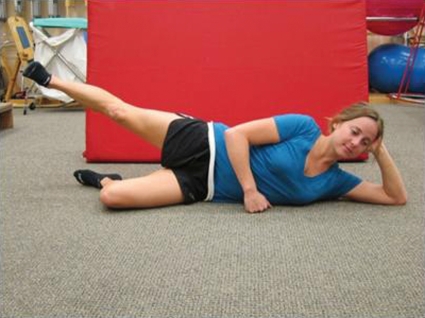
Sidelying abduction: Start with subject sidelying on non-dominant side. Subject flexes the hip and knee of the support side and then abducts the dominant leg to approximately 30 degrees while maintaining neutral or slight hip extension and knee extension with the toes pointed forward for a count of two beats up and two beats down. (Figure 5)
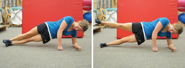
Side Plank with Abduction, dominant leg up: (Start with subject in a side plank position with dominant leg up. Subject is instructed to keep shoulders, hips, knees, and ankles in line bilaterally, and then to rise to plank position with hips lifted off ground to achieve neutral alignment of trunk, hips, and knees. The subject is allowed upper extremity support as seen on left. While balancing on elbows and feet, the subject raises the top leg into abduction (right) for one beat and then lowers leg for one beat. Subject maintains plank position throughout all repetitions (Figure 6).
Side Plank with Abduction, dominant leg down: Exercise position is identical to Exercise 4 except on the opposite side. Subject is instructed to abduct the non-dominant uppermost leg for two beats and lowers leg for two beats. Subject maintains plank position throughout all repetitions.
Front Plank with Hip Extension: Start with subject prone on elbows in plank with trunk, hips, and knees in neutral alignment (left). Subject lifts the dominant leg off of the ground, flexes the knee of the dominant leg, and extends the hip past neutral hip alignment by bringing the heel toward the ceiling (right) for one beat and then returns to parallel for one beat. (Figure 7)
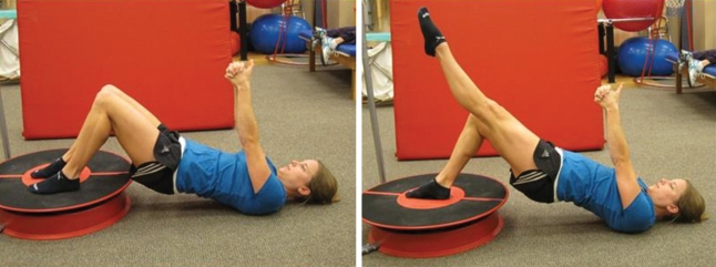
Single Limb Bridging on Stable Surface: Start with subject in hook-lying position (left). The subject is instructed to bridge on both legs by keeping the feet on the floor and raising hips off the ground to achieve neutral trunk, hip, and knee alignment for one beat. From this position, the subject extends the knee of the non-dominant leg to full knee extension while keeping the femurs parallel (right) for one beat, returns the non-dominant leg to the bridge position for one beat, and then lowers the body back to the ground for one beat (Figure 8).
Single Limb Bridge on Unstable Surface: Subject is positioned as in Exercise 7 and places the dominant foot in the center of the Core-Tex™ (left). The subject performs the same sequence as above (right) while maintaining the disc of the Core-Tex™ in the center. (Figure 9)
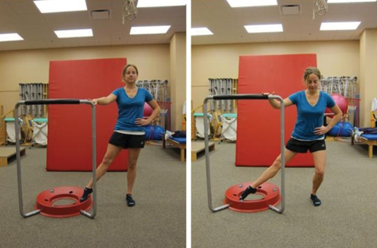
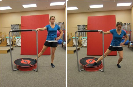
Hip Circumduction on Stable Surface: The subject places the non-dominant leg on the outside of the base of the Core-Tex™ and stands to the side of the Core-Tex™ on the dominant leg (left). The subject performs a single limb squat while tracing the toe of the non-dominant leg on the outside of the Core-Tex™ base (right) in an arc for three beats, then traces the toe back to the start, while returning to a standing position for three beats. Subjects were allowed two-finger unilateral upper extremity support on the frame of the Core-Tex™ for balance assist. (Figure 10).
Hip Circumduction on Unstable Surface: In standing, the subject places the non-dominant foot on the outer edge of the Core-Tex™ and stands to the side of the Core-Tex™ on the dominant leg (left). The subject then performs a single limb squat on the dominant leg while drawing an arc with the non-dominant foot, extending the arc away from the subject for three beats (right). The subject then returns the foot to the starting position by drawing the foot in, while returning to a standing position for three beats. Subjects were allowed upper extremity support as in Exercise 9. (Figure 11)
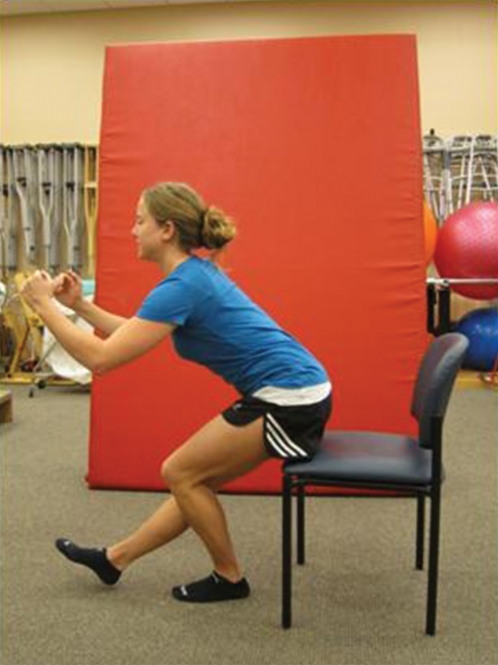
Single Limb Squat: Subject stands on the dominant leg, slowly lowering the buttocks to touch a chair 47cm in height for two beats and then extends back to standing for two beats. (Figure 12)
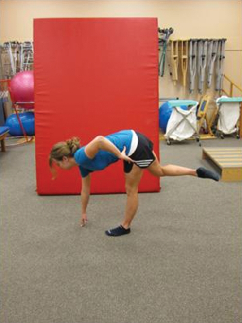
Single Limb Deadlift: Subject stands on the dominant leg and slowly flexes at the hip, keeping the back straight, to touch the floor with the opposite hand for two beats. Subject then extends at the hip to standing for two beats. Subjects were permitted to have knees either straight or slightly bent in the case that hamstring tightness limited subject's ability to touch the floor. (Figure 13)
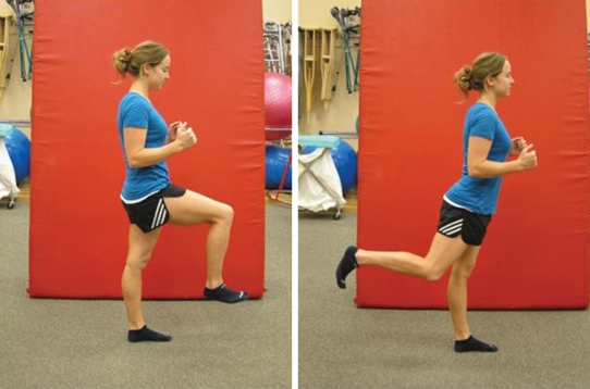
Dynamic Leg Swing: Subject is positioned in standing on the dominant leg, and then begins to swing the non-dominant leg (with the knee flexed) into hip flexion (left) and extension (right) at a rate of one beat forward and one beat backward. Subjects were instructed to move through a smooth range of hip motion and to not allow their trunk to move out of the upright position. (Figure 14)
Forward Step-up: Beginning with both feet on the ground, subject steps forward onto a 20cm step with the dominant leg for one beat. Subject then steps up with the non-dominant leg during the next beat. Subject then lowers the non-dominant leg back to the ground for one beat followed by the dominant leg during the next beat. (Figure 15)
Lateral Step-up: Subject stands on the edge of a 15cm box on the dominant leg and squats slowly to lower the heel of the non-dominant leg toward floor for one beat and then returns to start position during the next beat. (Figure 16)
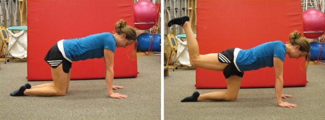
Quadruped Hip Extension: In quadruped (left) the subject extends the dominant leg at the hip, while keeping the knee flexed 90 degrees, to lift the foot toward the ceiling (right) to achieve neutral hip extension for two beats and then returns the dominant leg to the start position for two beats. This exercise was repeated with the non-dominant leg and EMG values were recorded in order to measure activity as both the stabilizing and moving leg. (Figure 17)
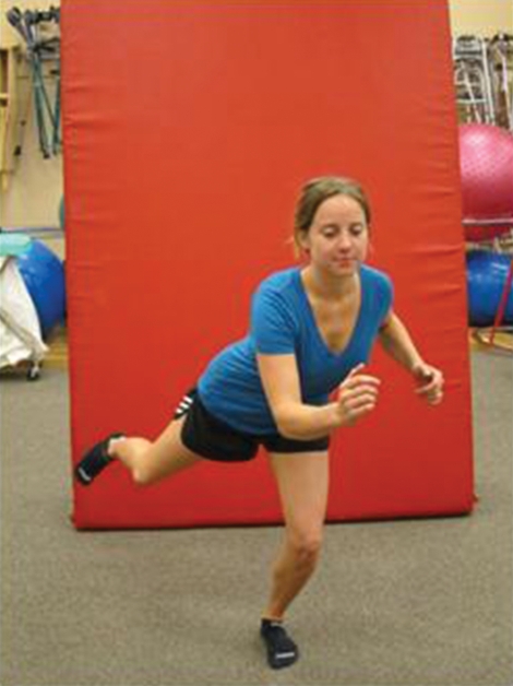
Skater Squat: Subject stands on the dominant leg and performs a squat to a comfortable knee flexion angle for two beats down and two beats up with non-dominant leg extended at the hip and flexed at the knee. The torso twists during the squat. The toe of the non-dominant leg was permitted to touch the ground between repetitions. (Figure 18)
Gluteal Squeeze: In standing with feet shoulder-width apart, subject squeezes gluteal muscles for two beats and then relaxes for two beats. Subjects were instructed to maximally contract the gluteal musculature during the exercise.
Figure 3.
Figure 4.
Figure 5.
Figure 6.
Figure 7.
Figure 8.
Figure 9.
Figure 10.
Figure 11.
Figure 12.
Figure 13.
Figure 14.
Figure 15.
Figure 16.
Figure 17.
Figure 18.
REFERENCES
- 1. Leetun D, Ireland M, Wilison J, et al. Core Stability Measures as Risk Factors for Lower Extremity Injury in Athletes. Med Sci Sports Exercise. 2004; 36: 926–934 [DOI] [PubMed] [Google Scholar]
- 2. Souza R, Powers C. Differences in Hip Kinematics, Muscle Strength, and Muscle Activation Between Subjects With and Without Patellofemoral Pain. J Orthop Sports Phys Ther. 2009; 39: 12–19 [DOI] [PubMed] [Google Scholar]
- 3. Distefano L, Blackburn J, Marshall S, et al. Gluteal Activation During Common Therapeutic Exercises. J Orthop Sports Phys Ther. 2009; 39: 532–540 [DOI] [PubMed] [Google Scholar]
- 4. Bolgla L, Uhl T. Electromyographic Analysis of Hip Rehabilitation Exercises in a Group of Healthy Subjects. J Orthop Sports Phyl Ther. 2005; 35: 488–494 [DOI] [PubMed] [Google Scholar]
- 5. Ayotte N, Stetts D, Keenan G, et al. Electromyographical Analysis of Selected Lower Extremity Muscles During 5 Unilateral Weight-Bearing Exercises. J Orthop Sports Phys Ther. 2007; 37: 48–55 [DOI] [PubMed] [Google Scholar]
- 6. Grimaldi A. Assessing Lateral Stability of the Hip and Pelvis. Manual Therapy. 2010, Pages 1–7 [DOI] [PubMed] [Google Scholar]
- 7. Levangie P. The Hip complex. In: Levangie P, Norkin C. Joint Structure and Function: A Comprehensive Analysis, 4th ed. Philadelphia, PA: F.A. Davis Company; 2005: 368–370 [Google Scholar]
- 8. Milner C, Hamill J, Davis I. Distinct Hip and Rearfoot Kinematics in Female Runners With a History of Tibial Stress Fracture. J Orthop Sports Phys Ther. 2010; 40: 59–66 [DOI] [PubMed] [Google Scholar]
- 9. Nelson-Wong E, Flynn T, Callaghan J. Development of Active Hip Abduction as a Screening Test for Identifying Occupational Low Back Pain. J Orthop Sports Phys Ther. 2009; 39: 649–657 [DOI] [PubMed] [Google Scholar]
- 10. Nelson-Wong E, Gregory D, Winter D, et al. Gluteus Medius Muscle Activation Patterns as a Predictor of Low Back Pain During Standing. Clin Bio. 2008; 23: 545–553 [DOI] [PubMed] [Google Scholar]
- 11. Ferber R, Noehren B, Hamill J, et al. Competitive Female Runners With a History of Iliotibial Band Syndrome Demonstrate Atypical Hip and Knee Kinematics. J Orthop Phys Ther. 2010; 40: 52–58 [DOI] [PubMed] [Google Scholar]
- 12. Hewett T, Myer G, Ford K, Anterior Cruciate Ligament Injuries in Female Athletes: Part 1, Mechanisms and Risk Factors. Am J Sports Med. 2006; 34: 299–311 [DOI] [PubMed] [Google Scholar]
- 13. Magalhaes E, Fukuda T, Sacramento S, et al. A Comparison of Hip Strength Between Sedentary Females With and Without Patellofemoral Pain Syndrome. J Orthop Sports Phys Ther. 2010; 40: 641–655 [DOI] [PubMed] [Google Scholar]
- 14. Souza R, Draper C, Fredericson M, et al. Femur Rotation and Patellofemoral Joint Kinematics: A Weight- Bearing Magnetic Resonance Imaging Analysis. J Orthop Sports Phys Ther. 2010; 40: 277–285 [DOI] [PubMed] [Google Scholar]
- 15. McKenzie K, Galea V, Wessel J, et al. Lower Extremity Kinematics of Females With Patellofemoral Pain Syndrome While Stair Stepping. J OrthopSports Phys Ther 2010; 40: 625–640 [DOI] [PubMed] [Google Scholar]
- 16. Fukuda T, Rossetto F, Magalhaes E, et al. Short-Term Effects of Hip Abductors and Lateral Rotators Strengthening in Females With Patellofemoral Pain Syndrome: A Randomized Controlled Clinical Trial. J Orthop Sports Phys Ther. 2010; 40: 736–742 [DOI] [PubMed] [Google Scholar]
- 17. Ireland M, Wilson J, Bellantyne B, et al. Hip Strength in Females With and Without Patellofemoral Pain. J Orthop Sports Phys Ther. 2003; 33: 671–676 [DOI] [PubMed] [Google Scholar]
- 18. Hislop H, Montgomery J. Daniels and Worthingham's Muscle Testing: Techniques of Manual Examination. St. Louis, MO; Elsevier Saunders; 2007 [Google Scholar]
- 19. Widler K, Glatthorn J, Bizzini M, et al. Assessment of Hip Abductor Muscle Strength. A Validity and Reliability Study. J Bone Joint Surg Am. 2009; 91: 2666–2672 [DOI] [PubMed] [Google Scholar]
- 20. Anderson L, Magnusson S, Nielsen M, et al. Neuromuscular Activation in Conventional Therapeutic Exercises and Heavy Resistance Exercises: Implications for Rehabilitation. Phys Ther. 2006; 86: 683–697 [PubMed] [Google Scholar]
- 21. Visser J, Mans E, van den Berg-Vos RM, et al. Comparison of Maximal Voluntary Isometric Contraction and Hand-held Dynamometry in Measuring Muscle Strength of Patients with Progressive Lower Motor Neuron Syndrome. Neuromuscul Disord. 2003; 13: 744–750 [DOI] [PubMed] [Google Scholar]
- 22. Fry A. The Role of Resistance Exercise Intensity on Muscle Fibre Adaptations. Sports Med. 2004; 34: Pages 663–679 [DOI] [PubMed] [Google Scholar]
- 23. Reimer R, Wikstrom E. Functional Fatigue of the Hip and Ankle Musculature Cause Similar Alterations In Single Leg Stance. J SMS. 2010; 13: 161–166 [DOI] [PubMed] [Google Scholar]
- 24.http://www.noraxon.com/products/accessories/bluesensor.php3 http://www.noraxon.com/products/accessories/bluesensor.php3
- 25.http://www.psychicscience.org/random http://www.psychicscience.org/random
- 26. Dowling J, Konert E, Ljucovic P, et al. Are Humans Able to Voluntarily Elicit Maximum Muscle Force. Neurosci Lett. 1994; 179: 25–28 [DOI] [PubMed] [Google Scholar]
- 27. Berger M, Watson B, Doherty T. Effect of maximal voluntary contraction on the amplitude of the compound muscle action potential: implications for the interpolated twitch technique. Muscle Nerve. 2010; 42: 498–503 [DOI] [PubMed] [Google Scholar]
- 28. Folland J, Williams A. Methodological Issues with the Interpolated Twitch Technique. J Electro Kinesiology. 2007; 17: 317–327 [DOI] [PubMed] [Google Scholar]
- 29. Shield A, Zhou S. Assessing Voluntary Muscle Activation with The Twitch Interpolation Technique. Sports Med. 2004; 34: 253–267 [DOI] [PubMed] [Google Scholar]


